
Article Figures & Data
Figures
- FIG 1.
Flow chart.
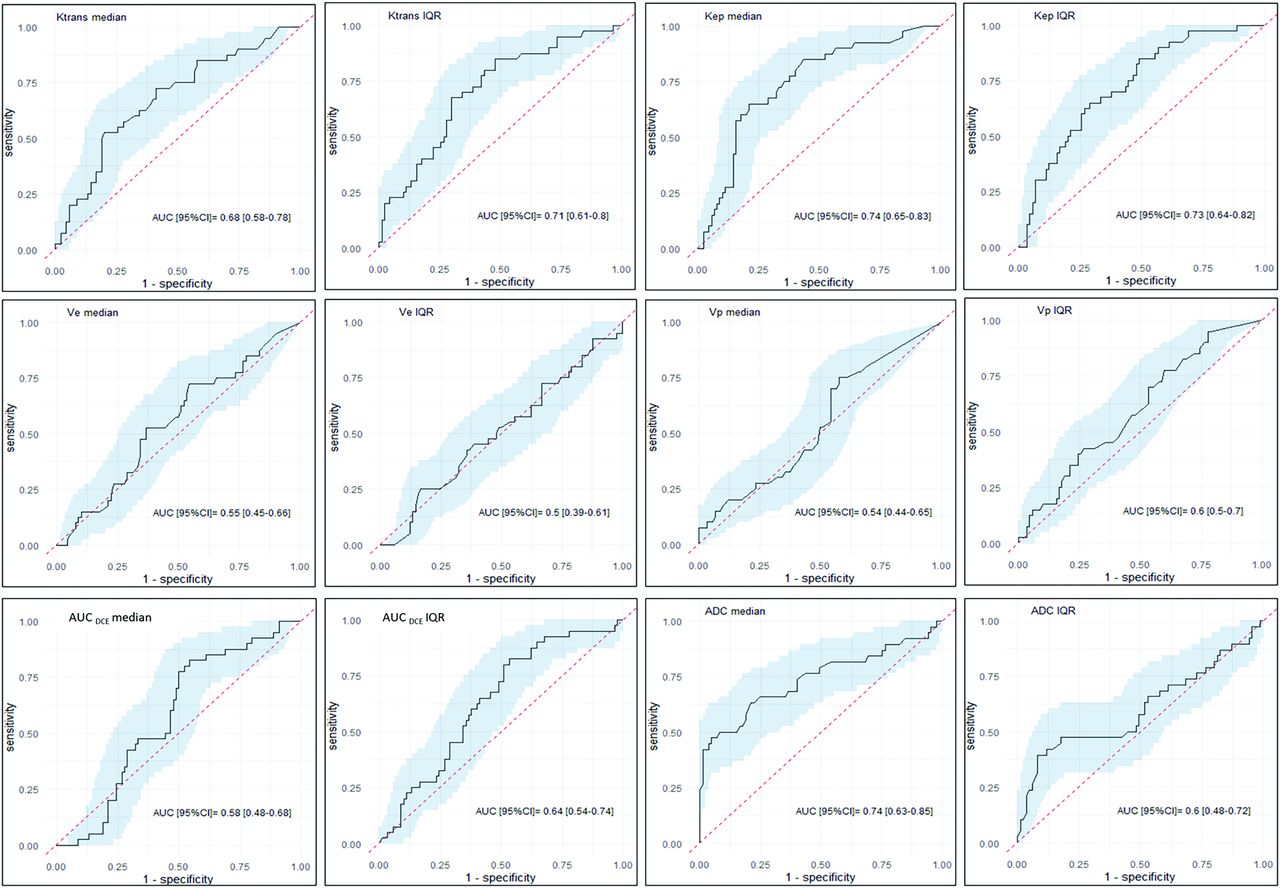
- FIG 2.
ROC curves of each DCE and ADC parameter when distinguishing malignant from benign lesions. The x-axis refers to 1-specificity, and the y-axis refers to the sensitivity. The black line represents the AUC; the blue area, the 95% confidence interval; and the dotted red line, the diagonal, ie, an AUC of 0.5.
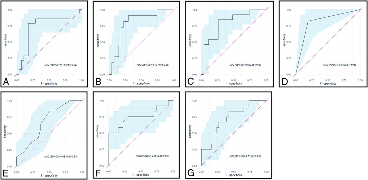
- FIG 3.
ROC curves of each model when distinguishing malignant from benign lesions. The x-axis refers to 1-specificity; the y-axis, to the sensitivity. The black line represents the AUC; the blue area, the 95% confidence interval; and the dotted red line, the diagonal, ie, an AUC of 0.5. A indicates model A with DCE parameters only; B, model B with DCE and ADC parameters combined; C, model C with DCE and morphologic imaging parameters combined; D, model D with DCE, ADC, and morphologic imaging parameters combined; E, model E with morphologic imaging parameters only; F, model F with ADC only; and G, model G with ADC and morphologic imaging parameters.
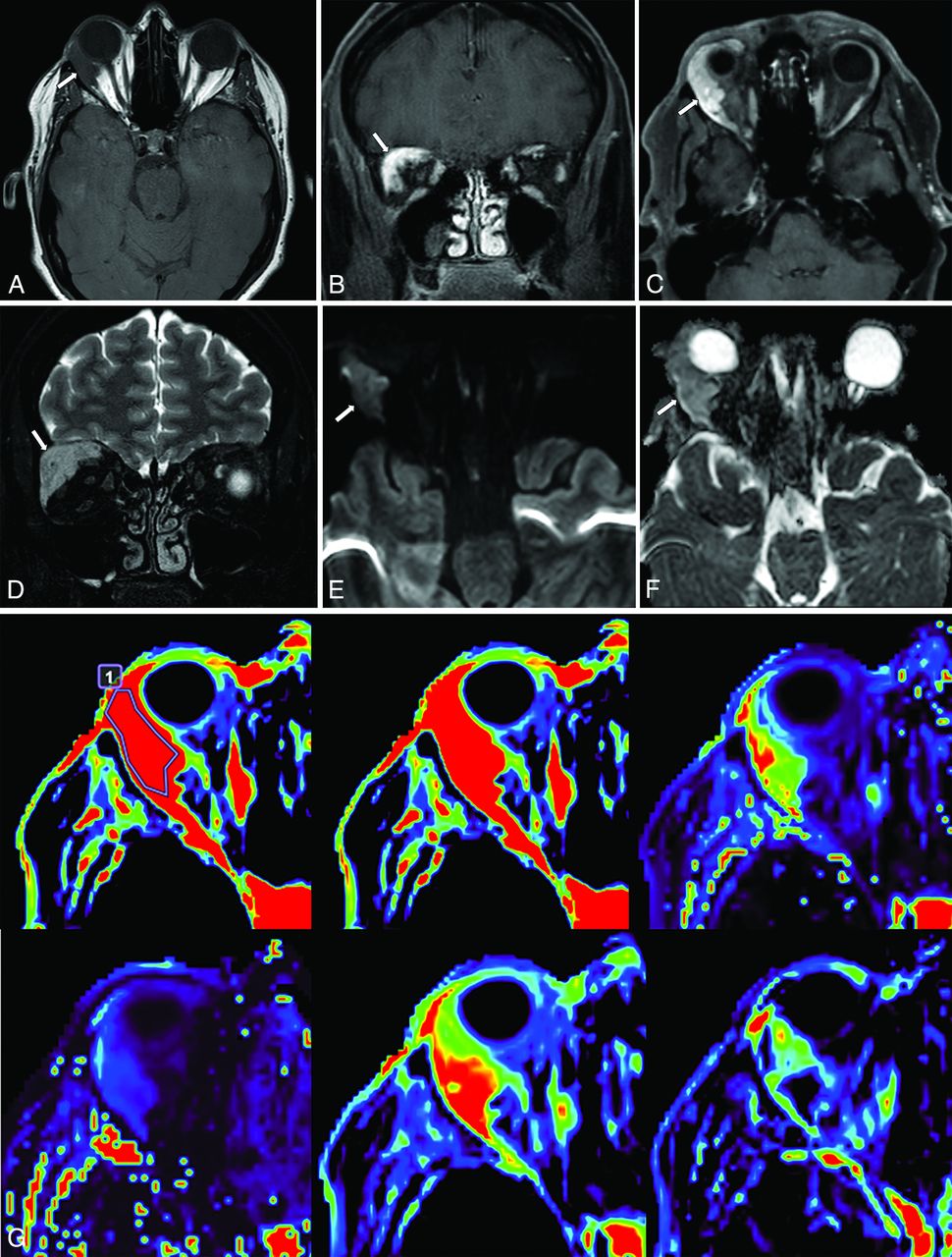
- FIG 4.
3T MR imaging in a 45-year-old female patient presenting with vertical diplopia. Axial T1WI (A), contrast-enhanced coronal (B) and axial (C) Dixon T1WI, coronal Dixon T2WI (D), axial DWI (E), axial ADC (F), and DCE MR imaging (G) showing a right orbital mass (arrow). DCE colorimetric maps are presented as follows from left to right and from top to bottom: AUCDCE with and without the region of interest drawn, Ktrans, Kep, Ve and Vp. Postsurgery, the final diagnosis was a carcinoma.


Tables
Mean (SD) or No. (%) Whole Sample (n = 131) Age 52 (17.1) (range, 19–88) Sex Female 66/131 (50.4%) Male 65/131 (49.6%) Histopathology Benign lesion Overall 90/131 (68.7%) Orbital inflammation 43/131 (32.8%) Orbital cavernous venous malformation 9/131 (6.9%) Other benign lesion 38/131 (29.0%) Malignant lesion Overall 41/131 (31.3%) Lymphoma 20/131 (15.3%) Primary solid malignant tumor 12/131 (6.9%) Solid tumor metastasis 9/131 (9.2%) Bilateral lesion 22/131 (16.8%) Ktrans (minute−1) Median (ICC) 0.99 IQR (ICC) 0.96 Kep (minute−1) Median (ICC) 0.99 IQR (ICC) 0.99 Ve (mL/100 mL of tissue; %) Median (ICC) 0.94 IQR (ICC) 0.73 Vp (mL/100 mL of tissue; %) Median (ICC) 0.87 IQR (ICC) 0.94 AUCDCE (mmol.min/L) Median (ICC) 0.98 IQR (ICC) 0.96 Type of the DCE TIC Cohen κ 0.67


Jump to section
Related Articles
Cited By...
- No citing articles found.