
Article Figures & Data
Figures
- FIG 1.
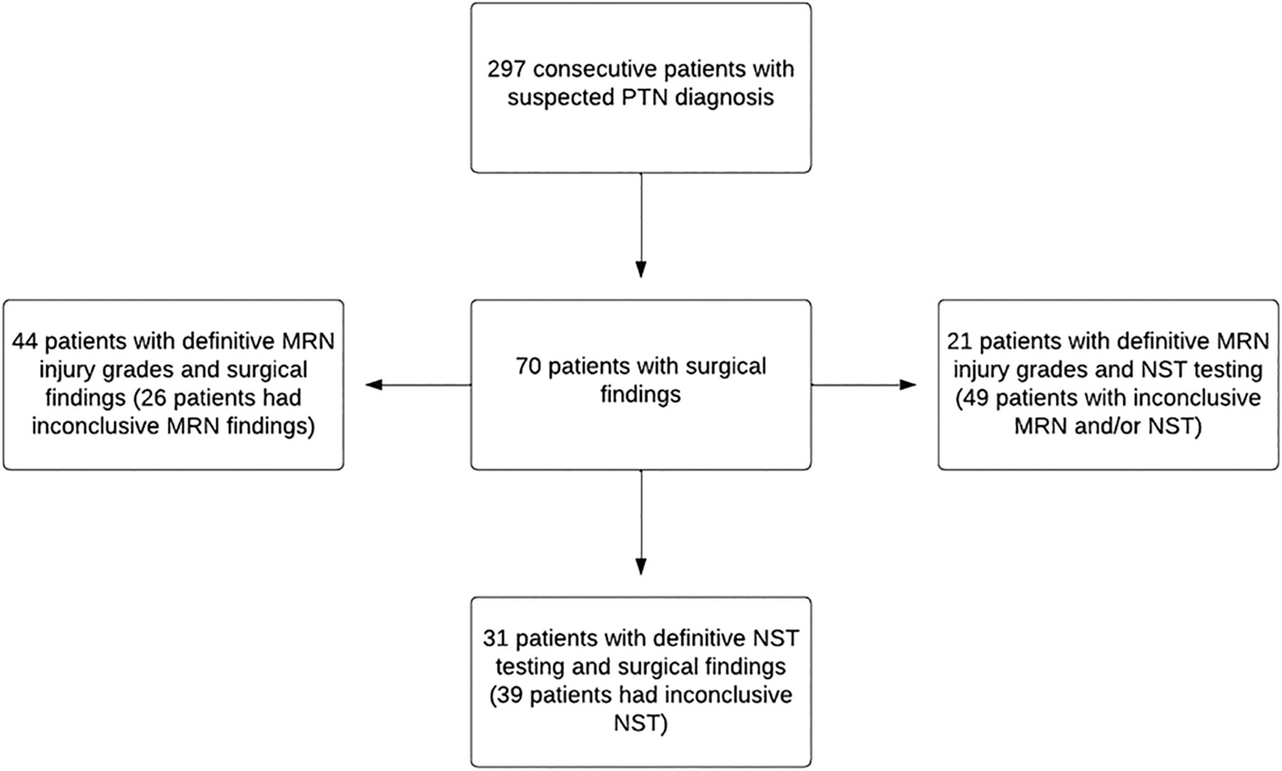
The 3 groups from the main cohort of 70 surgical patients were included in the statistical analysis. Because patients had inconclusive results in different modalities, the 3 groups had different sizes. For example, the 44 patients in the MRN versus surgical findings cohort were derived from the original 70 patients because 26 patients in the 70-patient surgical cohort had inconclusive MRN findings.
- FIG 2.
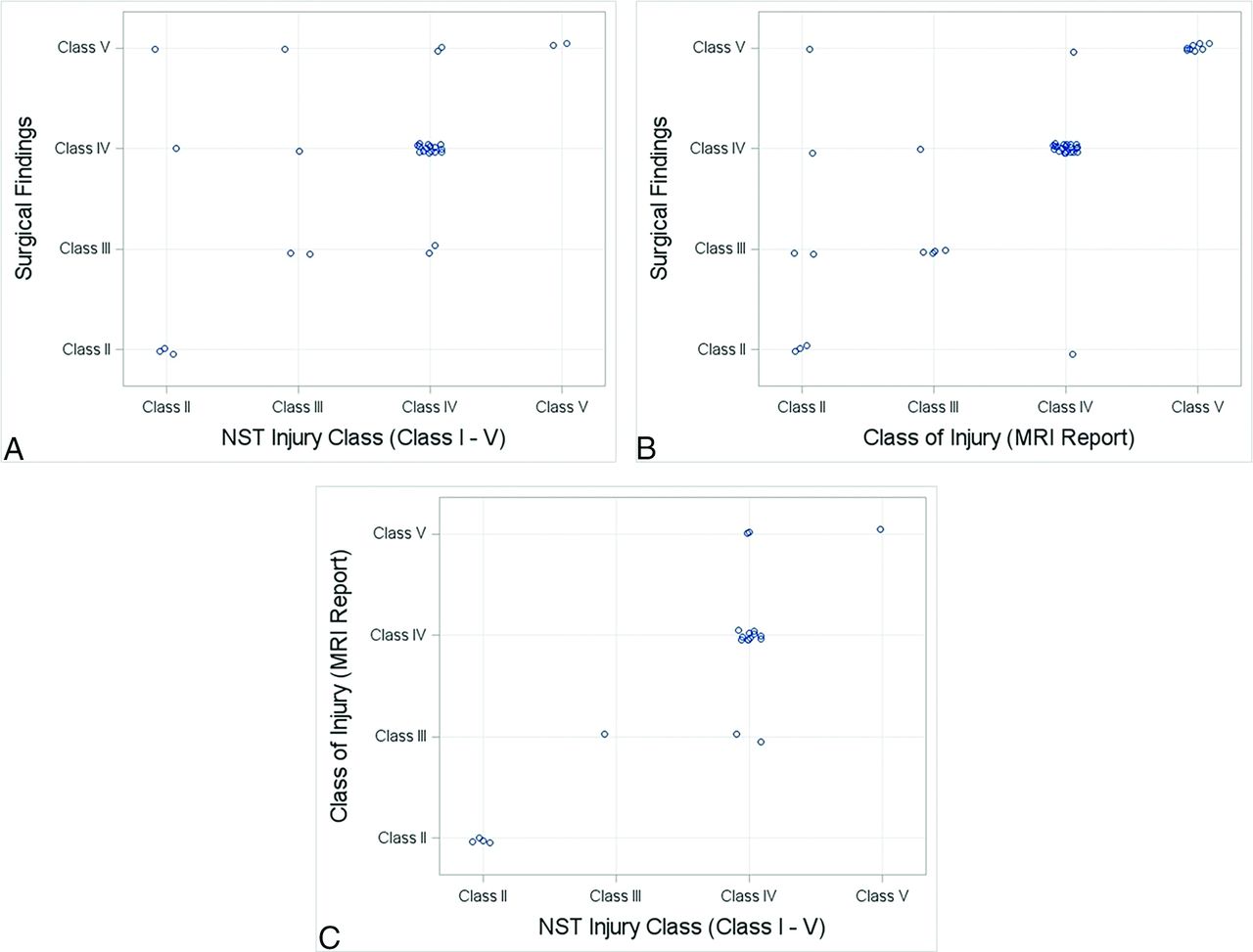
Jitter plots showing injury class distributions for (A) NST versus surgery, (B) MRN versus surgery, and (C) NST versus MRN.
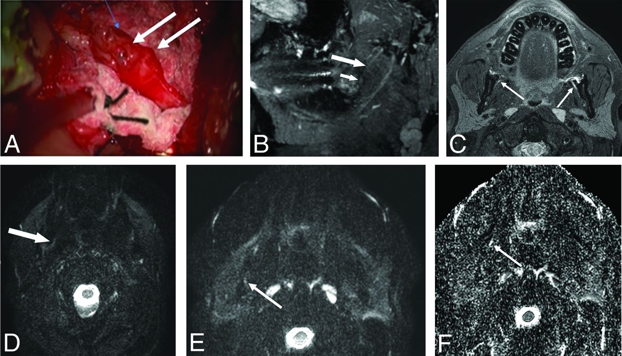
- FIG 3.
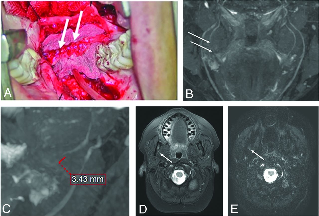
A 46-year-old woman with an injury to the right lingual nerve from molar teeth extraction. The NST yielded an injury grade of IV, but MRN yielded an injury grade of V, consistent with the surgical findings of a class V injury with amputation neuroma and a fibrous connection to the distal end. A, Intraoperative picture with amputation neuroma and foreign material highlighted by arrows. B, Coronal 3D PSIF MRN image of the lower face with arrows pointing to a gap in the right lingual nerve. C, Sagittal 3D PSIF MRN image reconstruction showing the neural gap in more detail, measuring 3.43 mm. D, Axial T2 SPAIR and (E) axial DTI showing the abnormal right lingual nerve (arrows). The nerve gap is best seen on 3D MRN images. PSIF indicates reversed fast imaging in steady state free precession; SPAIR, spectral attenuated inversion recovery.
- FIG 4.
A 33-year-old man with bilateral injuries to the lingual nerves caused by a third molar extraction. The NST was inconclusive, but MRN revealed a Sunderland grade IV injury, consistent with surgical findings. A, Intraoperative picture of the left lingual nerve showing a neuroma in continuity. B, 3D PSIF sagittal reconstructed 3D MRN image showing the focal nerve swelling (small arrow) in the abnormal nerve (large arrow) as a neuroma in continuity. C, Corresponding axial T2 SPAIR image showing abnormally hyperintense and enlarged lingual nerves bilaterally (arrows). D and E, Axial DTI and (F) axial ADC images showing the abnormally hyperintense right lingual nerve (arrows) with nonvisualization of the left lingual nerve on DTI and ADC images. PSIF indicates reversed fast imaging in steady state free precession; SPAIR, spectral attenuated inversion recovery.
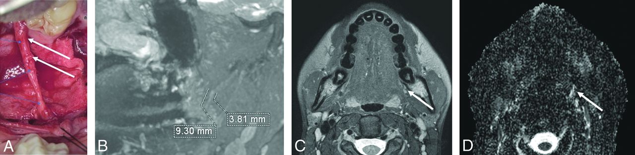
- FIG 5.
A 23-year-old woman with an injury to the right lingual nerve caused by a third molar extraction. Both MRN and NST revealed a Sunderland grade IV injury, consistent with surgical findings. A, Intraoperative picture showing neuroma in continuity. B, 3D PSIF sagittal reconstructed MRN image of the lower face showing a neuroma in continuity of 3.81 mm with a 9.3 mm overall abnormal hyperintense nerve. C, Focal nerve swelling can be appreciated in an axial T2-weighted SPAIR image and (D) the corresponding DTI, as outlined by the arrows. PSIF indicates reversed fast imaging in steady state free precession; SPAIR, spectral attenuated inversion recovery.


Tables
- Table 1.
Sunderland nerve injury classification with corresponding surgical findings, MRN findings, and surgical indications
Sunderland Injury Classification MRN Findings Recovery Potential Surgery Indication Surgical Findings I Homogeneous increased T2 signal of nerve with no change in caliber, usually resolve short of surgery Full None Intact with no internal or external fibrosis, normal neuroarchitecture II Homogeneous increased T2 signal of nerve and mild to moderate nerve thickening, less than 100% thickening than the adjacent or contralateral nerve Full None unless persistent pain for >3 months Intact with no internal fibrosis, with external fibrosis, restricted mobility with intact neuroarchitecture III Homogeneous increased T2 signal of nerve and moderate-marked nerve thickening, more than 100% thickening than the adjacent or contralateral nerve Slow/incomplete None or neurolysis Intact with internal and external fibrosis, restricted mobility, and disturbed neuroarchitecture IV Heterogeneous increased T2 signal of nerve and focal enlargement consistent with a neuroma-in-continuity in an otherwise continuous nerve Poor to none Nerve repair, graft, or transfer Partial transected nerve, some amount of distal nerve with or without lateral neuroma V Discontinuous nerve with end bulb neuroma and a complete nerve gap None Nerve repair, graft, or transfer Completely transected nerve - Table 2:
NST parameters. Present values exhibit comparable sensitivity within the normative range. Failed values are less than those of the control sites or the normative range. Elevated values are greater than those of the control sites. Absent values are greater than the maximum of the testing device
Injury Degree Level A: Spatiotemporal Sensory Perception Level B: Contact Detection with Monofilament Level C: Pain, Temperature, and Pressure Threshold and Tolerance Normal Present Present Present Mild Failed Present Present Moderate Failed Failed Present Severe Failed Failed Elevated Complete Failed Failed Absent Plane Sequence Coverage Slice Thickness/Gap (mm) Pixel Size (mm) FOV (mm) TR (ms) TE (ms) Comments 3 plane Scout Axial: Cover from skin to skin for R-L and A-P FOV Coronal: Cover from anterior nasal skin to back of the ear; R-L skin to skinSagittal: Cover both sides even if unilateral pain Axial 2D T2W TSE 4/0.4 0.3 × 0.4 170 × 180 3500–4500 50–65 Base of skull to C5 level Axial 2D T1W TSE 4/0.4 0.3 × 0.4 171 × 180 400–600 6–9 Base of skull to C5 level Coronal 3D DW-PSIF 0.9 ISO/0 Acquired ISO 172 × 180 12 3–4 Midskull to C5 level; b-value = 60/70 Axial 3D BFFE 0.9 ISO/0 Acquired ISO 173 × 180 5.2 3 Midskull to C2 level Axial DTI 4/0 1.5 × 1.5 174 × 180 5000–10,000 60–75 b-value = 0–600; 12 directions; echo spacing ≤ 0.7 ms Note:—T2W indicates T2-weighted; T1W, T1-weighted; BFFE, balanced fast field echo; R-L, right-left; A-P, anterior-posterior, DW, difffusion-weighted; PSIF, reversed fast imaging in steady state free precession.
- Table 4:
Distribution of injury grades for NST, MRN, and surgical findings among the 70 patients. Inconclusive results mean that the injury grade was unable to be narrowed down to just 1 class. For example, a grade of II/III being reported in the patient chart is recorded as inconclusive
Injury Grade NST MRN Surgery I 0 0 0 II 5 7 12 III 4 5 12 IV 20 24 33 V 2 8 13 Inconclusive 39 26 0 - Table 5:
Weighted Cohens kappa with 95% CIs for the NST grade versus MRN, surgical findings versus MRN, and surgical findings versus NST
Comparison Cohen Weighted Kappa Coefficient (quadratic) 95% CI NST versus surgical findings 0.51 (0.1–0.92) MRN versus surgical findings 0.7 (0.44–0.97) MRN versus NST 0.88 (0.76–1) Comparison Class Agreement Percentage NST versus surgical findings I to III 71.43% IV and V 75% Overall 74.19% MRN versus surgical findings I to III 70% IV and V 88.24% Overall 84.09% MRN versus NST Overall 80.95%


Jump to section
Related Articles
Cited By...
- No citing articles found.