
Article Figures & Data
Figures
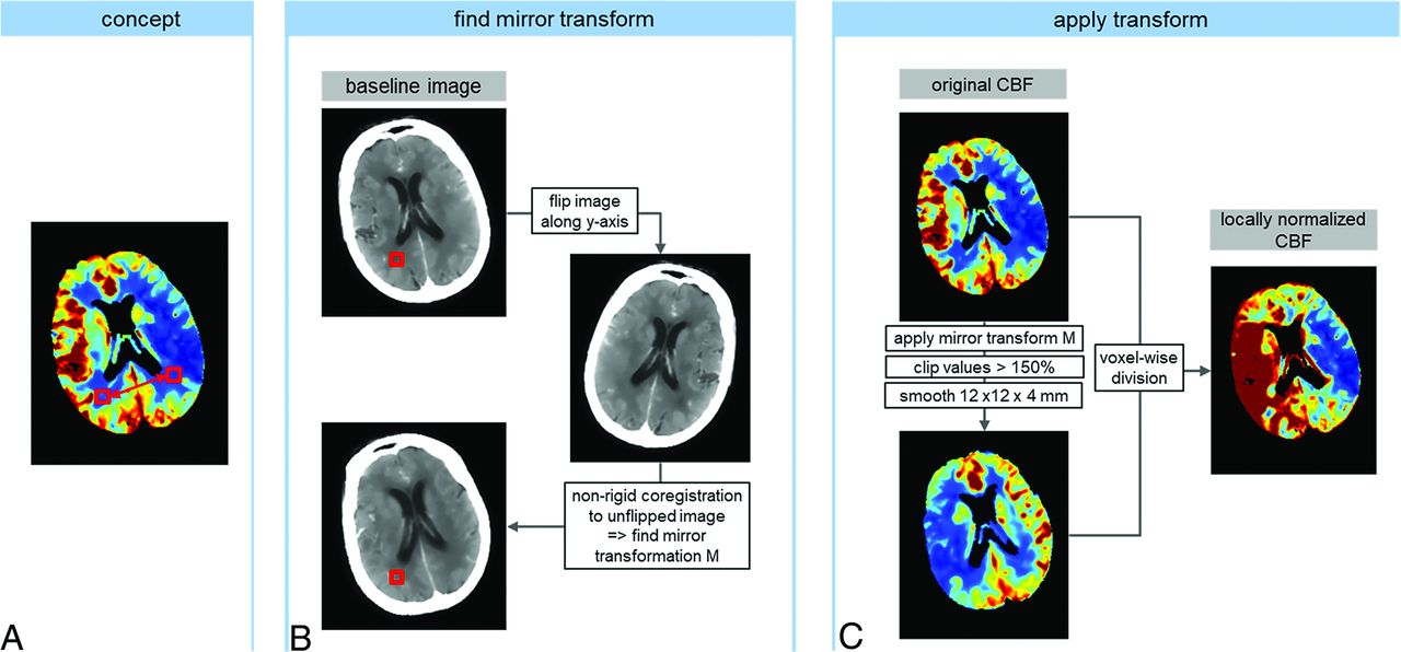
- FIG 1.
Schematic of the workflow for global and local normalization of CT perfusion-derived CBF with subsequent thresholding to 30% to obtain a segmentation of the ischemic core. In the global approach, each voxel is normalized to the entire contralateral hemisphere, whereas in the local approach, a voxel is compared with a local region on the contralateral side. This leads to an equalization between GM and WM and thus a more distinct contrast in the perfusion maps.
- FIG 2.
Correlation plot (A) and Bland-Altman plot (B) comparing core volumes derived with global and local normalization for the NoMa and MA groups (black and red, respectively). Dashed lines indicate linear regression in the correlation plot, and 2 SDs in the Bland-Altman plot.
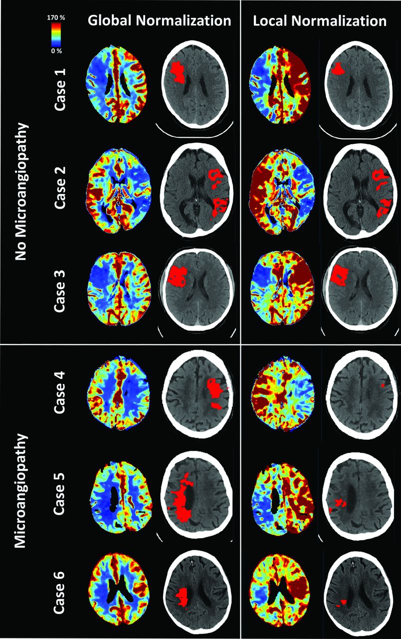
- FIG 3.
Comparison of global (left) and local (right) normalization for CBF maps and threshold-based ischemic core segmentations in 3 cases without and with WM small vessel disease.
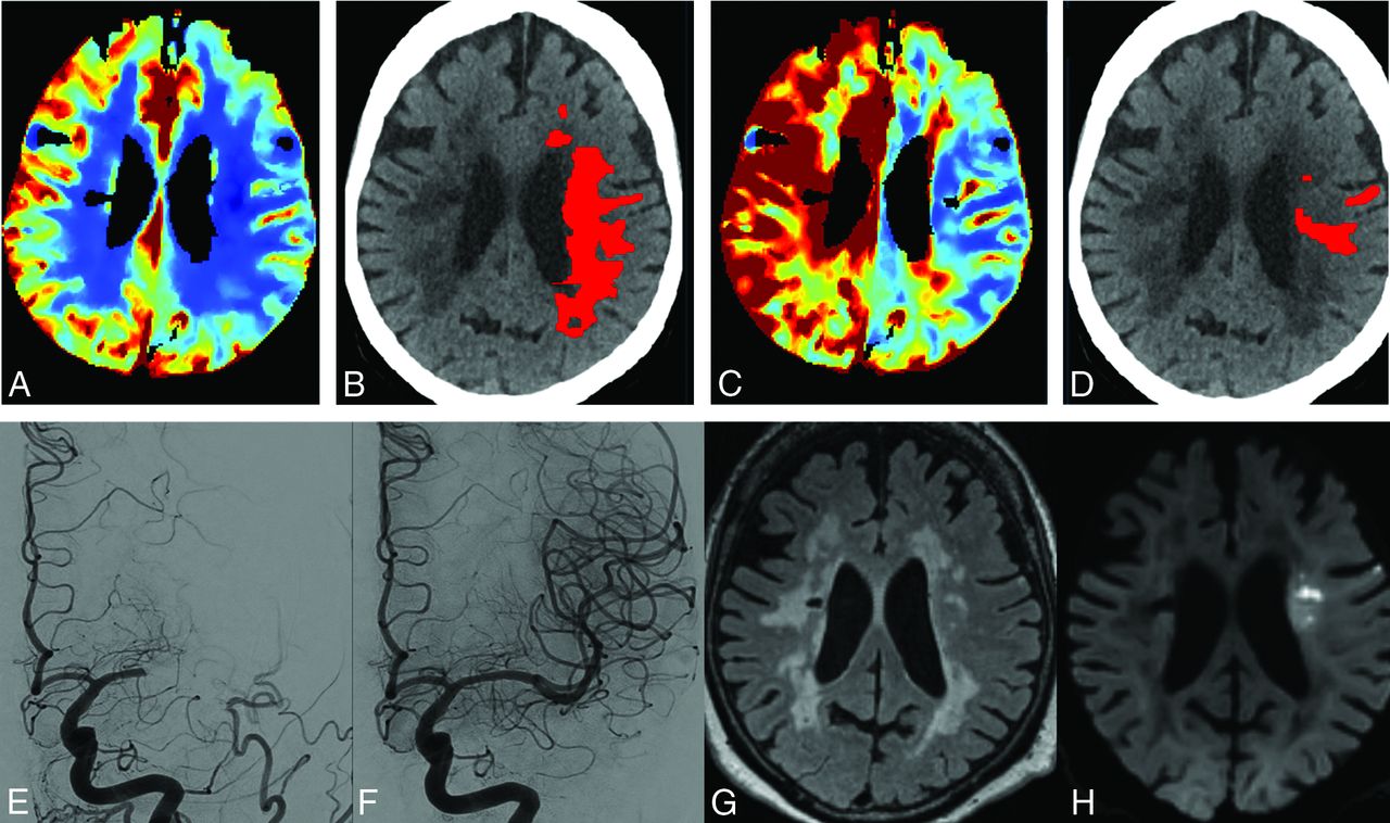
- FIG 4.
Image data of an 83-year-old woman 5 hours after onset of a left MCA occlusion. CTP with global contralateral normalization (A) suggested an infarct core involving the centrum semiovale (B). In contrast, CTP with local contralateral normalization (C) did not display the deep WM as infarct core (D). The M1 segment was recanalized following one thrombectomy maneuver by using a 4 × 40 mm Solitaire stent (E, F). Follow-up MR imaging shows distinct microangiopathy (G, FLAIR), but only scattered subacute centrum semiovale infarct (H, DWI).


Tables
Patient characteristics
n Sex (Female) Age (yr) Stroke Evident Mean Core Volume (mL) Global Normalization Local Normalization No microangiopathy 2830 49% 72.6 ± 15.2 1334 (47%) 41.0 ± 46.9 31.4 ± 41.1 Microangiopathy 335 59% 73.5 ± 17.1 175 (52%) 60.3 ± 49.0 33.6 ± 39.3


Jump to section
Related Articles
Cited By...
- No citing articles found.