Advertisement
  • Main menu
  • User menu
  • Search
  • English ▼
    • English
    • Afrikaans
    • Albanian
    • Amharic
    • Arabic
    • Armenian
    • Azerbaijani
    • Basque
    • Belarusian
    • Bengali
    • Bosnian
    • Bulgarian
    • Catalan
    • Cebuano
    • Chichewa
    • Chinese (Simplified)
    • Chinese (Traditional)
    • Corsican
    • Croatian
    • Czech
    • Danish
    • Dutch
    • Esperanto
    • Estonian
    • Filipino
    • Finnish
    • French
    • Frisian
    • Galician
    • Georgian
    • German
    • Greek
    • Gujarati
    • Haitian Creole
    • Hausa
    • Hawaiian
    • Hebrew
    • Hindi
    • Hmong
    • Hungarian
    • Icelandic
    • Igbo
    • Indonesian
    • Irish
    • Italian
    • Japanese
    • Javanese
    • Kannada
    • Kazakh
    • Khmer
    • Korean
    • Kurdish (Kurmanji)
    • Kyrgyz
    • Lao
    • Latin
    • Latvian
    • Lithuanian
    • Luxembourgish
    • Macedonian
    • Malagasy
    • Malay
    • Malayalam
    • Maltese
    • Maori
    • Marathi
    • Mongolian
    • Myanmar (Burmese)
    • Nepali
    • Norwegian
    • Pashto
    • Persian
    • Polish
    • Portuguese
    • Punjabi
    • Romanian
    • Russian
    • Samoan
    • Scottish Gaelic
    • Serbian
    • Sesotho
    • Shona
    • Sindhi
    • Sinhala
    • Slovak
    • Slovenian
    • Somali
    • Spanish
    • Sudanese
    • Swahili
    • Swedish
    • Tajik
    • Tamil
    • Telugu
    • Thai
    • Turkish
    • Ukrainian
    • Urdu
    • Uzbek
    • Vietnamese
    • Welsh
    • Xhosa
    • Yiddish
    • Yoruba
    • Zulu

Main menu

  • Home
  • Content
    • Current Issue
    • Accepted Manuscripts
    • Article Preview
    • Past Issue Archive
    • Video Articles
    • AJNR Case Collection
    • Case of the Week Archive
    • Case of the Month Archive
    • Classic Case Archive
  • Special Collections
    • AJNR Awards
    • Low-Field MRI
    • Alzheimer Disease
    • ASNR Foundation Special Collection
    • Photon-Counting CT
    • View All
  • Multimedia
    • AJNR Podcasts
    • AJNR SCANtastic
    • Trainee Corner
    • MRI Safety Corner
    • Imaging Protocols
  • For Authors
    • Submit a Manuscript
    • Submit a Video Article
    • Submit an eLetter to the Editor/Response
    • Manuscript Submission Guidelines
    • Statistical Tips
    • Fast Publishing of Accepted Manuscripts
    • Graphical Abstract Preparation
    • Imaging Protocol Submission
    • Author Policies
  • About Us
    • About AJNR
    • Editorial Board
    • Editorial Board Alumni
  • More
    • Become a Reviewer/Academy of Reviewers
    • Subscribers
    • Permissions
    • Alerts
    • Feedback
    • Advertisers
    • ASNR Home

User menu

  • Alerts
  • Log in

Search

  • Advanced search
American Journal of Neuroradiology

American Journal of Neuroradiology

ASHNR American Society of Functional Neuroradiology ASHNR American Society of Pediatric Neuroradiology ASSR
  • Alerts
  • Log in

Advanced Search

English ▼
  • English
  • Afrikaans
  • Albanian
  • Amharic
  • Arabic
  • Armenian
  • Azerbaijani
  • Basque
  • Belarusian
  • Bengali
  • Bosnian
  • Bulgarian
  • Catalan
  • Cebuano
  • Chichewa
  • Chinese (Simplified)
  • Chinese (Traditional)
  • Corsican
  • Croatian
  • Czech
  • Danish
  • Dutch
  • Esperanto
  • Estonian
  • Filipino
  • Finnish
  • French
  • Frisian
  • Galician
  • Georgian
  • German
  • Greek
  • Gujarati
  • Haitian Creole
  • Hausa
  • Hawaiian
  • Hebrew
  • Hindi
  • Hmong
  • Hungarian
  • Icelandic
  • Igbo
  • Indonesian
  • Irish
  • Italian
  • Japanese
  • Javanese
  • Kannada
  • Kazakh
  • Khmer
  • Korean
  • Kurdish (Kurmanji)
  • Kyrgyz
  • Lao
  • Latin
  • Latvian
  • Lithuanian
  • Luxembourgish
  • Macedonian
  • Malagasy
  • Malay
  • Malayalam
  • Maltese
  • Maori
  • Marathi
  • Mongolian
  • Myanmar (Burmese)
  • Nepali
  • Norwegian
  • Pashto
  • Persian
  • Polish
  • Portuguese
  • Punjabi
  • Romanian
  • Russian
  • Samoan
  • Scottish Gaelic
  • Serbian
  • Sesotho
  • Shona
  • Sindhi
  • Sinhala
  • Slovak
  • Slovenian
  • Somali
  • Spanish
  • Sudanese
  • Swahili
  • Swedish
  • Tajik
  • Tamil
  • Telugu
  • Thai
  • Turkish
  • Ukrainian
  • Urdu
  • Uzbek
  • Vietnamese
  • Welsh
  • Xhosa
  • Yiddish
  • Yoruba
  • Zulu
  • Home
  • Content
    • Current Issue
    • Accepted Manuscripts
    • Article Preview
    • Past Issue Archive
    • Video Articles
    • AJNR Case Collection
    • Case of the Week Archive
    • Case of the Month Archive
    • Classic Case Archive
  • Special Collections
    • AJNR Awards
    • Low-Field MRI
    • Alzheimer Disease
    • ASNR Foundation Special Collection
    • Photon-Counting CT
    • View All
  • Multimedia
    • AJNR Podcasts
    • AJNR SCANtastic
    • Trainee Corner
    • MRI Safety Corner
    • Imaging Protocols
  • For Authors
    • Submit a Manuscript
    • Submit a Video Article
    • Submit an eLetter to the Editor/Response
    • Manuscript Submission Guidelines
    • Statistical Tips
    • Fast Publishing of Accepted Manuscripts
    • Graphical Abstract Preparation
    • Imaging Protocol Submission
    • Author Policies
  • About Us
    • About AJNR
    • Editorial Board
    • Editorial Board Alumni
  • More
    • Become a Reviewer/Academy of Reviewers
    • Subscribers
    • Permissions
    • Alerts
    • Feedback
    • Advertisers
    • ASNR Home
  • Follow AJNR on Twitter
  • Visit AJNR on Facebook
  • Follow AJNR on Instagram
  • Join AJNR on LinkedIn
  • RSS Feeds

AJNR Awards, New Junior Editors, and more. Read the latest AJNR updates

Research ArticleNeurovascular/Stroke Imaging

Delayed Enhancement of Intracranial Atherosclerotic Plaque Can Better Differentiate Culprit Lesions: A Multiphase Contrast-Enhanced Vessel Wall MRI Study

Beibei Sun, Lingling Wang, Xiao Li, Jin Zhang, Jianjian Zhang, Jiaqi Tian, Mahmud Mossa-Basha, Jianrong Xu, Yan Zhou, Huilin Zhao and Chengcheng Zhu
American Journal of Neuroradiology March 2024, 45 (3) 262-270; DOI: https://doi.org/10.3174/ajnr.A8132
Beibei Sun
aFrom the Department of Radiology, Ren Ji Hospital (B.S., L.W., X.L., Jin Zhang, Jianjian Zhang, J.T., J.X., Y.Z., H.Z.), Shanghai Jiao Tong University School of Medicine, Shanghai China
bCollege of Health Science and Technology (B.S., L.W., X.L., Jin Zhang, Jianjian Zhang, J.T., J.X., Y.Z., H.Z.), Shanghai Jiao Tong University School of Medicine, Shanghai, China
cDepartment of Radiology (M.M., C.Z.), University of Washington, Seattle, Washington
  • Find this author on Google Scholar
  • Find this author on PubMed
  • Search for this author on this site
  • ORCID record for Beibei Sun
Lingling Wang
aFrom the Department of Radiology, Ren Ji Hospital (B.S., L.W., X.L., Jin Zhang, Jianjian Zhang, J.T., J.X., Y.Z., H.Z.), Shanghai Jiao Tong University School of Medicine, Shanghai China
bCollege of Health Science and Technology (B.S., L.W., X.L., Jin Zhang, Jianjian Zhang, J.T., J.X., Y.Z., H.Z.), Shanghai Jiao Tong University School of Medicine, Shanghai, China
  • Find this author on Google Scholar
  • Find this author on PubMed
  • Search for this author on this site
  • ORCID record for Lingling Wang
Xiao Li
aFrom the Department of Radiology, Ren Ji Hospital (B.S., L.W., X.L., Jin Zhang, Jianjian Zhang, J.T., J.X., Y.Z., H.Z.), Shanghai Jiao Tong University School of Medicine, Shanghai China
bCollege of Health Science and Technology (B.S., L.W., X.L., Jin Zhang, Jianjian Zhang, J.T., J.X., Y.Z., H.Z.), Shanghai Jiao Tong University School of Medicine, Shanghai, China
  • Find this author on Google Scholar
  • Find this author on PubMed
  • Search for this author on this site
  • ORCID record for Xiao Li
Jin Zhang
aFrom the Department of Radiology, Ren Ji Hospital (B.S., L.W., X.L., Jin Zhang, Jianjian Zhang, J.T., J.X., Y.Z., H.Z.), Shanghai Jiao Tong University School of Medicine, Shanghai China
bCollege of Health Science and Technology (B.S., L.W., X.L., Jin Zhang, Jianjian Zhang, J.T., J.X., Y.Z., H.Z.), Shanghai Jiao Tong University School of Medicine, Shanghai, China
  • Find this author on Google Scholar
  • Find this author on PubMed
  • Search for this author on this site
  • ORCID record for Jin Zhang
Jianjian Zhang
aFrom the Department of Radiology, Ren Ji Hospital (B.S., L.W., X.L., Jin Zhang, Jianjian Zhang, J.T., J.X., Y.Z., H.Z.), Shanghai Jiao Tong University School of Medicine, Shanghai China
bCollege of Health Science and Technology (B.S., L.W., X.L., Jin Zhang, Jianjian Zhang, J.T., J.X., Y.Z., H.Z.), Shanghai Jiao Tong University School of Medicine, Shanghai, China
  • Find this author on Google Scholar
  • Find this author on PubMed
  • Search for this author on this site
Jiaqi Tian
aFrom the Department of Radiology, Ren Ji Hospital (B.S., L.W., X.L., Jin Zhang, Jianjian Zhang, J.T., J.X., Y.Z., H.Z.), Shanghai Jiao Tong University School of Medicine, Shanghai China
bCollege of Health Science and Technology (B.S., L.W., X.L., Jin Zhang, Jianjian Zhang, J.T., J.X., Y.Z., H.Z.), Shanghai Jiao Tong University School of Medicine, Shanghai, China
  • Find this author on Google Scholar
  • Find this author on PubMed
  • Search for this author on this site
Mahmud Mossa-Basha
cDepartment of Radiology (M.M., C.Z.), University of Washington, Seattle, Washington
  • Find this author on Google Scholar
  • Find this author on PubMed
  • Search for this author on this site
  • ORCID record for Mahmud Mossa-Basha
Jianrong Xu
aFrom the Department of Radiology, Ren Ji Hospital (B.S., L.W., X.L., Jin Zhang, Jianjian Zhang, J.T., J.X., Y.Z., H.Z.), Shanghai Jiao Tong University School of Medicine, Shanghai China
bCollege of Health Science and Technology (B.S., L.W., X.L., Jin Zhang, Jianjian Zhang, J.T., J.X., Y.Z., H.Z.), Shanghai Jiao Tong University School of Medicine, Shanghai, China
  • Find this author on Google Scholar
  • Find this author on PubMed
  • Search for this author on this site
  • ORCID record for Jianrong Xu
Yan Zhou
aFrom the Department of Radiology, Ren Ji Hospital (B.S., L.W., X.L., Jin Zhang, Jianjian Zhang, J.T., J.X., Y.Z., H.Z.), Shanghai Jiao Tong University School of Medicine, Shanghai China
bCollege of Health Science and Technology (B.S., L.W., X.L., Jin Zhang, Jianjian Zhang, J.T., J.X., Y.Z., H.Z.), Shanghai Jiao Tong University School of Medicine, Shanghai, China
  • Find this author on Google Scholar
  • Find this author on PubMed
  • Search for this author on this site
Huilin Zhao
aFrom the Department of Radiology, Ren Ji Hospital (B.S., L.W., X.L., Jin Zhang, Jianjian Zhang, J.T., J.X., Y.Z., H.Z.), Shanghai Jiao Tong University School of Medicine, Shanghai China
bCollege of Health Science and Technology (B.S., L.W., X.L., Jin Zhang, Jianjian Zhang, J.T., J.X., Y.Z., H.Z.), Shanghai Jiao Tong University School of Medicine, Shanghai, China
  • Find this author on Google Scholar
  • Find this author on PubMed
  • Search for this author on this site
  • ORCID record for Huilin Zhao
Chengcheng Zhu
cDepartment of Radiology (M.M., C.Z.), University of Washington, Seattle, Washington
  • Find this author on Google Scholar
  • Find this author on PubMed
  • Search for this author on this site
  • ORCID record for Chengcheng Zhu
  • Article
  • Figures & Data
  • Supplemental
  • Info & Metrics
  • Responses
  • References
  • PDF
Loading

Article Figures & Data

Figures

  • Tables
  • FIG 1.
    • Download figure
    • Open in new tab
    • Download powerpoint
    FIG 1.

    Patient selection flow chart.
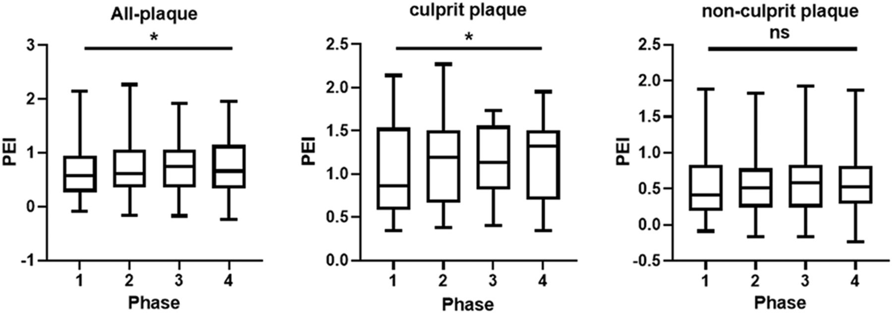
  • FIG 2.
    • Download figure
    • Open in new tab
    • Download powerpoint
    FIG 2.

    Plaque enhancement index (PEI; the mean signal intensity of plaque normalized by CSF) change of culprit and nonculprit plaques over time. The boxes were drawn with the median (line in the box) as well as the 25th and 75th percentiles. The bars above and below the box are the maximum and minimum values of the PEI, respectively. The PEI values of all intracranial plaques and culprit plaques increase over the 4 phases (P = .033, P = .034, respectively). The PEI values of the nonculprit plaques show no significant differences among the 4 postcontrast phases (P = .450). *, P < .05; ns, not significant.
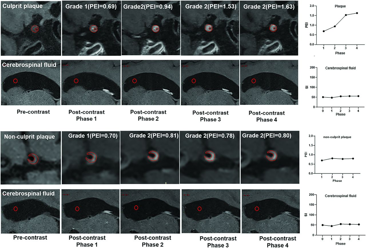
  • FIG 3.
    • Download figure
    • Open in new tab
    • Download powerpoint
    FIG 3.

    Cases of the culprit and nonculprit plaque signal intensity change during the 4 postcontrast phases.

  • FIG 4.
    • Download figure
    • Open in new tab
    • Download powerpoint
    FIG 4.

    The ROC of the plaque enhancement index in the 4 contrast-enhanced phases for differentiating between culprit and nonculprit plaques.

Tables

  • Figures
    • View popup
    Table 1:

    Demographic and intracranial plaque characteristics of 30 patients with acute stroke

    Patient DemographicsMean ± SD or Median (IQR) or n (%)
    Age (years)59.0 ± 9.7
    Body mass index (kg/cm2)24.6 ± 2.9
    Sex (male)18 (60.0%)
    Hypertension21 (70.0%)
    Diabetes11 (36.7%)
    Dyslipidemia6 (20.0%)
    Current smoking5 (16.7%)
    Plaque location
     Internal carotid artery (C4–7)25 (22.1%)
     Middle cerebral artery34 (30.1%)
     Anterior cerebral artery6 (5.3%)
     Basilar artery17 (15.0%)
     Vertebral artery21 (18.6%)
     Posterior cerebral artery10 (8.9%)
    Plaque stenosis (%)
     30% ≤ stenosis < 50% (grade I)74 (65.5%)
     50% ≤ stenosis < 70% (grade II)15 (13.3%)
     Stenosis ≥ 70% (grade III)24 (21.2%)
    Plaque numbers (n)3.0 (2.0–6.0)
    • View popup
    Table 2:

    Plaque enhancement index change of culprit and nonculprit plaques over time

    PhasePEI
    All Plaques (n = 113)aNonculprit Plaque (n = 83)aCulprit Plaque (n = 30)b
    1st0.66 ± 0.510.54 ± 0.441.02 ± 0.53
    2nd0.72 ± 0.520.55 ± 0.421.17 ± 0.50
    3rd0.73 ± 0.470.58 ± 0.391.16 ± 0.41
    4th0.76 ± 0.530.60 ± 0.441.20 ± 0.49
    Pc.033.450.034
    P 1st versus 2nd.014.578.007
    P 1st versus 3rd.049.446.057
    P 1st versus 4th.029.401.035
    P 2nd versus 3rd.879.640.869
    P 2nd versus 4th.502.572.680
    P 3rd versus 4th.458.771.477
    • ↵a A mixed-effects model repeated measures ANOVA for continuous variables was used to analyze the differences between all 4 postcontrast phases in all plaque groups and the nonculprit plaque group.

    • ↵b A 1-way repeated-measures ANOVA for continuous variables was used to analyze the differences between the 4 postcontrast phases in the culprit plaque group. This was followed by a pair-wise comparison post hoc analysis using Bonferroni correction to compare the differences between the PEI values of each 2 contrast phases.

    • ↵c P values of the comparisons of the 4 postcontrast phases.

    • View popup
    Table 3:

    Plaque enhancement index in different contrast-enhanced phases to differentiate between culprit and nonculprit plaques

    VariablesAUC (95% CI)Sensitivity/SpecificityCutoffP Value
    PEI 1st0.768 (0.680–0.843)59.0%/86.7%0.48<.001
    PEI 2nd0.829 (0.746–0.893)80.7%/70.0%0.83<.001
    PEI 3rd0.840 (0.759–0.902)83.1%/70.0%0.95<.001
    PEI 4th0.812 (0.727–0.879)84.3%/70.0%0.98<.001
    • View popup
    Table 4:

    Comparison of ROC curves of PEI to differentiate between culprit and nonculprit plaques between 4 postcontrast phases

    VariablesDifference between AUC area95% CIz statisticP Value
    PEI 1st versus PEI 2nd0.060 ± 0.0260.009–0.1122.293.022
    PEI 1st versus PEI 3rd0.071 ± 0.039−0.005–0.1481.824.068
    PEI 1st versus PEI 4th0.043 ± 0.038−0.032–0.1181.131.258
    PEI 2nd versus PEI 3rd0.011 ± 0.025−0.038–0.0600.444.657
    PEI 2nd versus PEI 4th0.017 ± 0.027−0.036–0.0700.628.530
    PEI 3rd versus PEI 4th0.028 ± 0.024−0.020–0.0761.151.249
PreviousNext
Back to top

In this issue

American Journal of Neuroradiology: 45 (3)
American Journal of Neuroradiology
Vol. 45, Issue 3
1 Mar 2024
  • Table of Contents
  • Index by author
  • Complete Issue (PDF)
Advertisement
Print
Download PDF
Email Article
Cite this article
0 Responses
Respond to this article
Share
Bookmark this article
Delayed Enhancement of Atherosclerotic Plaque
Beibei Sun, Lingling Wang, Xiao Li, Jin Zhang, Jianjian Zhang, Jiaqi Tian, Mahmud Mossa-Basha, Jianrong Xu, Yan Zhou, Huilin Zhao, Chengcheng Zhu
American Journal of Neuroradiology Mar 2024, 45 (3) 262-270; DOI: 10.3174/ajnr.A8132
del.icio.us logo Twitter logo Facebook logo Mendeley logo
  • Tweet Widget
Purchase

Jump to section

  • Article
    • Abstract
    • ABBREVIATIONS:
    • MATERIALS AND METHODS
    • RESULTS
    • DISCUSSION
    • CONCLUSIONS
    • Footnotes
    • References
  • Figures & Data
  • Supplemental
  • Info & Metrics
  • Responses
  • References
  • PDF

Related Articles

  • PubMed
  • Google Scholar

Cited By...

  • No citing articles found.
  • Crossref
  • Google Scholar

This article has not yet been cited by articles in journals that are participating in Crossref Cited-by Linking.

More in this TOC Section

  • 90-day Outcome Prediction in VBAO after EVT
  • P2-PCA VFR and BOLD-CVR in ICA Occlusion
  • Circle of Willis Variants and Stroke Outcomes
Show more Neurovascular/Stroke Imaging

Similar Articles

Advertisement

Indexed Content

  • Current Issue
  • Accepted Manuscripts
  • Article Preview
  • Past Issues
  • Editorials
  • Editor's Choice
  • Fellows' Journal Club
  • Letters to the Editor
  • Video Articles

Cases

  • Case Collection
  • Archive - Case of the Week
  • Archive - Case of the Month
  • Archive - Classic Case

More from AJNR

  • Trainee Corner
  • Imaging Protocols
  • MRI Safety Corner
  • Book Reviews

Multimedia

  • AJNR Podcasts
  • AJNR Scantastics

Resources

  • Turnaround Time
  • Submit a Manuscript
  • Submit a Video Article
  • Submit an eLetter to the Editor/Response
  • Manuscript Submission Guidelines
  • Statistical Tips
  • Fast Publishing of Accepted Manuscripts
  • Graphical Abstract Preparation
  • Imaging Protocol Submission
  • Evidence-Based Medicine Level Guide
  • Publishing Checklists
  • Author Policies
  • Become a Reviewer/Academy of Reviewers
  • News and Updates

About Us

  • About AJNR
  • Editorial Board
  • Editorial Board Alumni
  • Alerts
  • Permissions
  • Not an AJNR Subscriber? Join Now
  • Advertise with Us
  • Librarian Resources
  • Feedback
  • Terms and Conditions
  • AJNR Editorial Board Alumni

American Society of Neuroradiology

  • Not an ASNR Member? Join Now

© 2025 by the American Society of Neuroradiology All rights, including for text and data mining, AI training, and similar technologies, are reserved.
Print ISSN: 0195-6108 Online ISSN: 1936-959X

Powered by HighWire
Email this Article

Thank you for your interest in spreading the word on American Journal of Neuroradiology.

NOTE: We only request your email address so that the person you are recommending the page to knows that you wanted them to see it, and that it is not junk mail. We do not capture any email address.

Enter multiple addresses on separate lines or separate them with commas.
Delayed Enhancement of Intracranial Atherosclerotic Plaque Can Better Differentiate Culprit Lesions: A Multiphase Contrast-Enhanced Vessel Wall MRI Study
(Your Name) has sent you a message from American Journal of Neuroradiology
(Your Name) thought you would like to see the American Journal of Neuroradiology web site.
CAPTCHA
This question is for testing whether or not you are a human visitor and to prevent automated spam submissions.
Cite this article
Beibei Sun, Lingling Wang, Xiao Li, Jin Zhang, Jianjian Zhang, Jiaqi Tian, Mahmud Mossa-Basha, Jianrong Xu, Yan Zhou, Huilin Zhao, Chengcheng Zhu
Delayed Enhancement of Intracranial Atherosclerotic Plaque Can Better Differentiate Culprit Lesions: A Multiphase Contrast-Enhanced Vessel Wall MRI Study
American Journal of Neuroradiology Mar 2024, 45 (3) 262-270; DOI: 10.3174/ajnr.A8132

Citation Manager Formats

  • BibTeX
  • Bookends
  • EasyBib
  • EndNote (tagged)
  • EndNote 8 (xml)
  • Medlars
  • Mendeley
  • Papers
  • RefWorks Tagged
  • Ref Manager
  • RIS
  • Zotero

We use cookies on this site to enhance your user experience. By clicking any link on this page you are giving your consent for us to set cookies.