Advertisement
  • Main menu
  • User menu
  • Search
  • English ▼
    • English
    • Afrikaans
    • Albanian
    • Amharic
    • Arabic
    • Armenian
    • Azerbaijani
    • Basque
    • Belarusian
    • Bengali
    • Bosnian
    • Bulgarian
    • Catalan
    • Cebuano
    • Chichewa
    • Chinese (Simplified)
    • Chinese (Traditional)
    • Corsican
    • Croatian
    • Czech
    • Danish
    • Dutch
    • Esperanto
    • Estonian
    • Filipino
    • Finnish
    • French
    • Frisian
    • Galician
    • Georgian
    • German
    • Greek
    • Gujarati
    • Haitian Creole
    • Hausa
    • Hawaiian
    • Hebrew
    • Hindi
    • Hmong
    • Hungarian
    • Icelandic
    • Igbo
    • Indonesian
    • Irish
    • Italian
    • Japanese
    • Javanese
    • Kannada
    • Kazakh
    • Khmer
    • Korean
    • Kurdish (Kurmanji)
    • Kyrgyz
    • Lao
    • Latin
    • Latvian
    • Lithuanian
    • Luxembourgish
    • Macedonian
    • Malagasy
    • Malay
    • Malayalam
    • Maltese
    • Maori
    • Marathi
    • Mongolian
    • Myanmar (Burmese)
    • Nepali
    • Norwegian
    • Pashto
    • Persian
    • Polish
    • Portuguese
    • Punjabi
    • Romanian
    • Russian
    • Samoan
    • Scottish Gaelic
    • Serbian
    • Sesotho
    • Shona
    • Sindhi
    • Sinhala
    • Slovak
    • Slovenian
    • Somali
    • Spanish
    • Sudanese
    • Swahili
    • Swedish
    • Tajik
    • Tamil
    • Telugu
    • Thai
    • Turkish
    • Ukrainian
    • Urdu
    • Uzbek
    • Vietnamese
    • Welsh
    • Xhosa
    • Yiddish
    • Yoruba
    • Zulu

Main menu

  • Home
  • Content
    • Current Issue
    • Accepted Manuscripts
    • Article Preview
    • Past Issue Archive
    • Video Articles
    • AJNR Case Collection
    • Case of the Week Archive
    • Case of the Month Archive
    • Classic Case Archive
  • Special Collections
    • Advancing NeuroMRI with High-Relaxivity Contrast Agents
    • AJNR Awards
    • Low-Field MRI
    • Alzheimer Disease
    • ASNR Foundation Special Collection
    • Photon-Counting CT
    • View All
  • Multimedia
    • AJNR Podcasts
    • AJNR SCANtastic
    • Trainee Corner
    • MRI Safety Corner
    • Imaging Protocols
  • For Authors
    • Submit a Manuscript
    • Submit a Video Article
    • Submit an eLetter to the Editor/Response
    • Manuscript Submission Guidelines
    • Statistical Tips
    • Fast Publishing of Accepted Manuscripts
    • Graphical Abstract Preparation
    • Imaging Protocol Submission
    • Author Policies
  • About Us
    • About AJNR
    • Editorial Board
    • Editorial Board Alumni
  • More
    • Become a Reviewer/Academy of Reviewers
    • Subscribers
    • Permissions
    • Alerts
    • Feedback
    • Advertisers
    • ASNR Home

User menu

  • Alerts
  • Log in

Search

  • Advanced search
American Journal of Neuroradiology

American Journal of Neuroradiology

ASHNR American Society of Functional Neuroradiology ASHNR American Society of Pediatric Neuroradiology ASSR
  • Alerts
  • Log in

Advanced Search

English ▼
  • English
  • Afrikaans
  • Albanian
  • Amharic
  • Arabic
  • Armenian
  • Azerbaijani
  • Basque
  • Belarusian
  • Bengali
  • Bosnian
  • Bulgarian
  • Catalan
  • Cebuano
  • Chichewa
  • Chinese (Simplified)
  • Chinese (Traditional)
  • Corsican
  • Croatian
  • Czech
  • Danish
  • Dutch
  • Esperanto
  • Estonian
  • Filipino
  • Finnish
  • French
  • Frisian
  • Galician
  • Georgian
  • German
  • Greek
  • Gujarati
  • Haitian Creole
  • Hausa
  • Hawaiian
  • Hebrew
  • Hindi
  • Hmong
  • Hungarian
  • Icelandic
  • Igbo
  • Indonesian
  • Irish
  • Italian
  • Japanese
  • Javanese
  • Kannada
  • Kazakh
  • Khmer
  • Korean
  • Kurdish (Kurmanji)
  • Kyrgyz
  • Lao
  • Latin
  • Latvian
  • Lithuanian
  • Luxembourgish
  • Macedonian
  • Malagasy
  • Malay
  • Malayalam
  • Maltese
  • Maori
  • Marathi
  • Mongolian
  • Myanmar (Burmese)
  • Nepali
  • Norwegian
  • Pashto
  • Persian
  • Polish
  • Portuguese
  • Punjabi
  • Romanian
  • Russian
  • Samoan
  • Scottish Gaelic
  • Serbian
  • Sesotho
  • Shona
  • Sindhi
  • Sinhala
  • Slovak
  • Slovenian
  • Somali
  • Spanish
  • Sudanese
  • Swahili
  • Swedish
  • Tajik
  • Tamil
  • Telugu
  • Thai
  • Turkish
  • Ukrainian
  • Urdu
  • Uzbek
  • Vietnamese
  • Welsh
  • Xhosa
  • Yiddish
  • Yoruba
  • Zulu
  • Home
  • Content
    • Current Issue
    • Accepted Manuscripts
    • Article Preview
    • Past Issue Archive
    • Video Articles
    • AJNR Case Collection
    • Case of the Week Archive
    • Case of the Month Archive
    • Classic Case Archive
  • Special Collections
    • Advancing NeuroMRI with High-Relaxivity Contrast Agents
    • AJNR Awards
    • Low-Field MRI
    • Alzheimer Disease
    • ASNR Foundation Special Collection
    • Photon-Counting CT
    • View All
  • Multimedia
    • AJNR Podcasts
    • AJNR SCANtastic
    • Trainee Corner
    • MRI Safety Corner
    • Imaging Protocols
  • For Authors
    • Submit a Manuscript
    • Submit a Video Article
    • Submit an eLetter to the Editor/Response
    • Manuscript Submission Guidelines
    • Statistical Tips
    • Fast Publishing of Accepted Manuscripts
    • Graphical Abstract Preparation
    • Imaging Protocol Submission
    • Author Policies
  • About Us
    • About AJNR
    • Editorial Board
    • Editorial Board Alumni
  • More
    • Become a Reviewer/Academy of Reviewers
    • Subscribers
    • Permissions
    • Alerts
    • Feedback
    • Advertisers
    • ASNR Home
  • Follow AJNR on Twitter
  • Visit AJNR on Facebook
  • Follow AJNR on Instagram
  • Join AJNR on LinkedIn
  • RSS Feeds

AJNR Awards, New Junior Editors, and more. Read the latest AJNR updates


Improved Turnaround Times | Median time to first decision: 12 days

Research ArticlePediatric Neuroimaging

MRI-Based Brain Volume Scoring in Cerebral Malaria Is Externally Valid and Applicable to Lower-Resolution Images

Manu S. Goyal, Lorenna Vidal, Karen Chetcuti, Cowles Chilingulo, Khalid Ibrahim, Jeffrey Zhang, Dylan S. Small, Karl B. Seydel, Nicole O’Brien, Terrie E. Taylor and Douglas G. Postels
American Journal of Neuroradiology February 2024, 45 (2) 205-210; DOI: https://doi.org/10.3174/ajnr.A8098
Manu S. Goyal
aFrom the Mallinckrodt Institute of Radiology (M.S.G.), Washington University School of Medicine, St. Louis, Missouri
  • Find this author on Google Scholar
  • Find this author on PubMed
  • Search for this author on this site
  • ORCID record for Manu S. Goyal
Lorenna Vidal
bChildren’s Hospital of Philadelphia (L.V.), Philadelphia, Pennsylvania
  • Find this author on Google Scholar
  • Find this author on PubMed
  • Search for this author on this site
  • ORCID record for Lorenna Vidal
Karen Chetcuti
cDepartment of Radiology (K.C.), Kamuzu University of Health Sciences, Blantyre, Malawi
dDepartment of Radiology (K.C., C.C.), Queen Elizabeth Central Hospital, Blantyre, Malawi
  • Find this author on Google Scholar
  • Find this author on PubMed
  • Search for this author on this site
  • ORCID record for Karen Chetcuti
Cowles Chilingulo
dDepartment of Radiology (K.C., C.C.), Queen Elizabeth Central Hospital, Blantyre, Malawi
  • Find this author on Google Scholar
  • Find this author on PubMed
  • Search for this author on this site
  • ORCID record for Cowles Chilingulo
Khalid Ibrahim
fCollege of Osteopathic Medicine (K.B.S., T.E.T., K.I.), Michigan State University, East Lansing, Michigan
  • Find this author on Google Scholar
  • Find this author on PubMed
  • Search for this author on this site
Jeffrey Zhang
eDepartment of Statistics and Data Science (J.Z., D.S.S.), The Wharton School, University of Pennsylvania, Philadelphia, Pennsylvania
  • Find this author on Google Scholar
  • Find this author on PubMed
  • Search for this author on this site
Dylan S. Small
eDepartment of Statistics and Data Science (J.Z., D.S.S.), The Wharton School, University of Pennsylvania, Philadelphia, Pennsylvania
  • Find this author on Google Scholar
  • Find this author on PubMed
  • Search for this author on this site
Karl B. Seydel
fCollege of Osteopathic Medicine (K.B.S., T.E.T., K.I.), Michigan State University, East Lansing, Michigan
gBlantyre Malaria Project, (K.B.S., N.O., T.E.T., D.G.P.) Kamuzu University of Health Sciences, Blantyre, Malawi
  • Find this author on Google Scholar
  • Find this author on PubMed
  • Search for this author on this site
  • ORCID record for Karl B. Seydel
Nicole O’Brien
gBlantyre Malaria Project, (K.B.S., N.O., T.E.T., D.G.P.) Kamuzu University of Health Sciences, Blantyre, Malawi
hNationwide Children’s Hospital (N.O.), Division of Pediatric Critical Care, Columbus, Ohio
  • Find this author on Google Scholar
  • Find this author on PubMed
  • Search for this author on this site
Terrie E. Taylor
fCollege of Osteopathic Medicine (K.B.S., T.E.T., K.I.), Michigan State University, East Lansing, Michigan
gBlantyre Malaria Project, (K.B.S., N.O., T.E.T., D.G.P.) Kamuzu University of Health Sciences, Blantyre, Malawi
  • Find this author on Google Scholar
  • Find this author on PubMed
  • Search for this author on this site
  • ORCID record for Terrie E. Taylor
Douglas G. Postels
gBlantyre Malaria Project, (K.B.S., N.O., T.E.T., D.G.P.) Kamuzu University of Health Sciences, Blantyre, Malawi
iDivision of Neurology (D.G.P.), The George Washington University, Children’s National Medical Center, Washington, DC
  • Find this author on Google Scholar
  • Find this author on PubMed
  • Search for this author on this site
  • ORCID record for Douglas G. Postels
  • Article
  • Figures & Data
  • Info & Metrics
  • Responses
  • References
  • PDF
Loading

Article Figures & Data

Figures

  • Tables
  • FIG 1.
    • Download figure
    • Open in new tab
    • Download powerpoint
    FIG 1.

    Outcomes of children by the initial consensus BVS. Proportional outcomes of consensus BVS are demonstrated when trichotomized into 3 categories, ≤5, 6 or 6.5, and 7 or 8.
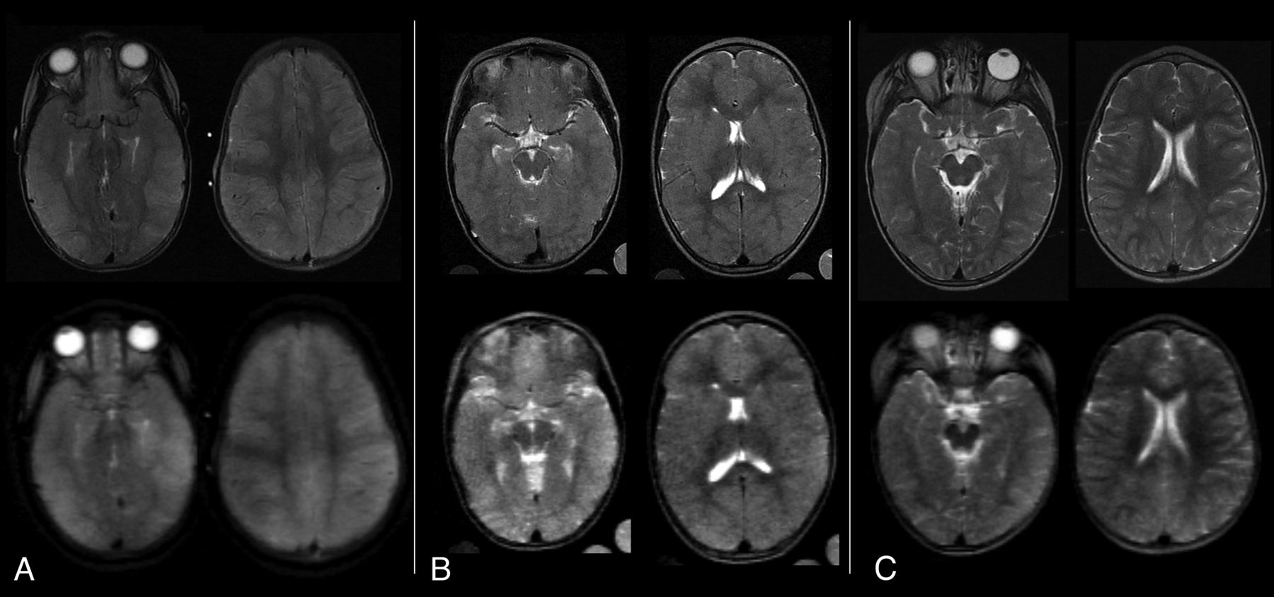
  • FIG 2.
    • Download figure
    • Open in new tab
    • Download powerpoint
    FIG 2.

    Sample T2w original and simulated MRIs from children with CM. The original brain MRIs were obtained on a Signa Ovation 0.35 T magnet (upper row) and corresponding axial images were obtained after image degradation to simulate the resolution of a very-low-field scanner (lower row). A, A BVS of 8 was assigned to the original scan as well as to the degraded images by all 3 radiologists. B, A BVS of 6 was assigned to the original scan, and 6 or 7, to the degraded images. C, A BVS of 4 was assigned to the original scan, and 5 or 3, to the degraded images.

Tables

  • Figures
    • View popup
    Table 1:

    MRI criteria for assignment of brain volume scores

    BVSBrief DescriptionFeatures
    1Severe atrophyMarkedly atypical for age with diffuse prominence of the cisternae, ventricles, and sulci
    2Mild atrophySubtle prominence of the cisternae, ventricles, and sulci for age
    3NormalTypical for age
    4Mild increased volumeMildly increased brain volume for age, but with normal cisternae, ventricles, and sulci
    5Mild swellingSome localized or regional loss of cisternae, ventricles, and sulci
    6Moderate swellingDiffuse involvement with incomplete effacement of the cisternae, ventricles, and sulci
    6.5aModerate-to-severe swellingFeatures between 6 and 7, eg, near-complete effacement of the sulci without cisternal/ventricular effacement or significant cisternal/ventricular effacement with partial loss of the sulci
    7Severe swellingLoss of all sulci with cisternal and ventricular effacement and decreased gray/white matter delineation
    8Severe swelling with herniationLoss of all sulci with cisternal and ventricular effacement with herniation
    • ↵a Score of 6.5 was not included in the original BVS.

    • View popup
    Table 2:

    ORs for mortality at a BVS threshold of 7 or 6

    Fisher TestOR95% CIP Value
    7–8 vs ≤6.516.174.3–69.7<.001
    6–8 vs ≤511.501.6–503.7.005
    Seydel et al7 (7–8 vs ≤6)13.75a4.3–58.6a<.001
    • ↵a The OR and CI reported in Seydel et al7 used logistic regression and was 14.0 (4.5–43.4). Here we use Fisher exact test instead because of its increased accuracy for sample sizes in this study, which was then also applied to Seydel et al7 to make these results comparable. Accordingly, the recalculated OR and CI are slightly different than that reported in Seydel et al7.

    • View popup
    Table 3:

    BVS assigned to degraded MRIs compared with original criterion standard BVS assigned to nondegraded scans

    Test StatisticR1R2R3Median
    Intraclass coefficient (95% CI)0.901 (0.830–0.944)0.878 (0.777–0.932)0.867 (0.762–0.926)0.928 (0.874–0.959)
    Mean bias+0.170–0.340+0.319+0.128
    Mean absolute deviation0.5530.5960.6170.468
PreviousNext
Back to top

In this issue

American Journal of Neuroradiology: 45 (2)
American Journal of Neuroradiology
Vol. 45, Issue 2
1 Feb 2024
  • Table of Contents
  • Index by author
  • Complete Issue (PDF)
Advertisement
Print
Download PDF
Email Article
Cite this article
0 Responses
Respond to this article
Share
Bookmark this article
MRI-Based Brain Volume Scoring in Cerebral Malaria
Manu S. Goyal, Lorenna Vidal, Karen Chetcuti, Cowles Chilingulo, Khalid Ibrahim, Jeffrey Zhang, Dylan S. Small, Karl B. Seydel, Nicole O’Brien, Terrie E. Taylor, Douglas G. Postels
American Journal of Neuroradiology Feb 2024, 45 (2) 205-210; DOI: 10.3174/ajnr.A8098
del.icio.us logo Twitter logo Facebook logo Mendeley logo
  • Tweet Widget
Purchase

Jump to section

  • Article
    • Abstract
    • ABBREVIATIONS:
    • MATERIALS AND METHODS
    • RESULTS
    • DISCUSSION
    • CONCLUSIONS
    • Acknowledgments
    • Footnotes
    • References
  • Figures & Data
  • Info & Metrics
  • Responses
  • References
  • PDF

Related Articles

  • No related articles found.
  • PubMed
  • Google Scholar

Cited By...

  • No citing articles found.
  • Crossref
  • Google Scholar

This article has not yet been cited by articles in journals that are participating in Crossref Cited-by Linking.

More in this TOC Section

  • Pediatric Glioma:Imaging Clusters,Molecular Traits
  • Clinical SVR of Fetal Brain MRI
  • FRACTURE MR in Congenital Vertebral Anomalies
Show more PEDIATRIC NEUROIMAGING

Similar Articles

Advertisement

Indexed Content

  • Current Issue
  • Accepted Manuscripts
  • Article Preview
  • Past Issues
  • Editorials
  • Editor's Choice
  • Fellows' Journal Club
  • Letters to the Editor
  • Video Articles

Cases

  • Case Collection
  • Archive - Case of the Week
  • Archive - Case of the Month
  • Archive - Classic Case

More from AJNR

  • Trainee Corner
  • Imaging Protocols
  • MRI Safety Corner
  • Book Reviews

Multimedia

  • AJNR Podcasts
  • AJNR Scantastics

Resources

  • Turnaround Time
  • Submit a Manuscript
  • Submit a Video Article
  • Submit an eLetter to the Editor/Response
  • Manuscript Submission Guidelines
  • Statistical Tips
  • Fast Publishing of Accepted Manuscripts
  • Graphical Abstract Preparation
  • Imaging Protocol Submission
  • Evidence-Based Medicine Level Guide
  • Publishing Checklists
  • Author Policies
  • Become a Reviewer/Academy of Reviewers
  • News and Updates

About Us

  • About AJNR
  • Editorial Board
  • Editorial Board Alumni
  • Alerts
  • Permissions
  • Not an AJNR Subscriber? Join Now
  • Advertise with Us
  • Librarian Resources
  • Feedback
  • Terms and Conditions
  • AJNR Editorial Board Alumni

American Society of Neuroradiology

  • Not an ASNR Member? Join Now

© 2025 by the American Society of Neuroradiology All rights, including for text and data mining, AI training, and similar technologies, are reserved.
Print ISSN: 0195-6108 Online ISSN: 1936-959X

Powered by HighWire
Email this Article

Thank you for your interest in spreading the word on American Journal of Neuroradiology.

NOTE: We only request your email address so that the person you are recommending the page to knows that you wanted them to see it, and that it is not junk mail. We do not capture any email address.

Enter multiple addresses on separate lines or separate them with commas.
MRI-Based Brain Volume Scoring in Cerebral Malaria Is Externally Valid and Applicable to Lower-Resolution Images
(Your Name) has sent you a message from American Journal of Neuroradiology
(Your Name) thought you would like to see the American Journal of Neuroradiology web site.
CAPTCHA
This question is for testing whether or not you are a human visitor and to prevent automated spam submissions.
Cite this article
Manu S. Goyal, Lorenna Vidal, Karen Chetcuti, Cowles Chilingulo, Khalid Ibrahim, Jeffrey Zhang, Dylan S. Small, Karl B. Seydel, Nicole O’Brien, Terrie E. Taylor, Douglas G. Postels
MRI-Based Brain Volume Scoring in Cerebral Malaria Is Externally Valid and Applicable to Lower-Resolution Images
American Journal of Neuroradiology Feb 2024, 45 (2) 205-210; DOI: 10.3174/ajnr.A8098

Citation Manager Formats

  • BibTeX
  • Bookends
  • EasyBib
  • EndNote (tagged)
  • EndNote 8 (xml)
  • Medlars
  • Mendeley
  • Papers
  • RefWorks Tagged
  • Ref Manager
  • RIS
  • Zotero

We use cookies on this site to enhance your user experience. By clicking any link on this page you are giving your consent for us to set cookies.