Advertisement

Main menu

  • Home
  • Content
    • Current Issue
    • Accepted Manuscripts
    • Article Preview
    • Past Issue Archive
    • Video Articles
    • AJNR Case Collection
    • Case of the Week Archive
    • Case of the Month Archive
    • Classic Case Archive
  • Special Collections
    • Advancing NeuroMRI with High-Relaxivity Contrast Agents
    • AJNR Awards
    • Low-Field MRI
    • Alzheimer Disease
    • ASNR Foundation Special Collection
    • Photon-Counting CT
    • View All
  • Multimedia
    • AJNR Podcasts
    • AJNR SCANtastic
    • Trainee Corner
    • MRI Safety Corner
    • Imaging Protocols
  • For Authors
    • Submit a Manuscript
    • Submit a Video Article
    • Submit an eLetter to the Editor/Response
    • Manuscript Submission Guidelines
    • Statistical Tips
    • Fast Publishing of Accepted Manuscripts
    • Graphical Abstract Preparation
    • Imaging Protocol Submission
    • Author Policies
  • About Us
    • About AJNR
    • Editorial Board
    • Editorial Board Alumni
  • More
    • Become a Reviewer/Academy of Reviewers
    • Subscribers
    • Permissions
    • Alerts
    • Feedback
    • Advertisers
    • ASNR Home

User menu

  • Alerts
  • Log in

Search

  • Advanced search
American Journal of Neuroradiology

American Journal of Neuroradiology

ASHNR American Society of Functional Neuroradiology ASHNR American Society of Pediatric Neuroradiology ASSR
  • Alerts
  • Log in

Advanced Search

  • Home
  • Content
    • Current Issue
    • Accepted Manuscripts
    • Article Preview
    • Past Issue Archive
    • Video Articles
    • AJNR Case Collection
    • Case of the Week Archive
    • Case of the Month Archive
    • Classic Case Archive
  • Special Collections
    • Advancing NeuroMRI with High-Relaxivity Contrast Agents
    • AJNR Awards
    • Low-Field MRI
    • Alzheimer Disease
    • ASNR Foundation Special Collection
    • Photon-Counting CT
    • View All
  • Multimedia
    • AJNR Podcasts
    • AJNR SCANtastic
    • Trainee Corner
    • MRI Safety Corner
    • Imaging Protocols
  • For Authors
    • Submit a Manuscript
    • Submit a Video Article
    • Submit an eLetter to the Editor/Response
    • Manuscript Submission Guidelines
    • Statistical Tips
    • Fast Publishing of Accepted Manuscripts
    • Graphical Abstract Preparation
    • Imaging Protocol Submission
    • Author Policies
  • About Us
    • About AJNR
    • Editorial Board
    • Editorial Board Alumni
  • More
    • Become a Reviewer/Academy of Reviewers
    • Subscribers
    • Permissions
    • Alerts
    • Feedback
    • Advertisers
    • ASNR Home
  • Follow AJNR on Twitter
  • Visit AJNR on Facebook
  • Follow AJNR on Instagram
  • Join AJNR on LinkedIn
  • RSS Feeds

AJNR Awards, New Junior Editors, and more. Read the latest AJNR updates


Improved Turnaround Times | Median time to first decision: 12 days

Research ArticleBrain Tumor Imaging

Quantification of T2-FLAIR Mismatch in Nonenhancing Diffuse Gliomas Using Digital Subtraction

Nicholas S. Cho, Francesco Sanvito, Viên Lam Le, Sonoko Oshima, Ashley Teraishi, Jingwen Yao, Donatello Telesca, Catalina Raymond, Whitney B. Pope, Phioanh L. Nghiemphu, Albert Lai, Timothy F. Cloughesy, Noriko Salamon and Benjamin M. Ellingson
American Journal of Neuroradiology February 2024, 45 (2) 188-197; DOI: https://doi.org/10.3174/ajnr.A8094
Nicholas S. Cho
aFrom the UCLA Brain Tumor Imaging Laboratory (N.S.C., F.S., V.L.L., S.O., A.T., J.Y., C.R., B.M.E.), Center for Computer Vision and Imaging Biomarkers, University of California, Los Angeles, Los Angeles, California
bDepartment of Radiological Sciences (N.S.C., F.S., V.L.L., S.O., A.T., J.Y., C.R., W.B.P., N.S., B.M.E.), David Geffen School of Medicine, University of California, Los Angeles, Los Angeles, California
cDepartment of Bioengineering (N.S.C., V.L.L., B.M.E.), Henry Samueli School of Engineering and Applied Science, University of California, Los Angeles, Los Angeles, California
dMedical Scientist Training Program (N.S.C.), David Geffen School of Medicine, University of California, Los Angeles, Los Angeles, California
  • Find this author on Google Scholar
  • Find this author on PubMed
  • Search for this author on this site
  • ORCID record for Nicholas S. Cho
Francesco Sanvito
aFrom the UCLA Brain Tumor Imaging Laboratory (N.S.C., F.S., V.L.L., S.O., A.T., J.Y., C.R., B.M.E.), Center for Computer Vision and Imaging Biomarkers, University of California, Los Angeles, Los Angeles, California
bDepartment of Radiological Sciences (N.S.C., F.S., V.L.L., S.O., A.T., J.Y., C.R., W.B.P., N.S., B.M.E.), David Geffen School of Medicine, University of California, Los Angeles, Los Angeles, California
  • Find this author on Google Scholar
  • Find this author on PubMed
  • Search for this author on this site
  • ORCID record for Francesco Sanvito
Viên Lam Le
aFrom the UCLA Brain Tumor Imaging Laboratory (N.S.C., F.S., V.L.L., S.O., A.T., J.Y., C.R., B.M.E.), Center for Computer Vision and Imaging Biomarkers, University of California, Los Angeles, Los Angeles, California
bDepartment of Radiological Sciences (N.S.C., F.S., V.L.L., S.O., A.T., J.Y., C.R., W.B.P., N.S., B.M.E.), David Geffen School of Medicine, University of California, Los Angeles, Los Angeles, California
cDepartment of Bioengineering (N.S.C., V.L.L., B.M.E.), Henry Samueli School of Engineering and Applied Science, University of California, Los Angeles, Los Angeles, California
  • Find this author on Google Scholar
  • Find this author on PubMed
  • Search for this author on this site
Sonoko Oshima
aFrom the UCLA Brain Tumor Imaging Laboratory (N.S.C., F.S., V.L.L., S.O., A.T., J.Y., C.R., B.M.E.), Center for Computer Vision and Imaging Biomarkers, University of California, Los Angeles, Los Angeles, California
bDepartment of Radiological Sciences (N.S.C., F.S., V.L.L., S.O., A.T., J.Y., C.R., W.B.P., N.S., B.M.E.), David Geffen School of Medicine, University of California, Los Angeles, Los Angeles, California
  • Find this author on Google Scholar
  • Find this author on PubMed
  • Search for this author on this site
Ashley Teraishi
aFrom the UCLA Brain Tumor Imaging Laboratory (N.S.C., F.S., V.L.L., S.O., A.T., J.Y., C.R., B.M.E.), Center for Computer Vision and Imaging Biomarkers, University of California, Los Angeles, Los Angeles, California
bDepartment of Radiological Sciences (N.S.C., F.S., V.L.L., S.O., A.T., J.Y., C.R., W.B.P., N.S., B.M.E.), David Geffen School of Medicine, University of California, Los Angeles, Los Angeles, California
  • Find this author on Google Scholar
  • Find this author on PubMed
  • Search for this author on this site
  • ORCID record for Ashley Teraishi
Jingwen Yao
aFrom the UCLA Brain Tumor Imaging Laboratory (N.S.C., F.S., V.L.L., S.O., A.T., J.Y., C.R., B.M.E.), Center for Computer Vision and Imaging Biomarkers, University of California, Los Angeles, Los Angeles, California
bDepartment of Radiological Sciences (N.S.C., F.S., V.L.L., S.O., A.T., J.Y., C.R., W.B.P., N.S., B.M.E.), David Geffen School of Medicine, University of California, Los Angeles, Los Angeles, California
  • Find this author on Google Scholar
  • Find this author on PubMed
  • Search for this author on this site
  • ORCID record for Jingwen Yao
Donatello Telesca
eDepartment of Biostatistics (D.T.), Fielding School of Public Health, University of California Los Angeles, Los Angeles, California
  • Find this author on Google Scholar
  • Find this author on PubMed
  • Search for this author on this site
Catalina Raymond
aFrom the UCLA Brain Tumor Imaging Laboratory (N.S.C., F.S., V.L.L., S.O., A.T., J.Y., C.R., B.M.E.), Center for Computer Vision and Imaging Biomarkers, University of California, Los Angeles, Los Angeles, California
bDepartment of Radiological Sciences (N.S.C., F.S., V.L.L., S.O., A.T., J.Y., C.R., W.B.P., N.S., B.M.E.), David Geffen School of Medicine, University of California, Los Angeles, Los Angeles, California
  • Find this author on Google Scholar
  • Find this author on PubMed
  • Search for this author on this site
  • ORCID record for Catalina Raymond
Whitney B. Pope
bDepartment of Radiological Sciences (N.S.C., F.S., V.L.L., S.O., A.T., J.Y., C.R., W.B.P., N.S., B.M.E.), David Geffen School of Medicine, University of California, Los Angeles, Los Angeles, California
  • Find this author on Google Scholar
  • Find this author on PubMed
  • Search for this author on this site
  • ORCID record for Whitney B. Pope
Phioanh L. Nghiemphu
fUCLA Neuro-Oncology Program (P.L.N., A.L., T.F.C.), David Geffen School of Medicine, University of California, Los Angeles, Los Angeles, California
gDepartment of Neurology (P.L.N., A.L., T.F.C.), David Geffen School of Medicine, University of California, Los Angeles, Los Angeles, California
  • Find this author on Google Scholar
  • Find this author on PubMed
  • Search for this author on this site
  • ORCID record for Phioanh L. Nghiemphu
Albert Lai
fUCLA Neuro-Oncology Program (P.L.N., A.L., T.F.C.), David Geffen School of Medicine, University of California, Los Angeles, Los Angeles, California
gDepartment of Neurology (P.L.N., A.L., T.F.C.), David Geffen School of Medicine, University of California, Los Angeles, Los Angeles, California
  • Find this author on Google Scholar
  • Find this author on PubMed
  • Search for this author on this site
  • ORCID record for Albert Lai
Timothy F. Cloughesy
fUCLA Neuro-Oncology Program (P.L.N., A.L., T.F.C.), David Geffen School of Medicine, University of California, Los Angeles, Los Angeles, California
gDepartment of Neurology (P.L.N., A.L., T.F.C.), David Geffen School of Medicine, University of California, Los Angeles, Los Angeles, California
  • Find this author on Google Scholar
  • Find this author on PubMed
  • Search for this author on this site
  • ORCID record for Timothy F. Cloughesy
Noriko Salamon
bDepartment of Radiological Sciences (N.S.C., F.S., V.L.L., S.O., A.T., J.Y., C.R., W.B.P., N.S., B.M.E.), David Geffen School of Medicine, University of California, Los Angeles, Los Angeles, California
  • Find this author on Google Scholar
  • Find this author on PubMed
  • Search for this author on this site
  • ORCID record for Noriko Salamon
Benjamin M. Ellingson
aFrom the UCLA Brain Tumor Imaging Laboratory (N.S.C., F.S., V.L.L., S.O., A.T., J.Y., C.R., B.M.E.), Center for Computer Vision and Imaging Biomarkers, University of California, Los Angeles, Los Angeles, California
bDepartment of Radiological Sciences (N.S.C., F.S., V.L.L., S.O., A.T., J.Y., C.R., W.B.P., N.S., B.M.E.), David Geffen School of Medicine, University of California, Los Angeles, Los Angeles, California
cDepartment of Bioengineering (N.S.C., V.L.L., B.M.E.), Henry Samueli School of Engineering and Applied Science, University of California, Los Angeles, Los Angeles, California
hDepartment of Neurosurgery (B.M.E.), David Geffen School of Medicine, University of California, Los Angeles, Los Angeles, California
iDepartment of Psychiatry and Biobehavioral Sciences (B.M.E.), David Geffen School of Medicine, University of California, Los Angeles, Los Angeles, California
  • Find this author on Google Scholar
  • Find this author on PubMed
  • Search for this author on this site
  • ORCID record for Benjamin M. Ellingson
  • Article
  • Figures & Data
  • Supplemental
  • Info & Metrics
  • Responses
  • References
  • PDF
Loading

Article Figures & Data

Figures

  • Tables
  • FIG 1.
    • Download figure
    • Open in new tab
    • Download powerpoint
    FIG 1.

    Representative true-positive and true-negative typical cases with T2-FLAIR subtraction maps and tumor segmentations. A, Patient A is a 38-year-old woman from the TCIA cohort diagnosed with a grade 2 IDHm-A demonstrating 59.6% T2-FLAIR mismatch volume (true-positive result). B, Patient B is a 23-year-old man from the institutional cohort diagnosed with grade 2 IDHm-A demonstrating 48.0% T2-FLAIR mismatch volume (true-positive result). C, Patient C is a 35-year-old woman from the TCIA cohort diagnosed with grade 2 IDHm-O demonstrating heterogeneous T2-weighted and T2-weighted FLAIR hyperintensity and no T2-FLAIR mismatch on the subtraction map (true-negative result). D, Patient D is a 59-year-old woman from the institutional cohort diagnosed with grade 4 IDHwt glioblastoma. The relative hypointense central T2-weighted FLAIR signal with a peripheral hyperintense rim may mimic a T2FM sign at first glance, but the corresponding absence of T2FM on the subtraction map from the heterogeneous T2-weighted hyperintensity clearly shows the lack of T2FM (true-negative result). Corresponding tumor segmentation VOIs of T2FM (pink) and T2FNM (green) subregions (A–D) as well as the T2FNM subregion external borders encoded with thickness (A/C) are shown. INST indicates institutional.

  • FIG 2.
    • Download figure
    • Open in new tab
    • Download powerpoint
    FIG 2.

    Representative false-negative and false-positive cases with T2-FLAIR subtraction maps and tumor segmentations. A, Patient A is a 38-year-old man from the TCIA cohort diagnosed with grade 2 IDHm-A with no T2-FLAIR mismatch on the subtraction map (false-negative result). B, Patient B is a 36-year-old woman from the institutional cohort diagnosed with grade 2 IDHm-A with no T2-FLAIR mismatch on the subtraction map excluding the cystic portion (false-negative result). C, Patient C is a 45-year-old man from the TCIA cohort diagnosed with grade 2 IDHm-O demonstrating 35.0% T2FM volume, which was assessed as a false-positive for the T2FM sign by 1 reader using subtraction maps (false-positive result). D, Patient D is a 35-year-old woman diagnosed with IDHwt glioblastoma demonstrating 12.5% T2FM volume, which was assessed as a false-positive for the T2-FLAIR mismatch sign by both readers using subtraction maps. Corresponding tumor-segmentation VOIs of T2FM (pink) and T2FNM (green) subregions are shown (A–D) (false-positive result).
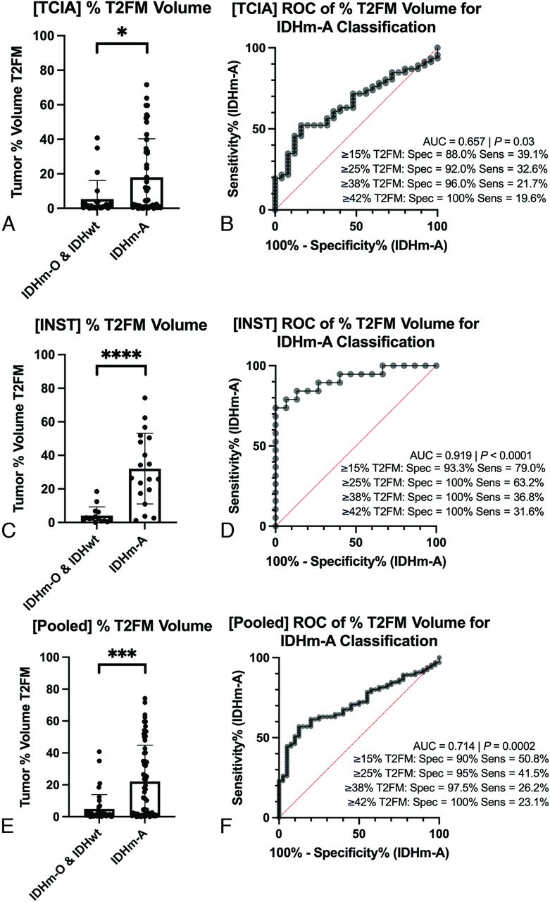
  • FIG 3.
    • Download figure
    • Open in new tab
    • Download powerpoint
    FIG 3.

    Tumor percentage of T2FM volume and diagnostic performance for IDHm-A classification. Results are shown for both the TCIA cohort (A and B) and the institutional cohort (C and D). IDHm-A had a higher percentage T2FM volume (% T2FM volume) compared with the other molecular signatures (A and C). ROC curves show that a specificity of 100% for the IDHm-A diagnosis could be achieved with a threshold of ≥42% T2FM volume in both cohorts (B and D), though in the validation cohort, a threshold of ≥25% T2FM volume sufficed (D). The summary results when pooling the 2 cohorts are also shown (E and F). INST indicates institutional; Spec, specificity; Sens, sensitivity; *, P < .05; ***, P < .001; ****, P < .0001.

  • FIG 4.
    • Download figure
    • Open in new tab
    • Download powerpoint
    FIG 4.

    T2FNM thickness, tumor grade, and tumor volume relationships based on tumor percentage T2FM volume. In both cohorts, tumor percentage T2FM volume (% T2FM volume) was significantly negatively correlated with T2FNM subregion thickness (A and C, P < .0001). Grade 3–4 IDHm-A had significantly higher percentage T2FM volume compared to grade 2 IDHm-A (B, TCIA, P = .008; E, institutional, P = .03), and tumor volumes were significantly greater in ≥15% T2FM volume IDHm-A compared with <15% T2FM volume IDHm-A (C, TCIA, P = .01; F, institutional, P = .04). INST indicates institutional; *, P < .05, **, P < .01.

Tables

  • Figures
    • View popup
    Table 1:

    Patient data

    CharacteristicTCIA Cohort (n = 70 Patients with n = 71 Lesions)Institutional Cohort (n = 34 Patients/Lesions)
    Age (mean) (range)43 (22–78)42 (22–79)
    Sex: male/female41/2918/16
    Diagnosis (No.) (%)
    IDHm-A46 (64.8%)19 (55.9%)
      Grade 23311
      Grade 3137
      Grade 401
    IDHm-O9 (12.7%)9 (26.5%)
     Grade 298
     Grade 301
    IDHwt glioma16 (22.5%)6 (17.6%)
    • View popup
    Table 2:

    Reader study results of T2FM sign: Sensitivity and specificity

    Classification Definition/ReaderWith Subtraction MapSensitivity (95% CI)Specificity (95% CI)
    Patel et al6
     Reader 1No35.4 (23.9–48.2)97.5 (86.8–99.9)
    Yes36.9 (25.3–49.8)92.5 (79.6–98.4)
     Reader 2No30.8 (19.9–43.5)92.5 (79.6–98.4)
    Yes38.5 (26.7–51.4)92.5 (79.6–98.4)
    Lasocki et al8
     Reader 1No50.8 (38.1–63.4)90.0 (76.3–97.2)
    Yes55.4 (42.5–67.7)85.0 (70.2–94.3)
     Reader 2No41.5 (29.4–54.4)92.5 (79.6–98.4)
    Yes49.2 (36.6–61.9)92.5 (79.6–98.4)
    • View popup
    Table 3:

    Reader study results of T2FM sign: Interreader agreement

    Classification Definition/with Subtraction MapCohen κ (95% CI)
    Patel et al6
     No0.70 (0.53–0.87)
     Yes0.78 (0.64–0.92)
    Lasocki et al8
     No0.76 (0.63–0.89)
     Yes0.78 (0.65–0.90)
PreviousNext
Back to top

In this issue

American Journal of Neuroradiology: 45 (2)
American Journal of Neuroradiology
Vol. 45, Issue 2
1 Feb 2024
  • Table of Contents
  • Index by author
  • Complete Issue (PDF)
Advertisement
Print
Download PDF
Email Article

Thank you for your interest in spreading the word on American Journal of Neuroradiology.

NOTE: We only request your email address so that the person you are recommending the page to knows that you wanted them to see it, and that it is not junk mail. We do not capture any email address.

Enter multiple addresses on separate lines or separate them with commas.
Quantification of T2-FLAIR Mismatch in Nonenhancing Diffuse Gliomas Using Digital Subtraction
(Your Name) has sent you a message from American Journal of Neuroradiology
(Your Name) thought you would like to see the American Journal of Neuroradiology web site.
CAPTCHA
This question is for testing whether or not you are a human visitor and to prevent automated spam submissions.
Cite this article
Nicholas S. Cho, Francesco Sanvito, Viên Lam Le, Sonoko Oshima, Ashley Teraishi, Jingwen Yao, Donatello Telesca, Catalina Raymond, Whitney B. Pope, Phioanh L. Nghiemphu, Albert Lai, Timothy F. Cloughesy, Noriko Salamon, Benjamin M. Ellingson
Quantification of T2-FLAIR Mismatch in Nonenhancing Diffuse Gliomas Using Digital Subtraction
American Journal of Neuroradiology Feb 2024, 45 (2) 188-197; DOI: 10.3174/ajnr.A8094

Citation Manager Formats

  • BibTeX
  • Bookends
  • EasyBib
  • EndNote (tagged)
  • EndNote 8 (xml)
  • Medlars
  • Mendeley
  • Papers
  • RefWorks Tagged
  • Ref Manager
  • RIS
  • Zotero
0 Responses
Respond to this article
Share
Bookmark this article
Quantifying T2-FLAIR Mismatch in Diffuse Gliomas
Nicholas S. Cho, Francesco Sanvito, Viên Lam Le, Sonoko Oshima, Ashley Teraishi, Jingwen Yao, Donatello Telesca, Catalina Raymond, Whitney B. Pope, Phioanh L. Nghiemphu, Albert Lai, Timothy F. Cloughesy, Noriko Salamon, Benjamin M. Ellingson
American Journal of Neuroradiology Feb 2024, 45 (2) 188-197; DOI: 10.3174/ajnr.A8094
del.icio.us logo Twitter logo Facebook logo Mendeley logo
  • Tweet Widget
  • Facebook Like
  • Google Plus One
Purchase

Jump to section

  • Article
    • Abstract
    • ABBREVIATIONS:
    • MATERIALS AND METHODS
    • RESULTS
    • DISCUSSION
    • CONCLUSIONS
    • Footnotes
    • References
  • Figures & Data
  • Supplemental
  • Info & Metrics
  • Responses
  • References
  • PDF

Related Articles

  • PubMed
  • Google Scholar

Cited By...

  • No citing articles found.
  • Crossref
  • Google Scholar

This article has not yet been cited by articles in journals that are participating in Crossref Cited-by Linking.

More in this TOC Section

  • T1WI Signal Distinguishes Brain SFT and Meningioma
  • Brain Tumor Subtyping Image-Based search via MRI
  • Neuroimaging of Erdheim-Chester Disease
Show more BRAIN TUMOR IMAGING

Similar Articles

Advertisement

Indexed Content

  • Current Issue
  • Accepted Manuscripts
  • Article Preview
  • Past Issues
  • Editorials
  • Editor's Choice
  • Fellows' Journal Club
  • Letters to the Editor
  • Video Articles

Cases

  • Case Collection
  • Archive - Case of the Week
  • Archive - Case of the Month
  • Archive - Classic Case

More from AJNR

  • Trainee Corner
  • Imaging Protocols
  • MRI Safety Corner
  • Book Reviews

Multimedia

  • AJNR Podcasts
  • AJNR Scantastics

Resources

  • Turnaround Time
  • Submit a Manuscript
  • Submit a Video Article
  • Submit an eLetter to the Editor/Response
  • Manuscript Submission Guidelines
  • Statistical Tips
  • Fast Publishing of Accepted Manuscripts
  • Graphical Abstract Preparation
  • Imaging Protocol Submission
  • Evidence-Based Medicine Level Guide
  • Publishing Checklists
  • Author Policies
  • Become a Reviewer/Academy of Reviewers
  • News and Updates

About Us

  • About AJNR
  • Editorial Board
  • Editorial Board Alumni
  • Alerts
  • Permissions
  • Not an AJNR Subscriber? Join Now
  • Advertise with Us
  • Librarian Resources
  • Feedback
  • Terms and Conditions
  • AJNR Editorial Board Alumni

American Society of Neuroradiology

  • Not an ASNR Member? Join Now

© 2025 by the American Society of Neuroradiology All rights, including for text and data mining, AI training, and similar technologies, are reserved.
Print ISSN: 0195-6108 Online ISSN: 1936-959X

Powered by HighWire