
Article Figures & Data
Figures
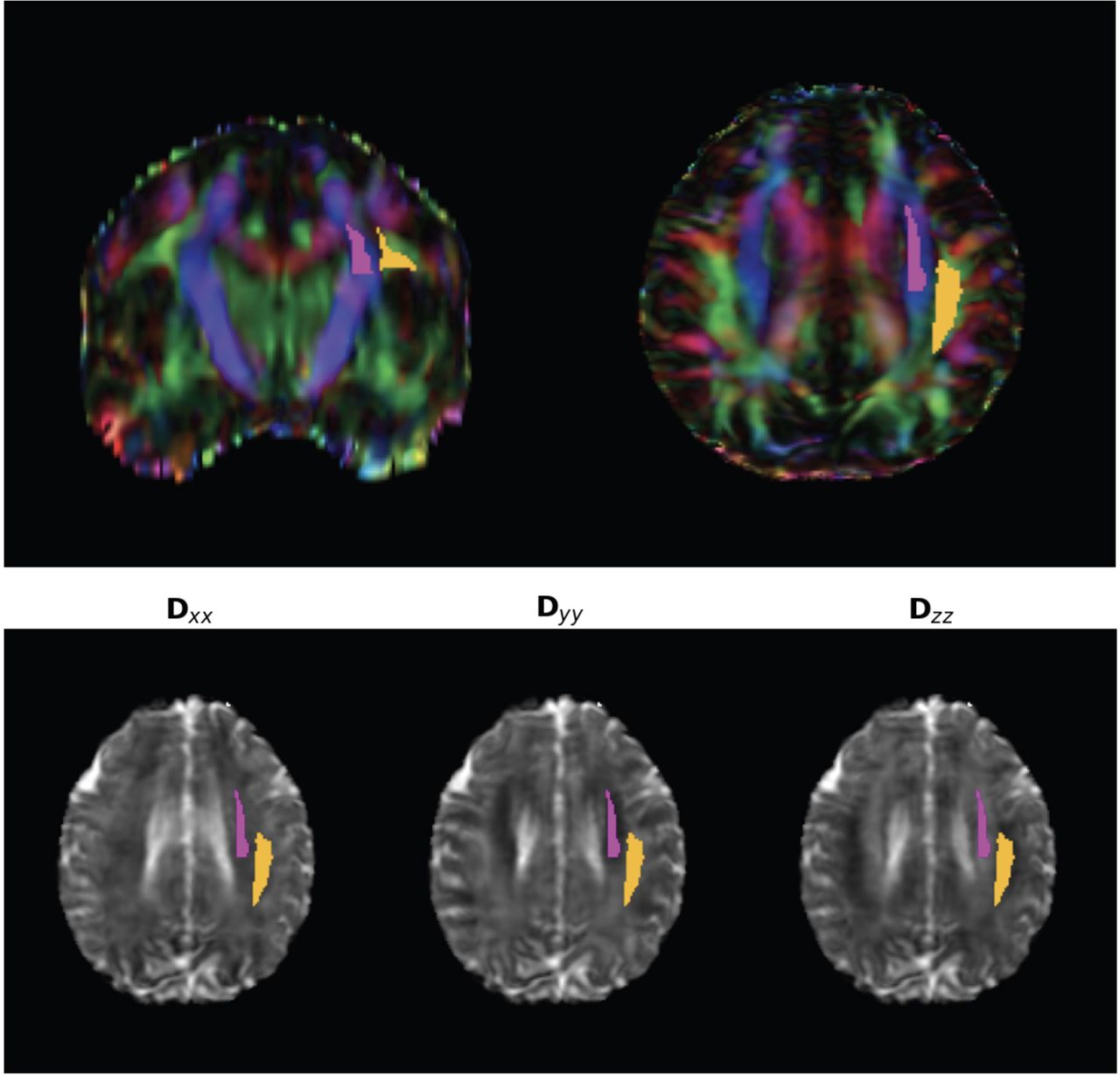
- FIG 1.
ROI by using atlas-based ALPS index. Top row: The projection (superior and posterior corona radiata, magenta) and association (superior longitudinal fasciculus, yellow) fibers were defined by the labels of the ICBM DTI-81 Atlas. Bottom row: Gray-scale maps of X, Y, and Z diffusion with ROIs.
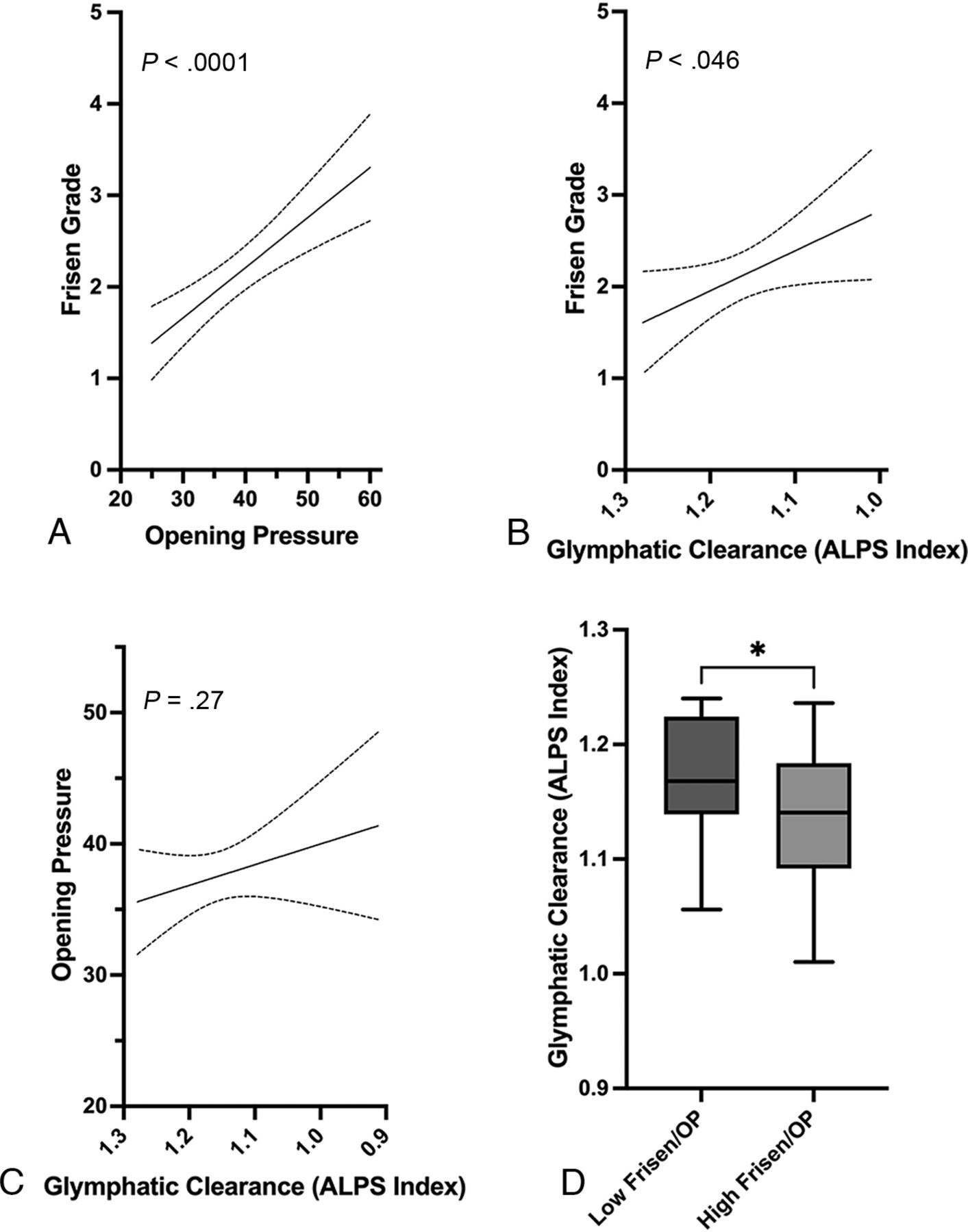
- FIG 2.
A, Association between lumbar puncture opening pressure and Frisen papilledema grade in patients with IIH. B, Association between glymphatic function (ALPS index) and Frisen papilledema grade. The glymphatic clearance on the x-axis is declining moving left to right. C, Association between glymphatic clearance and lumbar puncture opening pressure in patients with IIH. D, Box-and-whisker plot showing difference in glymphatic clearance between patients with IIH and low Frisen grade papilledema (0–2) and lower opening pressure (<50th percentile in cohort) compared with patients with IIH and high Frisen grade papilledema (3–5) and higher opening pressure (≥50th percentile). * indicates P < .05.
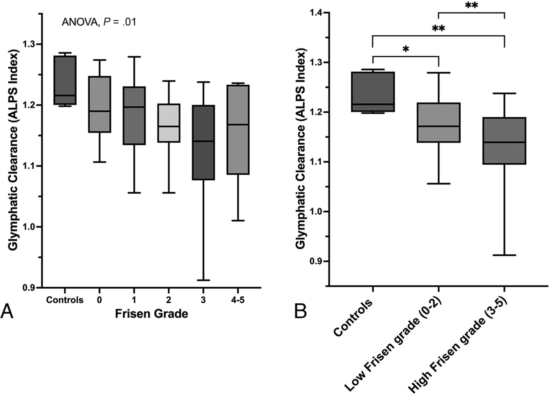
- FIG 3.
A, Box-and-whisker plot showing glymphatic clearance and how it relates between controls and patients with IIH and varying Frisen papilledema grades. B, Box-and-whisker plot showing difference in glymphatic clearance between controls and patients with IIH and low-grade papilledema (Frisen grade 0–2) and high-grade papilledema (Frisen grade 3–5). * indicates P < .05; ** indicates P < .01.
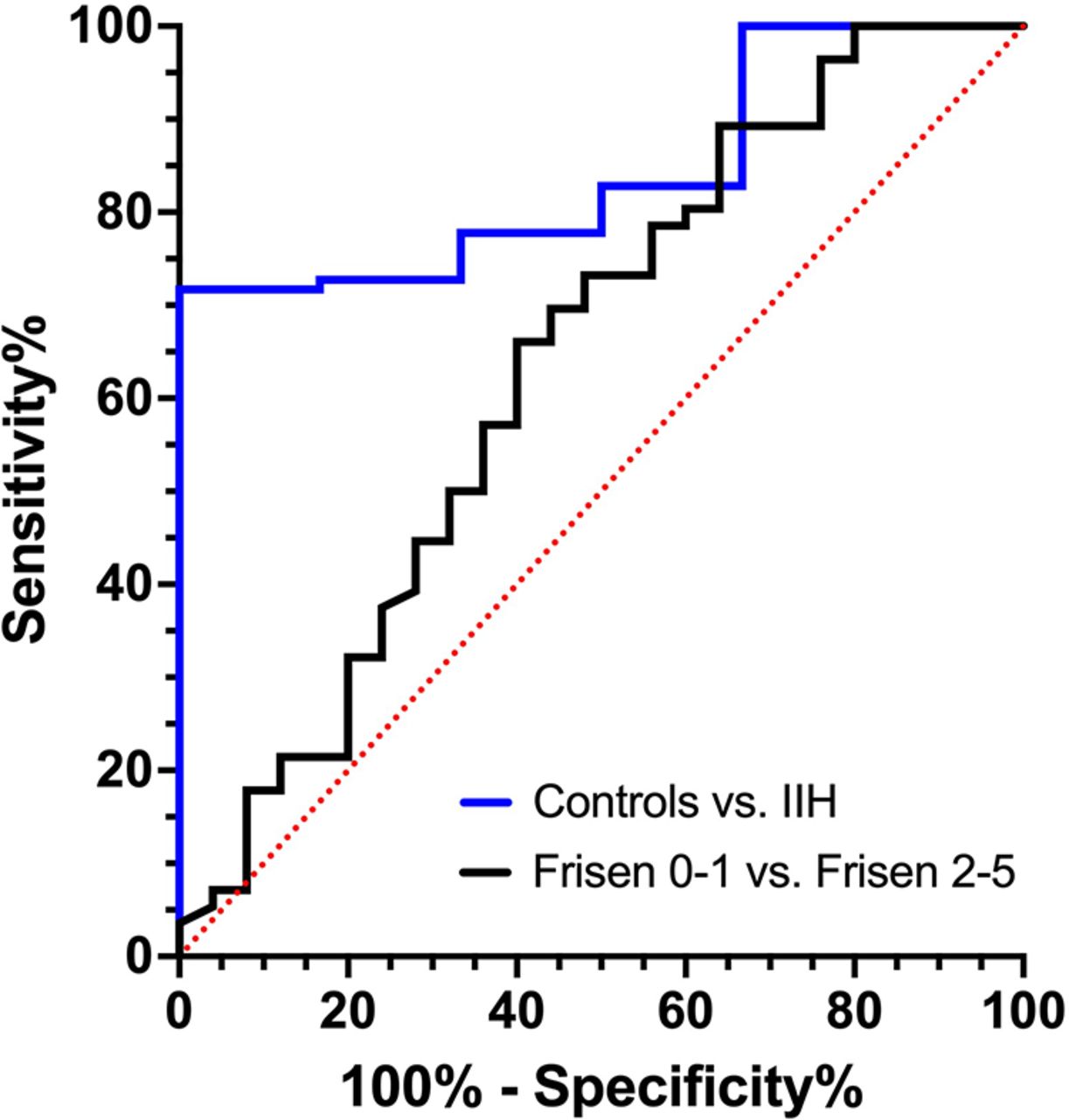
- FIG 4.
ROC curve analysis assessing the diagnostic performance of the ALPS index between patients with IIH versus controls and between patients with IIH and less-severe (Frisen papilledema grade 0–1) and more-severe (Frisen papilledema grade 2–5) disease.


