Advertisement

Main menu

  • Home
  • Content
    • Current Issue
    • Accepted Manuscripts
    • Article Preview
    • Past Issue Archive
    • Video Articles
    • AJNR Case Collection
    • Case of the Week Archive
    • Case of the Month Archive
    • Classic Case Archive
  • Special Collections
    • AJNR Awards
    • Low-Field MRI
    • Alzheimer Disease
    • ASNR Foundation Special Collection
    • Photon-Counting CT
    • View All
  • Multimedia
    • AJNR Podcasts
    • AJNR SCANtastic
    • Trainee Corner
    • MRI Safety Corner
    • Imaging Protocols
  • For Authors
    • Submit a Manuscript
    • Submit a Video Article
    • Submit an eLetter to the Editor/Response
    • Manuscript Submission Guidelines
    • Statistical Tips
    • Fast Publishing of Accepted Manuscripts
    • Graphical Abstract Preparation
    • Imaging Protocol Submission
    • Author Policies
  • About Us
    • About AJNR
    • Editorial Board
    • Editorial Board Alumni
  • More
    • Become a Reviewer/Academy of Reviewers
    • Subscribers
    • Permissions
    • Alerts
    • Feedback
    • Advertisers
    • ASNR Home

User menu

  • Alerts
  • Log in

Search

  • Advanced search
American Journal of Neuroradiology

American Journal of Neuroradiology

ASHNR American Society of Functional Neuroradiology ASHNR American Society of Pediatric Neuroradiology ASSR
  • Alerts
  • Log in

Advanced Search

  • Home
  • Content
    • Current Issue
    • Accepted Manuscripts
    • Article Preview
    • Past Issue Archive
    • Video Articles
    • AJNR Case Collection
    • Case of the Week Archive
    • Case of the Month Archive
    • Classic Case Archive
  • Special Collections
    • AJNR Awards
    • Low-Field MRI
    • Alzheimer Disease
    • ASNR Foundation Special Collection
    • Photon-Counting CT
    • View All
  • Multimedia
    • AJNR Podcasts
    • AJNR SCANtastic
    • Trainee Corner
    • MRI Safety Corner
    • Imaging Protocols
  • For Authors
    • Submit a Manuscript
    • Submit a Video Article
    • Submit an eLetter to the Editor/Response
    • Manuscript Submission Guidelines
    • Statistical Tips
    • Fast Publishing of Accepted Manuscripts
    • Graphical Abstract Preparation
    • Imaging Protocol Submission
    • Author Policies
  • About Us
    • About AJNR
    • Editorial Board
    • Editorial Board Alumni
  • More
    • Become a Reviewer/Academy of Reviewers
    • Subscribers
    • Permissions
    • Alerts
    • Feedback
    • Advertisers
    • ASNR Home
  • Follow AJNR on Twitter
  • Visit AJNR on Facebook
  • Follow AJNR on Instagram
  • Join AJNR on LinkedIn
  • RSS Feeds

AJNR Awards, New Junior Editors, and more. Read the latest AJNR updates

Research ArticleNeurovascular/Stroke Imaging

High-Resolution Head CTA: A Prospective Patient Study Comparing Image Quality of Photon-Counting Detector CT and Energy-Integrating Detector CT

Felix E. Diehn, Zhongxing Zhou, Jamison E. Thorne, Norbert G. Campeau, Alex A. Nagelschneider, Laurence J. Eckel, John C. Benson, Ajay A. Madhavan, Girish Bathla, Vance T. Lehman, Nathan R. Huber, Francis Baffour, Joel G. Fletcher, Cynthia H. McCollough and Lifeng Yu
American Journal of Neuroradiology October 2024, 45 (10) 1441-1449; DOI: https://doi.org/10.3174/ajnr.A8342
Felix E. Diehn
aFrom the Department of Radiology, Mayo Clinic, Rochester, Minnesota
  • Find this author on Google Scholar
  • Find this author on PubMed
  • Search for this author on this site
  • ORCID record for Felix E. Diehn
Zhongxing Zhou
aFrom the Department of Radiology, Mayo Clinic, Rochester, Minnesota
  • Find this author on Google Scholar
  • Find this author on PubMed
  • Search for this author on this site
Jamison E. Thorne
aFrom the Department of Radiology, Mayo Clinic, Rochester, Minnesota
  • Find this author on Google Scholar
  • Find this author on PubMed
  • Search for this author on this site
  • ORCID record for Jamison E. Thorne
Norbert G. Campeau
aFrom the Department of Radiology, Mayo Clinic, Rochester, Minnesota
  • Find this author on Google Scholar
  • Find this author on PubMed
  • Search for this author on this site
  • ORCID record for Norbert G. Campeau
Alex A. Nagelschneider
aFrom the Department of Radiology, Mayo Clinic, Rochester, Minnesota
  • Find this author on Google Scholar
  • Find this author on PubMed
  • Search for this author on this site
  • ORCID record for Alex A. Nagelschneider
Laurence J. Eckel
aFrom the Department of Radiology, Mayo Clinic, Rochester, Minnesota
  • Find this author on Google Scholar
  • Find this author on PubMed
  • Search for this author on this site
  • ORCID record for Laurence J. Eckel
John C. Benson
aFrom the Department of Radiology, Mayo Clinic, Rochester, Minnesota
  • Find this author on Google Scholar
  • Find this author on PubMed
  • Search for this author on this site
  • ORCID record for John C. Benson
Ajay A. Madhavan
aFrom the Department of Radiology, Mayo Clinic, Rochester, Minnesota
  • Find this author on Google Scholar
  • Find this author on PubMed
  • Search for this author on this site
  • ORCID record for Ajay A. Madhavan
Girish Bathla
aFrom the Department of Radiology, Mayo Clinic, Rochester, Minnesota
  • Find this author on Google Scholar
  • Find this author on PubMed
  • Search for this author on this site
  • ORCID record for Girish Bathla
Vance T. Lehman
aFrom the Department of Radiology, Mayo Clinic, Rochester, Minnesota
  • Find this author on Google Scholar
  • Find this author on PubMed
  • Search for this author on this site
  • ORCID record for Vance T. Lehman
Nathan R. Huber
aFrom the Department of Radiology, Mayo Clinic, Rochester, Minnesota
  • Find this author on Google Scholar
  • Find this author on PubMed
  • Search for this author on this site
  • ORCID record for Nathan R. Huber
Francis Baffour
aFrom the Department of Radiology, Mayo Clinic, Rochester, Minnesota
  • Find this author on Google Scholar
  • Find this author on PubMed
  • Search for this author on this site
  • ORCID record for Francis Baffour
Joel G. Fletcher
aFrom the Department of Radiology, Mayo Clinic, Rochester, Minnesota
  • Find this author on Google Scholar
  • Find this author on PubMed
  • Search for this author on this site
  • ORCID record for Joel G. Fletcher
Cynthia H. McCollough
aFrom the Department of Radiology, Mayo Clinic, Rochester, Minnesota
  • Find this author on Google Scholar
  • Find this author on PubMed
  • Search for this author on this site
  • ORCID record for Cynthia H. McCollough
Lifeng Yu
aFrom the Department of Radiology, Mayo Clinic, Rochester, Minnesota
  • Find this author on Google Scholar
  • Find this author on PubMed
  • Search for this author on this site
  • Article
  • Figures & Data
  • Supplemental
  • Info & Metrics
  • Responses
  • References
  • PDF
Loading

Article Figures & Data

Figures

  • Tables
  • FIG 1.
    • Download figure
    • Open in new tab
    • Download powerpoint
    FIG 1.

    Data preparation and training mechanism of a dedicated ultra-high resolution convolutional neural network (UHR-CNN) for denoising high resolution images from PCD-CT. The model used a modified 6-layer residual U-Net with 7 slices as input. The high-quality images were from iterative reconstruction (IR) images with a thick section thickness and the low-quality images were generated by adding noise patches from subtraction of filtered back-projection (FBP) and IR images followed by spatial decoupling.
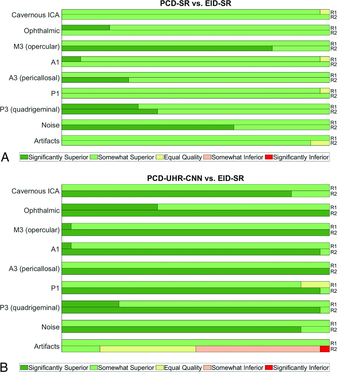
  • FIG 2.
    • Download figure
    • Open in new tab
    • Download powerpoint
    FIG 2.

    Reader evaluation, PCD-CT versus EID-CT. Horizontal percentage stacked bar chart plots demonstrate reader 1 and 2 (R1, R2) scores for PCD-SR versus EID-SR (A) and PCD-UHR-CNN versus EID-SR (B). Bars are split into segments and the length of each bar is 100%. Segments from left to right: significantly superior (+2), somewhat superior (+1), equal quality (0), somewhat inferior (−1), significantly inferior (−2).
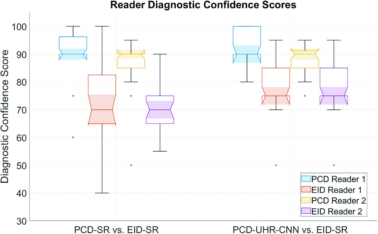
  • FIG 3.
    • Download figure
    • Open in new tab
    • Download powerpoint
    FIG 3.

    Data plot of diagnostic confidence of arterial findings. Grouped boxplot figure highlighting PCD-SR versus EID-SR and PCD-UHR-CNN versus EID-SR for diagnostic confidence scores of each reader. The bottom, top, and middle lines of each box indicate the 25th percentiles, 75th percentiles, and sample median of the data. Samples whose notches do not overlap are statistically significantly different at the 5% significance level. (Matlab 2021).

  • FIG 4.
    • Download figure
    • Open in new tab
    • Download powerpoint
    FIG 4.

    Iodine contrast, noise, and CNR quantitative evaluation. Grouped boxplots for (A) iodine contrast, (B) noise, and (C) iodine CNR results measured in the bilateral distal ICA and midcervical ICA for EID-SR, PCD-SR, and PCD-UHR-CNN. The bottom, top, and middle lines of each box indicate the 25th percentiles, 75th percentiles, and sample median of the data. Samples whose notches do not overlap are significantly different at the 5% significance level. (Matlab 2021).

  • FIG 5.
    • Download figure
    • Open in new tab
    • Download powerpoint
    FIG 5.

    Improved visualization of distal intracranial arterial branches on PCD-CT head CTA compared with EID-CT. Thin section oblique axial head CTA images highlighting the right M3 segment (arrows), by using EID-SR with 0.6-mm section thickness (A) and PCD-UHR-CNN CT with 0.2-mm section thickness (B) techniques. This distal intracranial arterial segment is best visualized by using PCD-CT rather than EID-CT, particularly with PCD-UHR-CNN–but also PCD-SR CT (the latter not shown). The right M3 segment is indeed difficult to visualize on EID-CT compared with PCD-CT. The insets demonstrate the improved visualization of the M3 segment (arrows) in the oblique sagittal plane.

  • FIG 6.
    • Download figure
    • Open in new tab
    • Download powerpoint
    FIG 6.

    Improved confidence for diagnosing infundibula on PCD-CT head CTA compared with EID-CT. Thin section oblique axial head CTA images, by using EID-SR with 0.6-mm section thickness (A) and PCD-UHR-CNN CT with 0.2-mm section thickness (B) techniques. The diagnostic confidence for diagnosing this infundibulum rather than possible aneurysm improves with PCD-SR matched to EID technique (not shown) and even more so with PCD-UHR-CNN CT at the thinnest section thickness. An ∼2-mm left supraclinoid ICA outpouching (infundibulum or aneurysm) is seen in an oblique axial plane on EID-CT (solid arrow in A), but better delineated on PCD-CT (arrow in B). The anterior choroidal artery arising from the apex of the outpouching is much better visualized on PCD-CT (arrowhead in B) than EID-CT (dashed arrow in A), thereby confidently diagnosing an infundibulum rather than an aneurysm. The insets demonstrate the improved visualization of the anterior choroidal artery arising from the apex of the outpouching in the oblique sagittal plane.

  • FIG 7.
    • Download figure
    • Open in new tab
    • Download powerpoint
    FIG 7.

    Improved characterization of arterial pathology on PCD-CT head CTA compared with EID-CT. Thin section oblique axial head CTA images by using EID-SR with 0.6-mm section thickness (A) and PCD-UHR-CNN CT with 0.2-mm section thickness (B) techniques. The diagnostic confidence for arterial pathology improves with PCD-SR matched to EID technique (not shown) and even more so with PCD-UHR-CNN CT at the thinnest section thickness. A right P2 segment stenosis has a relatively high-grade appearance on EID-CT (arrow in A). On PCD-CT, the stenosis is better demonstrated (arrow in B). The PCD-CT image depicts the stenosis as only mild to at most moderate in degree. The insets demonstrate the improved visualization of the right P2 stenosis in the oblique sagittal plane.

Tables

  • Figures
    • View popup
    Table 1:

    PCD-CT versus EID-CT scan and reconstruction parameters

    ParameterEIDPCD
    Scanner modelForceAlpha
    kV100/Sn150 kV120 kV (manual kV)
    QRM/Effective mAs/IQ-levelHead/Neck CTA: 360/225 (AEC on), Head CTA: 280/175 (AEC off)230 (CAREkeV IQ Level)
    Collimation (mm)192 × 0.6120 × 0.2
    CTDIvol (mGy)37.1 ± 4.736.1 ± 4.0
    Section/Increment (mm)0.6/0.4 (mixing ratio 0.5)0.6/0.4; 0.2/0.2
    KernelQr54Qr56; Qr89
    Matrix10241024
    Iterative reconstruction and noise reductionADMIRE-3Quantum iterative reconstruction Strength 3; UHR-CNN
    Contrast injectionOminipaque 350, 100 mL at 4 mL/s, followed by 35 mL saline at 4 mL/sOminipaque 350, 100 mL at 4 mL/s, followed by 35 mL saline at 4 mL/s
    • Note:—IQ indicates image quality (a PCD setting); AEC, automatic exposure control.

    • View popup
    Table 2:

    Overall image quality and diagnostic confidence of arterial findings, PCD-CT versus EID-CT

    Session 1: PCD-SR vs EID-SRPCD-SR (1–5)EID-SR (1–5)P ValueB vs. A (−2 to 2)
    Reader 1
     Overall image quality4.0 ± 0.03.0 ± 0.01.0 ± 0.0
     Diagnostic confidence (0–100)90.4 ± 9.174.2 ± 15.9<.0010.8 ± 0.8
    Reader 2    
     Overall image quality4.9 ± 0.43.5 ± 0.5<.0011.0 ± 0.0
     Diagnostic confidence (0–100)86.4 ± 8.870.4 ± 7.9<.0010.5 ± 0.8
    Reader 1 & 2 combined
     Overall image quality4.5 ± 0.53.3 ± 0.4<.0011.0 ± 0.0
     Diagnostic confidence (0–100)88.4 ± 9.172.3 ± 12.6<.0010.7 ± 0.8
    Session 2: PCD-UHR-CNN vs EID-SRPCD-UHR-CNN (1–5)EID-SR (1–5)P valueB vs. A (−2 to 2)
    Reader 1
     Overall image quality4.0 ± 0.03.0 ± 0.01.0 ± 0.0
     Diagnostic confidence (0–100)93 ± 5.878.2 ± 9.3<.0010.6 ± 1.4
    Reader 2    
     Overall image quality4.9 ± 0.33.0 ± 0.0<.0011.9 ± 0.3
     Diagnostic confidence (0–100)88.6 ± 5.970.4 ± 5<.0011.0 ± 1.5
    Reader 1 & 2 combined    
     Overall image quality4.5 ± 0.53.0 ± 0.0<.0011.5 ± 0.5
     Diagnostic confidence (0–100)90.8 ± 6.274.3 ± 8.4<.0010.8 ± 1.4
PreviousNext
Back to top

In this issue

American Journal of Neuroradiology: 45 (10)
American Journal of Neuroradiology
Vol. 45, Issue 10
1 Oct 2024
  • Table of Contents
  • Index by author
  • Complete Issue (PDF)
Advertisement
Print
Download PDF
Email Article

Thank you for your interest in spreading the word on American Journal of Neuroradiology.

NOTE: We only request your email address so that the person you are recommending the page to knows that you wanted them to see it, and that it is not junk mail. We do not capture any email address.

Enter multiple addresses on separate lines or separate them with commas.
High-Resolution Head CTA: A Prospective Patient Study Comparing Image Quality of Photon-Counting Detector CT and Energy-Integrating Detector CT
(Your Name) has sent you a message from American Journal of Neuroradiology
(Your Name) thought you would like to see the American Journal of Neuroradiology web site.
CAPTCHA
This question is for testing whether or not you are a human visitor and to prevent automated spam submissions.
Cite this article
Felix E. Diehn, Zhongxing Zhou, Jamison E. Thorne, Norbert G. Campeau, Alex A. Nagelschneider, Laurence J. Eckel, John C. Benson, Ajay A. Madhavan, Girish Bathla, Vance T. Lehman, Nathan R. Huber, Francis Baffour, Joel G. Fletcher, Cynthia H. McCollough, Lifeng Yu
High-Resolution Head CTA: A Prospective Patient Study Comparing Image Quality of Photon-Counting Detector CT and Energy-Integrating Detector CT
American Journal of Neuroradiology Oct 2024, 45 (10) 1441-1449; DOI: 10.3174/ajnr.A8342

Citation Manager Formats

  • BibTeX
  • Bookends
  • EasyBib
  • EndNote (tagged)
  • EndNote 8 (xml)
  • Medlars
  • Mendeley
  • Papers
  • RefWorks Tagged
  • Ref Manager
  • RIS
  • Zotero
0 Responses
Respond to this article
Share
Bookmark this article
High-Res Head CTA: Photon vs. Energy CT
Felix E. Diehn, Zhongxing Zhou, Jamison E. Thorne, Norbert G. Campeau, Alex A. Nagelschneider, Laurence J. Eckel, John C. Benson, Ajay A. Madhavan, Girish Bathla, Vance T. Lehman, Nathan R. Huber, Francis Baffour, Joel G. Fletcher, Cynthia H. McCollough, Lifeng Yu
American Journal of Neuroradiology Oct 2024, 45 (10) 1441-1449; DOI: 10.3174/ajnr.A8342
del.icio.us logo Twitter logo Facebook logo Mendeley logo
  • Tweet Widget
  • Facebook Like
  • Google Plus One
Purchase

Jump to section

  • Article
    • Abstract
    • ABBREVIATIONS:
    • MATERIALS AND METHODS
    • RESULTS
    • DISCUSSION
    • CONCLUSIONS
    • Footnotes
    • References
  • Figures & Data
  • Supplemental
  • Info & Metrics
  • Responses
  • References
  • PDF

Related Articles

  • PubMed
  • Google Scholar

Cited By...

  • No citing articles found.
  • Crossref (2)
  • Google Scholar

This article has been cited by the following articles in journals that are participating in Crossref Cited-by Linking.

  • Editorial Comment: Optimizing Imaging With Photon-Counting Detector CTA of the Head and Neck
    Sheng-Che Hung
    American Journal of Roentgenology 2025 224 1
  • Neuro applications of photon-counting detector CT
    Akio Hiwatashi, Masahiro Nakashima, Kazuhisa Matsumoto, Takatsune Kawaguchi, Misugi Urano, Tatsuya Kawai
    British Journal of Radiology 2025

More in this TOC Section

  • DMV relationship with SVD and DTI measures in RSSI
  • Role of Hypoperfusion Intensity Ratio in Vessel Occlusions: A Review on Safety and Clinical Outcomes
  • DMV, IFS and CSVD burden
Show more Neurovascular/Stroke Imaging

Similar Articles

Advertisement

Indexed Content

  • Current Issue
  • Accepted Manuscripts
  • Article Preview
  • Past Issues
  • Editorials
  • Editor's Choice
  • Fellows' Journal Club
  • Letters to the Editor
  • Video Articles

Cases

  • Case Collection
  • Archive - Case of the Week
  • Archive - Case of the Month
  • Archive - Classic Case

More from AJNR

  • Trainee Corner
  • Imaging Protocols
  • MRI Safety Corner

Multimedia

  • AJNR Podcasts
  • AJNR Scantastics

Resources

  • Turnaround Time
  • Submit a Manuscript
  • Submit a Video Article
  • Submit an eLetter to the Editor/Response
  • Manuscript Submission Guidelines
  • Statistical Tips
  • Fast Publishing of Accepted Manuscripts
  • Graphical Abstract Preparation
  • Imaging Protocol Submission
  • Evidence-Based Medicine Level Guide
  • Publishing Checklists
  • Author Policies
  • Become a Reviewer/Academy of Reviewers
  • News and Updates

About Us

  • About AJNR
  • Editorial Board
  • Editorial Board Alumni
  • Alerts
  • Permissions
  • Not an AJNR Subscriber? Join Now
  • Advertise with Us
  • Librarian Resources
  • Feedback
  • Terms and Conditions
  • AJNR Editorial Board Alumni

American Society of Neuroradiology

  • Not an ASNR Member? Join Now

© 2025 by the American Society of Neuroradiology All rights, including for text and data mining, AI training, and similar technologies, are reserved.
Print ISSN: 0195-6108 Online ISSN: 1936-959X

Powered by HighWire