Advertisement

Main menu

  • Home
  • Content
    • Current Issue
    • Accepted Manuscripts
    • Article Preview
    • Past Issue Archive
    • Video Articles
    • AJNR Case Collection
    • Case of the Week Archive
    • Case of the Month Archive
    • Classic Case Archive
  • Special Collections
    • AJNR Awards
    • Low-Field MRI
    • Alzheimer Disease
    • ASNR Foundation Special Collection
    • Photon-Counting CT
    • View All
  • Multimedia
    • AJNR Podcasts
    • AJNR SCANtastic
    • Trainee Corner
    • MRI Safety Corner
    • Imaging Protocols
  • For Authors
    • Submit a Manuscript
    • Submit a Video Article
    • Submit an eLetter to the Editor/Response
    • Manuscript Submission Guidelines
    • Statistical Tips
    • Fast Publishing of Accepted Manuscripts
    • Graphical Abstract Preparation
    • Imaging Protocol Submission
    • Author Policies
  • About Us
    • About AJNR
    • Editorial Board
    • Editorial Board Alumni
  • More
    • Become a Reviewer/Academy of Reviewers
    • Subscribers
    • Permissions
    • Alerts
    • Feedback
    • Advertisers
    • ASNR Home

User menu

  • Alerts
  • Log in

Search

  • Advanced search
American Journal of Neuroradiology

American Journal of Neuroradiology

ASHNR American Society of Functional Neuroradiology ASHNR American Society of Pediatric Neuroradiology ASSR
  • Alerts
  • Log in

Advanced Search

  • Home
  • Content
    • Current Issue
    • Accepted Manuscripts
    • Article Preview
    • Past Issue Archive
    • Video Articles
    • AJNR Case Collection
    • Case of the Week Archive
    • Case of the Month Archive
    • Classic Case Archive
  • Special Collections
    • AJNR Awards
    • Low-Field MRI
    • Alzheimer Disease
    • ASNR Foundation Special Collection
    • Photon-Counting CT
    • View All
  • Multimedia
    • AJNR Podcasts
    • AJNR SCANtastic
    • Trainee Corner
    • MRI Safety Corner
    • Imaging Protocols
  • For Authors
    • Submit a Manuscript
    • Submit a Video Article
    • Submit an eLetter to the Editor/Response
    • Manuscript Submission Guidelines
    • Statistical Tips
    • Fast Publishing of Accepted Manuscripts
    • Graphical Abstract Preparation
    • Imaging Protocol Submission
    • Author Policies
  • About Us
    • About AJNR
    • Editorial Board
    • Editorial Board Alumni
  • More
    • Become a Reviewer/Academy of Reviewers
    • Subscribers
    • Permissions
    • Alerts
    • Feedback
    • Advertisers
    • ASNR Home
  • Follow AJNR on Twitter
  • Visit AJNR on Facebook
  • Follow AJNR on Instagram
  • Join AJNR on LinkedIn
  • RSS Feeds

AJNR Awards, New Junior Editors, and more. Read the latest AJNR updates

Research ArticleAdult Brain
Open Access

Longitudinal Changes in Cerebral Perfusion, Perivascular Space Volume, and Ventricular Volume in a Healthy Cohort Undergoing a Spaceflight Analog

J.B. Tidwell, J.A. Taylor, H.R. Collins, J.H. Chamberlin, G. Barisano, F. Sepehrband, M.D. Turner, G. Gauthier, E.R. Mulder, D.A. Gerlach and D.R. Roberts
American Journal of Neuroradiology September 2023, 44 (9) 1026-1031; DOI: https://doi.org/10.3174/ajnr.A7949
J.B. Tidwell
aFrom the Department of Radiology and Radiological Science (J.B.T., J.A.T., H.R.C., J.H.C., M.D.T., G.G., D.R.R.), Medical University of South Carolina; Charleston, South Carolina
  • Find this author on Google Scholar
  • Find this author on PubMed
  • Search for this author on this site
  • ORCID record for J.B. Tidwell
J.A. Taylor
aFrom the Department of Radiology and Radiological Science (J.B.T., J.A.T., H.R.C., J.H.C., M.D.T., G.G., D.R.R.), Medical University of South Carolina; Charleston, South Carolina
  • Find this author on Google Scholar
  • Find this author on PubMed
  • Search for this author on this site
  • ORCID record for J.A. Taylor
H.R. Collins
aFrom the Department of Radiology and Radiological Science (J.B.T., J.A.T., H.R.C., J.H.C., M.D.T., G.G., D.R.R.), Medical University of South Carolina; Charleston, South Carolina
  • Find this author on Google Scholar
  • Find this author on PubMed
  • Search for this author on this site
  • ORCID record for H.R. Collins
J.H. Chamberlin
aFrom the Department of Radiology and Radiological Science (J.B.T., J.A.T., H.R.C., J.H.C., M.D.T., G.G., D.R.R.), Medical University of South Carolina; Charleston, South Carolina
  • Find this author on Google Scholar
  • Find this author on PubMed
  • Search for this author on this site
  • ORCID record for J.H. Chamberlin
G. Barisano
bLaboratory of Neuroimaging (F.S.), University of Southern California, Los Angeles, California
  • Find this author on Google Scholar
  • Find this author on PubMed
  • Search for this author on this site
  • ORCID record for G. Barisano
F. Sepehrband
cDepartment of Neurosurgery (G.B.), Stanford University, Stanford, California
  • Find this author on Google Scholar
  • Find this author on PubMed
  • Search for this author on this site
M.D. Turner
aFrom the Department of Radiology and Radiological Science (J.B.T., J.A.T., H.R.C., J.H.C., M.D.T., G.G., D.R.R.), Medical University of South Carolina; Charleston, South Carolina
  • Find this author on Google Scholar
  • Find this author on PubMed
  • Search for this author on this site
  • ORCID record for M.D. Turner
G. Gauthier
aFrom the Department of Radiology and Radiological Science (J.B.T., J.A.T., H.R.C., J.H.C., M.D.T., G.G., D.R.R.), Medical University of South Carolina; Charleston, South Carolina
  • Find this author on Google Scholar
  • Find this author on PubMed
  • Search for this author on this site
  • ORCID record for G. Gauthier
E.R. Mulder
cDepartment of Neurosurgery (G.B.), Stanford University, Stanford, California
  • Find this author on Google Scholar
  • Find this author on PubMed
  • Search for this author on this site
  • ORCID record for E.R. Mulder
D.A. Gerlach
cDepartment of Neurosurgery (G.B.), Stanford University, Stanford, California
  • Find this author on Google Scholar
  • Find this author on PubMed
  • Search for this author on this site
  • ORCID record for D.A. Gerlach
D.R. Roberts
aFrom the Department of Radiology and Radiological Science (J.B.T., J.A.T., H.R.C., J.H.C., M.D.T., G.G., D.R.R.), Medical University of South Carolina; Charleston, South Carolina
  • Find this author on Google Scholar
  • Find this author on PubMed
  • Search for this author on this site
  • ORCID record for D.R. Roberts
  • Article
  • Figures & Data
  • Info & Metrics
  • Responses
  • References
  • PDF
Loading

Abstract

BACKGROUND AND PURPOSE: A global decrease in brain perfusion has recently been reported during exposure to a ground-based spaceflight analog. Considering that CSF and glymphatic flow are hypothesized to be propelled by arterial pulsations, it is unknown whether a change in perfusion would impact these CSF compartments. The aim of the current study was to evaluate the relationship among changes in cerebral perfusion, ventricular volume, and perivascular space volume before, during, and after a spaceflight analog.

MATERIALS AND METHODS: Eleven healthy participants underwent 30 days of bed rest at 6° head-down tilt with 0.5% atmospheric CO2 as a spaceflight analog. For each participant, 6 MR imaging brain scans, including perfusion and anatomic-weighted T1 sequences, were obtained before, during, and after the analog period. Global perfusion, ventricular volume, and perivascular space volume time courses were constructed and evaluated with repeated measures ANOVAs.

RESULTS: Global perfusion followed a divergent time trajectory from ventricular and perivascular space volume, with perfusion decreasing during the analog, whereas ventricular and perivascular space volume increased (P < .001). These patterns subsequently reversed during the 2-week recovery period.

CONCLUSIONS: The patterns of change in brain physiology observed in healthy participants suggest a relationship between cerebral perfusion and CSF homeostasis. Further study is warranted to determine whether a causal relationship exists and whether similar neurophysiologic responses occur during spaceflight.

ABBREVIATIONS:

BDC
baseline data collection
HDT
head-down tilt
ICP
intracranial pressure
PVS
perivascular spaces
R
recovery
VaPER
Visual Impairment Intracranial Pressure and Psychological :envihab Research

Spaceflight is associated with various environmental stressors including the absence of normal gravity, chronic exposure to altered atmospheric compositions, and reduced sensory input. Study of the body's physiologic responses to these challenges will advance future space exploration and may provide insight into normal function on Earth. Following spaceflight, brain MR imaging has revealed structural changes such as upward shift of the brain,1,2 narrowing of the vertex CSF spaces,3 increased ventricular volume,3⇓⇓-6 enlargement of perivascular spaces (PVS),7,8, and redistribution of free water.9

Modeling the spaceflight environment on Earth is challenging. Space agencies have commonly used a 6° head-down tilt (HDT) bed rest as an earth-based analog to study the effects of microgravity on the body. By reversing the gravitational vector in the z-direction toward the head compared with toward the feet in the normal daily upright position, HDT simulates certain physiologic changes of spaceflight including the following: unloading of the lower body, altered sensory input, and cephalad fluid shifts.10,11 However, many have noted shortcomings of HDT as a direct spaceflight analog.10,12 In 2017, the National Aeronautics and Space Administration (NASA) and the German Space Agency performed a 30-day bed rest study, known as the Visual Impairment Intracranial Pressure and Psychological :envihab Research (VaPER) study, which improved on past bed rest protocols by exposing participants to elevated CO2 levels to mimic the International Space Station conditions and enforcing a “strict” HDT position throughout the intervention.13 In the multi-investigator VaPER bed rest study, several researchers have documented alterations in brain function and behavioral performance.14⇓⇓-17 Of relevance to the current study, Roberts et al16 documented a mean decrease in global relative brain perfusion during the bed rest period.

However, the relationship between reduced brain perfusion and other physiologic variables such as ventricular volume and PVS has not been examined in participants in VaPER. Previous work has shown that both parameters are sensitive to spaceflight3,5,7,8 and additionally, that PVS has evidence of links to decreased cerebral perfusion.18,19 Therefore, the purpose of this study was to expand on previous findings by examining concurrent changes in perfusion, ventricular volume, and PVS during HDT and recovery. We hypothesized that decreased perfusion would be accompanied by an increase in ventricular and PVS volumes, like that seen in astronauts. Understanding changes in cerebral physiology that occur in response to the unique physiologic stressor of altered gravity is vital for ensuring optimal performance and safety for continued space exploration, while also providing insight to better understand fundamental cerebral structure and function in patients on Earth.

MATERIALS AND METHODS

Participants

Eleven healthy participants (6 men, 5 women; median age, 33 years; median absolute deviation = 6) participated in the VaPER study conducted in Cologne, Germany, at the :envihab facility of the German Aerospace Center (Deutsches Zentrum für Luft-und Raumfahrt). Participants provided written informed consent, and the study was approved by the ethics commission of the local medical association (Ärztekammer Nordrhein) and institutional review boards at NASA and the Medical University of South Carolina. All participants underwent routine health screening as previously reported, and all were nonsmokers for at least 6 months before the start of the study.14

Study Protocol

A detailed protocol of the multi-investigator study has been described previously.13,16,17 Briefly, participants began their stay at the :envihab facility 14 days before the bed rest portion of the study for baseline data collection (BDC), while remaining ambulatory under normal atmospheric conditions. Next, they underwent 30 days of 6° HDT bed rest in a 0.5% CO2 environment (HDT + CO2). Participants maintained the HDT position at all times, including while eating, and were continually monitored via video to ensure compliance. Finally, they recovered (R) in the facility for 14 days postbed rest, returning to normal atmospheric and ambulatory conditions. MR imaging was performed at 6 time points: 13 (BDC-13) and 7 (BDC-7) days before bed rest, on days 7 (HDT7) and 29 (HDT29) during bed rest, and 5 (R + 5) and 12 (R + 12) days after bed rest during recovery. Throughout the study, participants were given standardized meals to maintain body weight and standardized daily water consumption levels based on their weight.20 Participants were not allowed to have caffeinated beverages.20 For scans during the analog period, participants were placed on a foam wedge on the MR imaging table to strictly maintain the HDT position and supplied CO2 at 0.5% via a mask to maintain the same CO2 exposure throughout the MR imaging examinations. Following the precedent of prior reports, BDC-13 was considered an acclimation time point, and BDC-7 was considered the pre-bed rest time point.14,15,17

MR Imaging Protocol

MR imaging was performed at 3T (Biograph mMR, software, Version VE11P; Siemens). The protocol included a 3D T1-weighted gradient-echo pulse sequence for anatomy (192 slices, 0.94 × 0.94 × 0.90 mm, FOV = 270 × 270 mm, TR = 1.9 seconds, TE = 2.49 ms, flip angle = 9°) and pulsed ASL using 3D gradient/spin-echo sequences with background suppression, flow-sensitive alternating inversion recovery (FAIR) labeling, and quantitative imaging of perfusion with a single subtraction with thin-section TI1 periodic saturation (Q2-TIPS) bolus saturation (40 slices, 1.5 × 1.5 × 3 mm voxel resolution, FOV = 192 × 192, TR = 4600 ms, TE = 16.38 ms, flip angle = 180°). Four control-label pairs were acquired with a 700-ms pulse duration and a 1990-ms postlabeling delay. This sequence, which was the only ASL perfusion sequence available on the :envihab MR scanner, did not include calibration imaging needed for CBF quantification. Thus, perfusion-weighted maps were globally scaled with an arbitrary value of M0 = 1000. Therefore, as previously, relative perfusion values are reported.16

Image Processing

Detailed image-processing methods are included with Online Supplemental Data and briefly described here.

PWIs were created from ASL data using the FMRIB Software Library (FSL, Version 6.0.3; (http://www.fmrib.ox.ac.uk/fsl) as previously described.16 Global whole-brain PWI values were extracted from the masked perfusion maps to statistically evaluate the mean perfusion of each subject. Segmentation and calculation of ventricular volumes were performed using FreeSurfer Recon-all (Version 6.0.0; http://surfer.nmr.mgh.harvard.edu) on T1-weighted structural brain images.21 A sum of the lateral and third ventricle volumes was calculated and hereafter is referred to as our measure of ventricle volume. The fourth ventricle was omitted due to previous work showing that its volume was unchanged by spaceflight.5,6 WM perivascular space (WM-PVS) segmentation was performed on the parcellations in native space previously obtained from FreeSurfer via an automated pipeline,22 as in previous studies.22⇓-24 In this study, we focused solely on WM-PVS and not the basal ganglia PVS because the widespread orientation of WM-PVS aligned more closely with our interest in global perfusion.

Statistical Analyses

Age is described with median and median absolute deviation. Z-scores [z = (x – mean) / (SD)] were calculated across time for each subject to account for individual differences and facilitate comparisons between metrics measured on different scales. A repeated measures ANOVA was performed on the subjects' z-scores for each time point for global relative perfusion, ventricular volume, and PVS volume. Partial eta-squared (η2p) effect sizes were reported. We evaluated 5 time points: BDC-7, HDT7, HDT29, R + 5, and R + 12. Pair-wise comparisons were used to evaluate changes in brain metrics for the following a priori comparisons of interest: from baseline to the end of bed rest (BDC-7 versus HDT29), from baseline to recovery (BDC-7 to R + 12), and between brain metrics at these times (BDC-7, HDT29, R + 12). Pair-wise effect sizes were calculated with the Hedges' g and were interpreted as very small (g = 0.01), small (g = 0.20), medium (g = 0.50), large (g = 0.80), very large (g = 1.20), and huge (g = 2.00)25 and were reported with 95% CIs. Because arterial pulsations are thought to propel CSF along the PVS, a Spearman rank correlation was performed between individual changes in perfusion and changes in PVS z-scores from baseline (BDC-7) to the end of bed rest (HDT29). Statistical significance was set at the α < .05 threshold, and analyses were conducted with R statistical and computing software, Version 4.1.2 (http://www.r-project.org/) and SPSS, Version 27 (IBM).

RESULTS

Mean raw perfusion, ventricle volume, and PVS values are provided in Table 1 and participant examples are provided in Figures 1⇓–3. A significant interaction revealed differing trajectories for perfusion, PVS volume, and ventricular volume as time progressed from before HDT + CO2 to recovery, (F(8, 80) = 11.08; P < .001; η2P = 0.53) (Table 2 and Fig 4).

View this table:
  • View inline
  • View popup
Table 1:

Raw values for brain metrics at each time pointa

View this table:
  • View inline
  • View popup
Table 2:

Z-score scaled brain metrics at each time pointa

Baseline Differences among Perfusion, Ventricular Volume, and PVS

Relative to the mean level across time for each metric, at baseline, perfusion was greater than ventricular volume (P < .001; g = 1.49; 95% CI, 0.61–2.33) and PVS volume (P < .001; g = 1.57; 95% CI, 0.67–2.43), and PVS volume did not differ from ventricular volume (P = .440; g = 0.23; 95% CI, −0.35–0.81).

Brain Changes during HDT

By the end of the HDT + CO2 period, there was an increase in ventricular volume (P < .001; g = 1.92; 95% CI, 0.90–2.92; Fig 2) and PVS volume (P < .001; g = 2.64; 95% CI, 1.36–3.90; Fig 3), whereas perfusion decreased (P < .001; g = 1.78; 95% CI, 0.81–2.72; Fig 1) compared with baseline. A negative change-change correlation with perfusion and PVS was observed from BDC-7 to HDT29, indicating that a larger increase in PVS volume was associated with a larger decrease in perfusion (rs[9]) = −0.77, P = .008).

FIG 1.
  • Download figure
  • Open in new tab
  • Download powerpoint
FIG 1.

Mean perfusion-weighted images using masks of all participants at each time point throughout the study. Yellow/red indicates greater perfusion on arbitrary scaled units.

FIG 2.
  • Download figure
  • Open in new tab
  • Download powerpoint
FIG 2.

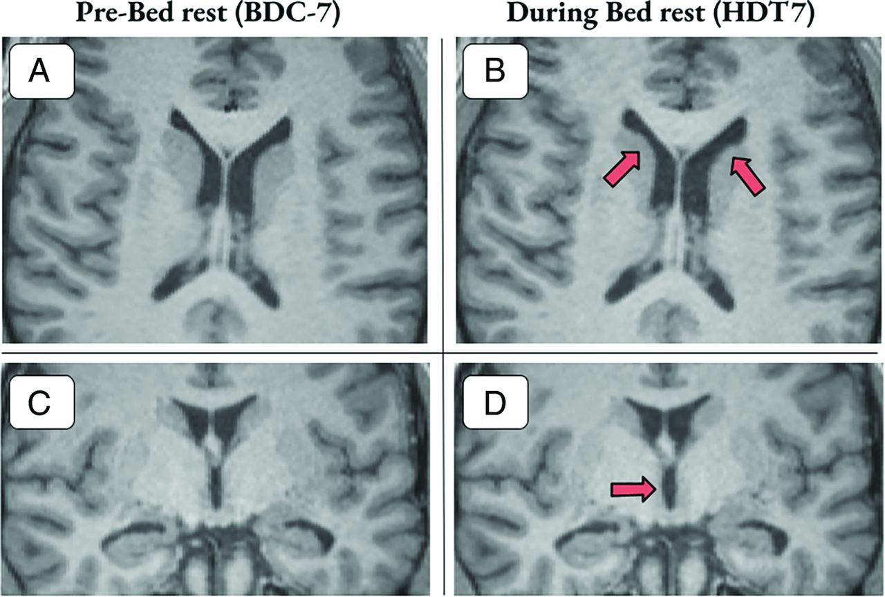
T1 images with examples of ventricular volume enlargement for 2 participants 7 days into bed rest. A and B, Axial section with arrows highlighting areas of gross ventricular enlargement in the lateral ventricles. C and D, Coronal section with arrows highlighting enlargement of the third ventricle. Participants shown experienced a 9.12% and 12.31% increase in ventricular volume, respectively.

FIG 3.
  • Download figure
  • Open in new tab
  • Download powerpoint
FIG 3.

3D mask representation of WM-PVS of a sample participant from pre-bed rest (BDC-7) to the end of bed rest (HDT29). The following participant experienced a 12% increase in PVS volume.

Brain Recovery from HDT

The divergent trajectories of brain metrics continued during recovery as ventricular volume (P < .001; g = 2.67; 95% CI, 1.38–3.95) and PVS volume (P = .012; g = 0.89; 95% CI, 0.19–1.56) decreased from HDT29 to R + 12, whereas global perfusion increased during recovery (P = .006; g = 1.01; 95% CI, 0.28–1.71; Table 2 and Fig 4). At the end of recovery (R + 12), their relative positions shifted with global perfusion significantly greater than both PVS volume (P = .025; g = 0.76; 95% CI, 0.09–1.41) and ventricular volume (P = .004; g = 1.07; 95% CI, 0.32–1.78) with no difference between PVS and ventricular volume (P = .146; g = 0.46; 95% CI, −0.15–1.05).

FIG 4.
  • Download figure
  • Open in new tab
  • Download powerpoint
FIG 4.

Global relative perfusion, ventricular volume, and PVS time courses. Gray shaded area represents the duration of the HDT + CO2 intervention. Data points for scanning days include pre- (BDC-7), during (HDT7 & HDT29), and post- (R + 5 & R + 12) HDT + CO2 period and are represented with mean z-scores (standard error of the mean). A divergent trajectory is observed with perfusion decreasing while ventricular and perivascular space volume increase during the HDT + CO2 period.

HDT Differences between Perfusion, Ventricular Volume, and PVS

At HDT29, ventricular volume was greater, relative to its mean across time, than both PVS volume (P = .017; g = 0.83; 95% CI, 0.14–1.49) and perfusion (P < .001; g = 2.05; 95% CI, 0.99–3.09), and PVS volume was greater than perfusion (P < .001; g = 1.47; 95% CI, 0.60–2.30).

Recovery versus Baseline Differences

At the end of recovery, we found no differences compared with the baseline values for ventricular volume (P = .619; g = 0.15; 95% CI, −0.43–0.72), PVS volume (P = .275; g = 0.33; 95% CI, −0.26–0.91), or global perfusion (P = .503; g = 0.20; 95% CI, −0.20 to −0.38). The brain metrics revealed a double dissociation in which ventricular volume and PVS volume increased during HDT+CO2 with a decrease to baseline levels during recovery, whereas global perfusion decreased during the HDT + CO2 period and increased to baseline levels during recovery, suggesting distinct neurophysiological responses to simulated microgravity.

DISCUSSION

The purpose of this study was to investigate changes in cerebral perfusion, ventricular volume, and PVS volume in healthy participants in response to 30 days of HDT + CO2. The main finding was statistically significant changes in brain metrics of opposite directionality, with global perfusion decreasing and ventricular/PVS volumes increasing during HDT + CO2 and subsequent reversal during recovery.

While 2 recent studies found alterations in PVS volumes in astronauts postspaceflight,7,8 this is the first study to report a change in PVS volumes in an HDT microgravity analog setting, and in general, transient PVS dilation and reversal in a cohort of healthy participants. Furthermore, decreased global perfusion in VaPER participants presented by Roberts et al16 is the only previous study examining longitudinal changes in perfusion by MR imaging in a prolonged microgravity analog. A short-duration study found a 17%–20% decrease in CBF after 26.5 hours of 12° HDT measuring carotid and vertebral artery blood flow using a cine phase-contrast MR image, but it did not extrapolate to multiple time points.26 Other HDT studies have investigated measurements of CBF velocity via transcranial Doppler. However, the results of these studies are inconclusive, likely due to limitations of this technique based on the assumption that the cross-sectional area of the interrogated vessel is fixed, which might not be the case, particularly in an environment with altered CO2 levels.16,27 Although a perfusion-PVS link has not been explored in a longitudinal study, the inverse relationship observed in this study is in line with previous results that showed that greater PVS volumes may be associated with decreased CBF in healthy participants and patients with intracranial and extracranial atheroscleroisis.18,19 Moreover, the ventricular volume changes presented here align with previous findings after HDT (a 2%–3% increase in lateral ventricle volume was found after 26.5 hours of HDT)26 and long-duration spaceflight (increases of ∼11% have been found in both astronauts and cosmonauts.) 2,3,5

The mechanisms driving decreased cerebral perfusion and ventricular/PVS enlargement in healthy participants during the 30 days of HDT + CO2 are unknown. However, it is becoming clear that real and simulated microgravity elicit changes in cerebral hemodynamics and CSF dynamics.27,28 In spaceflight and HDT, normal hydrostatic pressure gradients, usually experienced along the head-to-foot axis during upright posture, are reduced, resulting in the observed cephalad fluid shifts.27⇓-29 It has been hypothesized that this upward fluid shift may lead to venous congestion in the head and possibly elevated intracranial pressure (ICP).5,16,28 From a hemodynamic perspective, venous congestion could lead to venous hypertension, elevated ICP, and decreased cerebral perfusion. Indeed, impaired venous outflow has been linked to reduced cerebral perfusion in patients with chronic cerebrospinal venous insufficiency.30⇓-32 Following surgical restoration of normal internal jugular venous flow, patients demonstrated improved brain perfusion and a 9.6% reduction in ventricular size.30 Most interesting, this change in ventricular volume related to venous insufficiency is similar to the ventricular volume change previously reported in astronauts following long-duration spaceflight.1,25 In an astronaut population, a recent article showed postflight increases in superior sagittal, transverse, and sigmoid sinus volumes.33 The authors suggest improper venous drainage due to the absence of gravitational gradients and that this may explain thrombosis and abnormal internal jugular venous flow reported in astronauts.34,35 As an additional contributing factor, cardiac output and stroke volume are likely affected by microgravity. Reviewing the current literature, Bateman and Bateman36 surmised that both are reduced in HDT but increased during spaceflight, which may point to a key physiologic difference between the environments.

Similarly, it is likely CSF homeostasis is altered by reversal of the gravitational gradient. Upward shift of the brain itself has been suggested to impair CSF resorption by compression of the superior sagittal sinus.5,33 In this theory, the ventricular system may then act as a buffer for excess CSF resulting in ventricular enlargement. Similarly, an increase in PVS volume may reflect obstruction or inefficiency in the exchange of CSF and interstitial fluid that occurs in perivascular channels. CSF flow is intrinsically connected to hemodynamics in that transmission of arterial pulsations are a driving force for CSF movement.37 In this view, decreased perfusion could reduce the ability to circulate CSF, with resulting consequences for the perivascular and ventricular compartments. Indeed, cross-sectional studies showing an inverse relationship between cerebral perfusion and PVS size have hypothesized that decreased blood flow leads to increased interstitial fluid around the PVS and subsequent PVS dilation.18 This hypothesis is in line with the negative correlation between perfusion and PVS volume seen in this study. Measurements of ICP could help elucidate the interplay between perfusion and CSF changes, but no direct measurement of ICP during long-duration HDT or microgravity has been performed. However, several studies have reported elevated ICP in the acute stages of HDT or in the transition from upright to supine.38,39 Additionally, several astronauts presented with mildly elevated opening pressures via lumbar puncture (21–28.5 cm H20) months after spaceflight.40

A study limitation was the necessary use of an ASL sequence that did not provide absolute CBF values. The small sample size (n = 11) and large number of relevant study variables reduced our statistical power, though several group-level findings had robust effect sizes. Additionally, various methods for PVS quantification exist, and the automated method used here based on PVS morphologic features has potential limitations, such as partial voluming effects of the enclosed vessel.41 Most important, it is not clear that HDT bed rest represents an accurate terrestrial analog for spaceflight.12

Here, we document an association among patterns of cerebral perfusion, ventricular volume, and PVS volume that occur over a HDT + CO2 intervention and recovery. The findings contribute to our understanding of the relationships among the circulatory, glymphatic, and ventricular systems of the brain, specifically, revealing a possible direct link between PVS volumes and cerebral perfusion demonstrated by a prolonged perturbation of CBF in healthy participants. The results also provide additional evidence of changes in cerebral physiology in response to simulated microgravity. The significance of these changes, if any, should be further explored. For example, alterations in vision and ophthalmologic findings following spaceflight have been reported in astronauts, known as the spaceflight-associated neuro-ocular syndrome and thought to be linked to cephalad fluid shifts and venous congestion.40 Altered cerebral physiology may also play a role in cognitive and performance decrements previously reported in astronauts.3,14,42 Most important, given the increasing number of commercial spaceflight participants who may not be as physically fit as career astronauts and who may have mild forms of cerebrovascular disease, a pronounced decrease in cerebral perfusion without adequate reserve raises the possibility of spaceflight-induced ischemic events. While these data represent one step forward in developing a comprehensive model of the neurophysiologic response to microgravity, ultimately inflight evaluation of cerebral perfusion and ICP in astronauts is greatly needed.

ACKNOWLEDGMENTS

Research reported in this study was facilitated by the Spaceflight Standard Measures Cross-Cutting Project of the Human Research Program of the National Aeronautics and Space Administration. We also thank the entire VaPER study staff.

Footnotes

  • This study was funded by the National Aeronautics and Space Administration (NASA), grant No. NNX13AJ92G.

  • Disclosure forms provided by the authors are available with the full text and PDF of this article at www.ajnr.org.

Indicates open access to non-subscribers at www.ajnr.org

References

  1. 1.↵
    1. Koppelmans V,
    2. Bloomberg JJ,
    3. Mulavara AP, et al
    . Brain structural plasticity with spaceflight. NPI Microgravity 2016;2:2–8 doi:10.1038/s41526-016-0001-9 pmid:28649622
    CrossRefPubMed
  2. 2.↵
    1. Roberts DR,
    2. Albrecht MH,
    3. Collins HR, et al
    . Effects of spaceflight on astronaut brain structure as indicated on MRI. N Engl J Med 2017;377:1746–53 doi:10.1056/NEJMoa1705129 pmid:29091569
    CrossRefPubMed
  3. 3.↵
    1. Roberts DR,
    2. Asemani D,
    3. Nietert PJ, et al
    . Prolonged microgravity affects human brain structure and function. AJNR Am J Neuroradiol 2019;40:1878–85 doi:10.3174/ajnr.A6249 pmid:31624117
    Abstract/FREE Full Text
  4. 4.↵
    1. Van Ombergen A,
    2. Jillings S,
    3. Jeurissen B, et al
    . Brain tissue–volume changes in cosmonauts. N Engl J Med 2018;379:1678–80 doi:10.1056/NEJMc1809011 pmid:30354959
    CrossRefPubMed
  5. 5.↵
    1. Van Ombergen A,
    2. Jillings S,
    3. Jeurissen B, et al
    . Brain ventricular volume changes induced by long-duration spaceflight. Proc Natl Acad Sci U S A 2019;116:10531–36 doi:10.1073/pnas.1820354116 pmid:31061119
    Abstract/FREE Full Text
  6. 6.↵
    1. Roberts DR,
    2. Inglesby DC,
    3. Brown TR, et al
    . Longitudinal change in ventricular volume is accelerated in astronauts undergoing long-duration spaceflight. Aging Brain 2021;1:100017 doi:10.1016/j.nbas.2021.100017 pmid:36911514
    CrossRefPubMed
  7. 7.↵
    1. Barisano G,
    2. Sepehrband F,
    3. Collins HR, et al
    . The effect of prolonged spaceflight on cerebrospinal fluid and perivascular spaces of astronauts and cosmonauts. Proc Natl Acad Sci U S A 2022;119:e2120439119 doi:10.1073/pnas.2120439119 pmid:35412862
    CrossRefPubMed
  8. 8.↵
    1. Hupfeld KE,
    2. Richmond SB,
    3. McGregor HR, et al
    . Longitudinal MRI-visible perivascular space (PVS) changes with long-duration spaceflight. Sci Rep 2022;12:7238 doi:10.1038/s41598-022-11593-y pmid:35513698
    CrossRefPubMed
  9. 9.↵
    1. Lee JK,
    2. Koppelmans V,
    3. Riascos RF, et al
    . Spaceflight-associated brain white matter microstructural changes and intracranial fluid redistribution. JAMA Neurol 2019;76:412–19 doi:10.1001/jamaneurol.2018.4882 pmid:30673793
    CrossRefPubMed
  10. 10.↵
    1. Hargens AR,
    2. Vico L
    . Long-duration bed rest as an analog to microgravity. J Appl Physiol (1985) 2016;120:891–903 doi:10.1152/japplphysiol.00935.2015 pmid:26893033
    CrossRefPubMed
  11. 11.↵
    1. Traon PL,
    2. Heer M,
    3. Narici MV, et al
    . From space to Earth: advances in human physiology from 20 years of bed rest studies (1986–2006). Eur J Appl Physiol 2007;101:143–94 doi:10.1007/s00421-007-0474-z pmid:17661073
    CrossRefPubMed
  12. 12.↵
    1. Ong J,
    2. Lee AG,
    3. Moss HE
    . Head-down tilt bed rest studies as a terrestrial analog for spaceflight associated neuro-ocular syndrome. Front Neurol 2021;12:648958 doi:10.3389/fneur.2021.648958 pmid:33841315
    CrossRefPubMed
  13. 13.↵
    1. Koppelmans V,
    2. Erdeniz B,
    3. De Dios YE, et al
    . Study protocol to examine the effects of spaceflight and a spaceflight analog on neurocognitive performance: extent, longevity, and neural bases. BMC Neurol 2013;13:205 doi:10.1186/1471-2377-13-205 pmid:24350728
    CrossRefPubMed
  14. 14.↵
    1. Lee JK,
    2. De Dios Y,
    3. Kofman I, et al
    . Head down tilt bed rest plus elevated CO2 as a spaceflight analog: effects on cognitive and sensorimotor performance. Front Hum Neurosci 2019;13:355 doi:10.3389/fnhum.2019.00355 pmid:31680909
    CrossRefPubMed
  15. 15.↵
    1. Hupfeld KE,
    2. Lee JK,
    3. Gadd NE, et al
    . Neural correlates of vestibular processing during a spaceflight analog with elevated carbon dioxide (CO2): a pilot study. Front Syst Neurosci 2020;13;80 doi:10.3389/fnsys,2019.00080 pmid:31998084
    CrossRefPubMed
  16. 16.↵
    1. Roberts DR,
    2. Collins HR,
    3. Lee JK, et al
    . Altered cerebral perfusion in response to chronic mild hypercapnia and head-down tilt bed rest as an analog for spaceflight. Neuroradiology 2021;63:1271–81 doi:10.1007/s00234-021-02660-8 pmid:33587162
    CrossRefPubMed
  17. 17.↵
    1. McGregor HR,
    2. Lee JK,
    3. Mulder ER, et al
    . Brain connectivity and behavioral changes in a spaceflight analog environment with elevated CO2. Neuroimage 2021;225:117450 doi:10.1016/j.neuroimage.2020.117450 pmid:33075558
    CrossRefPubMed
  18. 18.↵
    1. Liu S,
    2. Hou B,
    3. You H, et al
    . The association between perivascular spaces and cerebral blood flow, brain volume, and cardiovascular risk. Front Aging Neurosci 2021;13:599724 doi:10.3389/fnagi.2021.599724 pmid:34531732
    CrossRefPubMed
  19. 19.↵
    1. Wang H,
    2. Nie ZY,
    3. Liu M, et al
    . Clinical characteristics of perivascular space and brain CT perfusion in stroke-free patients with intracranial and extracranial atherosclerosis of different extents. Ann Transl Med 2020;8:215 doi:10.21037/atm.2020.01.35 pmid:32309362
    CrossRefPubMed
  20. 20.↵
    1. Laurie SS,
    2. Christian K,
    3. Kysar J, et al
    . Unchanged cerebrovascular CO2 reactivity and hypercapnic ventilatory response during strict head-down tilt bed rest in a mild hypercapnic environment. J Physiol 2020;598:2491–2505 doi:10.1113/JP279383 pmid:32196672
    CrossRefPubMed
  21. 21.↵
    1. Fischl B
    . FreeSurfer. Neuroimage 2012;62:774–81 doi:10.1016/j.neuroimage.2012.01.021 pmid:22248573
    CrossRefPubMed
  22. 22.↵
    1. Sepehrband F,
    2. Barisano G,
    3. Sheikh-Bahaei N, et al
    . Image processing approaches to enhance perivascular space visibility and quantification using MRI. Sci Rep 2019;9:1–12 doi:10.1038/s41598-019-48910-x pmid:31451792
    CrossRefPubMed
  23. 23.↵
    1. Barisano G,
    2. Sheikh-Bahaei N,
    3. Law M, et al
    . Body mass index, time of day and genetics affect perivascular spaces in the white matter. J Cereb Blood Flow Metab 2021;41:1563–78 doi:10.1177/0271678X20972856 pmid:33183133
    CrossRefPubMed
  24. 24.↵
    1. Donahue EK,
    2. Murdos A,
    3. Jakowec MW, et al
    . Global and regional changes in perivascular space in idiopathic and familial Parkinson's disease. Mov Disord 2021;36:1126–36 doi:10.1002/mds.28473 pmid:33470460
    CrossRefPubMed
  25. 25.↵
    1. Sawilowsky SS
    . New effect size rules of thumb. Journal of Modern Applied Statistical Methods 2009;8:597–99 doi:10.22237/jmasm/1257035100
    CrossRef
  26. 26.↵
    1. Kramer LA,
    2. Hasan KM,
    3. Sargsyan AE, et al
    ; SPACECOT Investigators Group. Quantitative MRI volumetry, diffusivity, cerebrovascular flow, and cranial hydrodynamics during head-down tilt and hypercapnia: the SPACECOT study. J Appl Physiol (1985) 2017;122:1155–66 doi:10.1152/japplphysiol.00887.2016 pmid:28209740
    CrossRefPubMed
  27. 27.↵
    1. Kawai Y,
    2. Doi M,
    3. Setogawa A, et al
    . Effects of microgravity on cerebral hemodynamics. Yonago Acta Med 2003;46:1–8
  28. 28.↵
    1. Nelson ES,
    2. Mulugeta L,
    3. Myers JG
    . Microgravity-induced fluid shift and ophthalmic changes. Life (Basel) 2014;4:621–65 doi:10.3390/Klife4040621 pmid:25387162
    CrossRefPubMed
  29. 29.↵
    1. Hans P
    . Hydrostatic pressure and neurosurgical pathology [in French]. Ann Fr Anesth Reanim 1994;13:123–26 doi:10.1016/S0750-7658(94)80195-9 pmid:8092568
    CrossRefPubMed
  30. 30.↵
    1. Zamboni P,
    2. Menegatti E,
    3. Cittanti C, et al
    . Fixing the jugular flow reduces ventricle volume and improves brain perfusion. J Vasc Surg Venous Lymphat Disord 2016;4:434–45 doi:10.1016/j.jvsv.2016.06.006 pmid:27638998
    CrossRefPubMed
  31. 31.↵
    1. Zamboni P,
    2. Menegatti E,
    3. Weinstock-Guttman B, et al
    . Hypoperfusion of brain parenchyma is associated with the severity of chronic cerebrospinal venous insufficiency in patients with multiple sclerosis: a cross-sectional preliminary report. BMC Med 2011;9:22 doi:10.1186/1741-7015-9-22 pmid:21385345
    CrossRefPubMed
  32. 32.↵
    1. Zhou D,
    2. Ding J,
    3. Asmaro K, et al
    . Clinical characteristics and neuroimaging findings in internal jugular venous outflow disturbance. Thromb Haemost 2019;119:308318 doi:10.1055/s-0038-1676815 pmid:30605919
    CrossRefPubMed
  33. 33.↵
    1. Rosenberg MJ,
    2. Coker MA,
    3. Taylor JA, et al
    . Comparison of dural venous sinus volumes before and after flight in astronauts with and without spaceflight-associated neuro-ocular syndrome. JAMA Netw Open 2021;4:e2131465 doi:10.1001/jamanetworkopen.2021.31465 pmid:34705011
    CrossRefPubMed
  34. 34.↵
    1. Marshall-Goebel K,
    2. Laurie SS,
    3. Alferova IV, et al
    . Assessment of jugular venous blood flow stasis and thrombosis during spaceflight. JAMA Netw Open 2019;2:e1915011 doi:10.1001/jamanetworkopen.2019.15011 pmid:31722025
    CrossRefPubMed
  35. 35.↵
    1. Auñón-Chancellor SM,
    2. Pattarini JM,
    3. Moll S, et al
    . Venous thrombosis during spaceflight. N Engl J Med 2020;382:89–90 doi:10.1056/NEJMc1905875 pmid:31893522
    CrossRefPubMed
  36. 36.↵
    1. Bateman GA,
    2. Bateman AR
    . A perspective on spaceflight associated neuro-ocular syndrome causation secondary to elevated venous sinus pressure. NPJ Microgravity 2022;8:3 doi:10.1038/s41526-022-00188-6 pmid:35169156
    CrossRefPubMed
  37. 37.↵
    1. Khasawneh AH,
    2. Garling RJ,
    3. Harris CA
    . Cerebrospinal fluid circulation: what do we know and how do we know it? Brain Circ 2018;4:14–18 doi:10.4103/bc.bc_3_18 pmid:30276331
    CrossRefPubMed
  38. 38.↵
    1. Murthy G,
    2. Marchbanks RJ,
    3. Watenpaugh DE, et al
    . Increased intracranial pressure in humans during simulated microgravity. Physiologist 1992;35:S184–85 pmid:1589495
    PubMed
  39. 39.↵
    1. Petersen LG,
    2. Petersen JC,
    3. Andresen M, et al
    . Postural influence on intracranial and cerebral perfusion pressure in ambulatory neurosurgical patients. Am J Physiol Regul Integr Comp Physiol 2016;310:R100–04 doi:10.1152/ajpregu.00302.2015 pmid:26468260
    CrossRefPubMed
  40. 40.↵
    1. Mader TH,
    2. Gibson CR,
    3. Pass AF, et al
    . Optic disc edema, globe flattening, choroidal folds, and hyperopic shifts observed in astronauts after long-duration space flight. Ophthalmology 2011;118:2058–69 doi:10.1016/j.ophtha.2011.06.021 pmid:21849212
    CrossRefPubMed
  41. 41.↵
    1. Barisano G,
    2. Lynch KM,
    3. Sibilia F, et al
    . Imaging perivascular space structure and function using brain MRI. Neuroimage 2022;257:119329 doi:10.1016/j.neuroimage.2022.119329 pmid:35609770
    CrossRefPubMed
  42. 42.↵
    1. Strangman G,
    2. Bevan G
    . Review of human cognitive performance in spaceflight. In: Proceedings of the 84th Annual Scientific Meeting of the Aerospace Medical Association, Chicago, Illinois. May 12–16, 2013
  • Received September 16, 2022.
  • Accepted after revision June 25, 2023.
  • © 2023 by American Journal of Neuroradiology
PreviousNext
Back to top

In this issue

American Journal of Neuroradiology: 44 (9)
American Journal of Neuroradiology
Vol. 44, Issue 9
1 Sep 2023
  • Table of Contents
  • Index by author
  • Complete Issue (PDF)
Advertisement
Print
Download PDF
Email Article

Thank you for your interest in spreading the word on American Journal of Neuroradiology.

NOTE: We only request your email address so that the person you are recommending the page to knows that you wanted them to see it, and that it is not junk mail. We do not capture any email address.

Enter multiple addresses on separate lines or separate them with commas.
Longitudinal Changes in Cerebral Perfusion, Perivascular Space Volume, and Ventricular Volume in a Healthy Cohort Undergoing a Spaceflight Analog
(Your Name) has sent you a message from American Journal of Neuroradiology
(Your Name) thought you would like to see the American Journal of Neuroradiology web site.
CAPTCHA
This question is for testing whether or not you are a human visitor and to prevent automated spam submissions.
Cite this article
J.B. Tidwell, J.A. Taylor, H.R. Collins, J.H. Chamberlin, G. Barisano, F. Sepehrband, M.D. Turner, G. Gauthier, E.R. Mulder, D.A. Gerlach, D.R. Roberts
Longitudinal Changes in Cerebral Perfusion, Perivascular Space Volume, and Ventricular Volume in a Healthy Cohort Undergoing a Spaceflight Analog
American Journal of Neuroradiology Sep 2023, 44 (9) 1026-1031; DOI: 10.3174/ajnr.A7949

Citation Manager Formats

  • BibTeX
  • Bookends
  • EasyBib
  • EndNote (tagged)
  • EndNote 8 (xml)
  • Medlars
  • Mendeley
  • Papers
  • RefWorks Tagged
  • Ref Manager
  • RIS
  • Zotero
0 Responses
Respond to this article
Share
Bookmark this article
Spaceflight Analog: Longitudinal Brain Changes
J.B. Tidwell, J.A. Taylor, H.R. Collins, J.H. Chamberlin, G. Barisano, F. Sepehrband, M.D. Turner, G. Gauthier, E.R. Mulder, D.A. Gerlach, D.R. Roberts
American Journal of Neuroradiology Sep 2023, 44 (9) 1026-1031; DOI: 10.3174/ajnr.A7949
del.icio.us logo Twitter logo Facebook logo Mendeley logo
  • Tweet Widget
  • Facebook Like
  • Google Plus One
Purchase

Jump to section

  • Article
    • Abstract
    • ABBREVIATIONS:
    • MATERIALS AND METHODS
    • RESULTS
    • DISCUSSION
    • ACKNOWLEDGMENTS
    • Footnotes
    • References
  • Figures & Data
  • Info & Metrics
  • Responses
  • References
  • PDF

Related Articles

  • No related articles found.
  • PubMed
  • Google Scholar

Cited By...

  • Reply:
  • Reply:
  • Spaceflight-Associated Neuro-Ocular Syndrome and Idiopathic Intracranial Hypertension: Can Anemia and Hyperemia Underlie Both?
  • Enlargement of Perivascular Spaces as a Downstream Consequence of Spaceflight Analog-Induced Alterations in Cerebral Venous Hemodynamics
  • Crossref (10)
  • Google Scholar

This article has been cited by the following articles in journals that are participating in Crossref Cited-by Linking.

  • A perspective on the evidence for glymphatic obstruction in spaceflight associated neuro-ocular syndrome and fatigue
    Grant Alexander Bateman, Alexander Robert Bateman
    npj Microgravity 2024 10 1
  • Neurocognitive Adaptations for Spatial Orientation and Navigation in Astronauts
    Ford Burles, Giuseppe Iaria
    Brain Sciences 2023 13 11
  • Enlargement of Perivascular Spaces as a Downstream Consequence of Spaceflight Analog-Induced Alterations in Cerebral Venous Hemodynamics
    Peter Wostyn, Thomas H. Mader, C. Robert Gibson, Maiken Nedergaard
    American Journal of Neuroradiology 2024 45 5
  • Oxidative stress, neuroinflammation, and the blood-brain barrier biomarkers on the brain response to spaceflight
    Xiao Wen Mao, Michael J Pecaut, Seta Stanbouly, Gregory Nelson
    Life Sciences in Space Research 2024 43
  • Development and validation of a perivascular space segmentation method in multi-center datasets
    Peiyu Huang, Lingyun Liu, Yao Zhang, Siyan Zhong, Peng Liu, Hui Hong, Shuyue Wang, Linyun Xie, Miao Lin, Yeerfan Jiaerken, Xiao Luo, Kaicheng Li, Qingze Zeng, Lei Cui, Jixuan Li, Yanxing Chen, Ruiting Zhang
    NeuroImage 2024 298
  • Spaceflight-Associated Neuro-Ocular Syndrome and Idiopathic Intracranial Hypertension: Can Anemia and Hyperemia Underlie Both?
    G.A. Bateman, A.R. Bateman
    American Journal of Neuroradiology 2024 45 5
  • Artificial gravity: an effective countermeasure for microgravity-induced headward fluid shift?
    Larry A. Kramer, Khader M. Hasan, Xu Zhang, Edwin Mulder, Darius A. Gerlach, Karina Marshall-Goebel, Brandon R. Macias, Steven S. Laurie, Gary Strangman, Raj Iyer, Eric M. Bershad
    Journal of Applied Physiology 2024 137 5
  • Neuroscience Research in Short-Duration Human Spaceflight
    Bader Shirah, Peter Zu Eulenburg, Christopher E. Mason, Shawna Pandya, Yvette Gonzalez, Aaron H. Persad, Mohammed M. Ahmed, Mishaal N. Ashemimry, Matthias Ertl, Jon Sen
    2025
  • Reply:
    Donna R. Roberts, Joe Tidwell, J. Andrew Taylor
    American Journal of Neuroradiology 2024 45 5
  • Reply:
    Donna R. Roberts, Joe Tidwell, J. Andrew Taylor
    American Journal of Neuroradiology 2024 45 5

More in this TOC Section

  • Diagnostic Neuroradiology of Monoclonal Antibodies
  • Cerebral ADC Changes in Fabry Disease
  • ML for Glioma Molecular Subtype Prediction
Show more ADULT BRAIN

Similar Articles

Advertisement

Indexed Content

  • Current Issue
  • Accepted Manuscripts
  • Article Preview
  • Past Issues
  • Editorials
  • Editor's Choice
  • Fellows' Journal Club
  • Letters to the Editor
  • Video Articles

Cases

  • Case Collection
  • Archive - Case of the Week
  • Archive - Case of the Month
  • Archive - Classic Case

More from AJNR

  • Trainee Corner
  • Imaging Protocols
  • MRI Safety Corner

Multimedia

  • AJNR Podcasts
  • AJNR Scantastics

Resources

  • Turnaround Time
  • Submit a Manuscript
  • Submit a Video Article
  • Submit an eLetter to the Editor/Response
  • Manuscript Submission Guidelines
  • Statistical Tips
  • Fast Publishing of Accepted Manuscripts
  • Graphical Abstract Preparation
  • Imaging Protocol Submission
  • Evidence-Based Medicine Level Guide
  • Publishing Checklists
  • Author Policies
  • Become a Reviewer/Academy of Reviewers
  • News and Updates

About Us

  • About AJNR
  • Editorial Board
  • Editorial Board Alumni
  • Alerts
  • Permissions
  • Not an AJNR Subscriber? Join Now
  • Advertise with Us
  • Librarian Resources
  • Feedback
  • Terms and Conditions
  • AJNR Editorial Board Alumni

American Society of Neuroradiology

  • Not an ASNR Member? Join Now

© 2025 by the American Society of Neuroradiology All rights, including for text and data mining, AI training, and similar technologies, are reserved.
Print ISSN: 0195-6108 Online ISSN: 1936-959X

Powered by HighWire