Advertisement

Main menu

  • Home
  • Content
    • Current Issue
    • Accepted Manuscripts
    • Article Preview
    • Past Issue Archive
    • Video Articles
    • AJNR Case Collection
    • Case of the Week Archive
    • Case of the Month Archive
    • Classic Case Archive
  • Special Collections
    • AJNR Awards
    • Low-Field MRI
    • Alzheimer Disease
    • ASNR Foundation Special Collection
    • Photon-Counting CT
    • View All
  • Multimedia
    • AJNR Podcasts
    • AJNR SCANtastic
    • Trainee Corner
    • MRI Safety Corner
    • Imaging Protocols
  • For Authors
    • Submit a Manuscript
    • Submit a Video Article
    • Submit an eLetter to the Editor/Response
    • Manuscript Submission Guidelines
    • Statistical Tips
    • Fast Publishing of Accepted Manuscripts
    • Graphical Abstract Preparation
    • Imaging Protocol Submission
    • Author Policies
  • About Us
    • About AJNR
    • Editorial Board
    • Editorial Board Alumni
  • More
    • Become a Reviewer/Academy of Reviewers
    • Subscribers
    • Permissions
    • Alerts
    • Feedback
    • Advertisers
    • ASNR Home

User menu

  • Alerts
  • Log in

Search

  • Advanced search
American Journal of Neuroradiology

American Journal of Neuroradiology

ASHNR American Society of Functional Neuroradiology ASHNR American Society of Pediatric Neuroradiology ASSR
  • Alerts
  • Log in

Advanced Search

  • Home
  • Content
    • Current Issue
    • Accepted Manuscripts
    • Article Preview
    • Past Issue Archive
    • Video Articles
    • AJNR Case Collection
    • Case of the Week Archive
    • Case of the Month Archive
    • Classic Case Archive
  • Special Collections
    • AJNR Awards
    • Low-Field MRI
    • Alzheimer Disease
    • ASNR Foundation Special Collection
    • Photon-Counting CT
    • View All
  • Multimedia
    • AJNR Podcasts
    • AJNR SCANtastic
    • Trainee Corner
    • MRI Safety Corner
    • Imaging Protocols
  • For Authors
    • Submit a Manuscript
    • Submit a Video Article
    • Submit an eLetter to the Editor/Response
    • Manuscript Submission Guidelines
    • Statistical Tips
    • Fast Publishing of Accepted Manuscripts
    • Graphical Abstract Preparation
    • Imaging Protocol Submission
    • Author Policies
  • About Us
    • About AJNR
    • Editorial Board
    • Editorial Board Alumni
  • More
    • Become a Reviewer/Academy of Reviewers
    • Subscribers
    • Permissions
    • Alerts
    • Feedback
    • Advertisers
    • ASNR Home
  • Follow AJNR on Twitter
  • Visit AJNR on Facebook
  • Follow AJNR on Instagram
  • Join AJNR on LinkedIn
  • RSS Feeds

AJNR Awards, New Junior Editors, and more. Read the latest AJNR updates

Research ArticleAdult Brain
Open Access

Parieto-Occipital Injury on Diffusion MRI Correlates with Poor Neurologic Outcome following Cardiac Arrest

E. Calabrese, S. Gandhi, J. Shih, M. Otero, D. Randazzo, C. Hemphill, R. Huie, J.F. Talbott and E. Amorim
American Journal of Neuroradiology March 2023, 44 (3) 254-260; DOI: https://doi.org/10.3174/ajnr.A7779
E. Calabrese
aFrom the Department of Radiology and Biomedical Imaging (E.C., S.G., J.F.T.)
  • Find this author on Google Scholar
  • Find this author on PubMed
  • Search for this author on this site
  • ORCID record for E. Calabrese
S. Gandhi
aFrom the Department of Radiology and Biomedical Imaging (E.C., S.G., J.F.T.)
dDepartment of Radiology and Biomedical Imaging (S.G., J.F.T., E.A.), Zuckerberg San Francisco General Hospital, San Francisco, California
  • Find this author on Google Scholar
  • Find this author on PubMed
  • Search for this author on this site
J. Shih
bDepartment of Neurology (J.S., M.O., D.R., C.H., E.A.), Weill Institute for Neurosciences
  • Find this author on Google Scholar
  • Find this author on PubMed
  • Search for this author on this site
  • ORCID record for J. Shih
M. Otero
bDepartment of Neurology (J.S., M.O., D.R., C.H., E.A.), Weill Institute for Neurosciences
  • Find this author on Google Scholar
  • Find this author on PubMed
  • Search for this author on this site
D. Randazzo
bDepartment of Neurology (J.S., M.O., D.R., C.H., E.A.), Weill Institute for Neurosciences
  • Find this author on Google Scholar
  • Find this author on PubMed
  • Search for this author on this site
  • ORCID record for D. Randazzo
C. Hemphill
bDepartment of Neurology (J.S., M.O., D.R., C.H., E.A.), Weill Institute for Neurosciences
  • Find this author on Google Scholar
  • Find this author on PubMed
  • Search for this author on this site
  • ORCID record for C. Hemphill
R. Huie
cDepartment of Neurological Surgery (R.H.), University of California, San Francisco, San Francisco, California
  • Find this author on Google Scholar
  • Find this author on PubMed
  • Search for this author on this site
  • ORCID record for R. Huie
J.F. Talbott
aFrom the Department of Radiology and Biomedical Imaging (E.C., S.G., J.F.T.)
dDepartment of Radiology and Biomedical Imaging (S.G., J.F.T., E.A.), Zuckerberg San Francisco General Hospital, San Francisco, California
  • Find this author on Google Scholar
  • Find this author on PubMed
  • Search for this author on this site
  • ORCID record for J.F. Talbott
E. Amorim
bDepartment of Neurology (J.S., M.O., D.R., C.H., E.A.), Weill Institute for Neurosciences
dDepartment of Radiology and Biomedical Imaging (S.G., J.F.T., E.A.), Zuckerberg San Francisco General Hospital, San Francisco, California
  • Find this author on Google Scholar
  • Find this author on PubMed
  • Search for this author on this site
  • ORCID record for E. Amorim
  • Article
  • Figures & Data
  • Info & Metrics
  • Responses
  • References
  • PDF
Loading

Article Figures & Data

Figures

  • Tables
  • FIG 1.
    • Download figure
    • Open in new tab
    • Download powerpoint
    FIG 1.

    Frequency of hypoxic-ischemic brain injury in both outcome groups. Colorized 3D brain renderings show the frequency of injury (as defined by ADC < 650 × 10–6 mm2/s) for the poor-outcome group (A) and the good outcome group (B), respectively. The color bar (right) indicates the frequency of injury across the whole brain. Note that regions with <5% injury frequency are transparent to allow better visualization of more frequently injured areas.
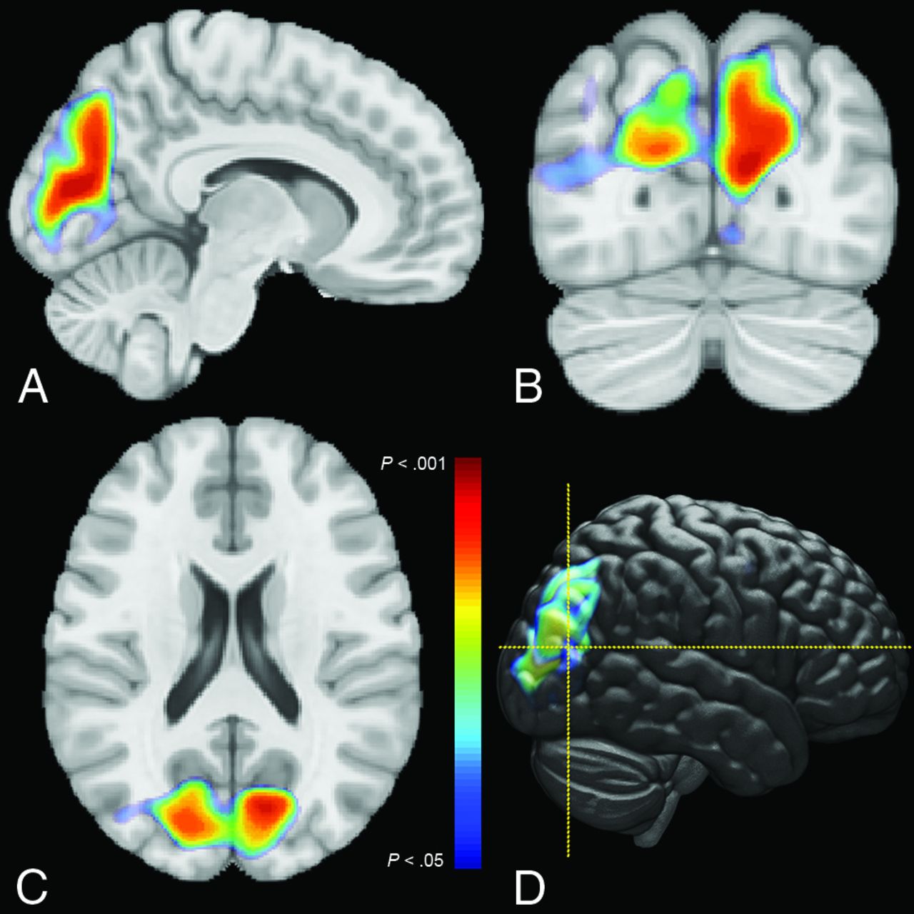
  • FIG 2.
    • Download figure
    • Open in new tab
    • Download powerpoint
    FIG 2.

    Results of voxelwise comparisons between outcome groups. Statistically significant voxels (family-wise error rate–corrected, P < .05) are shown as color overlays on orthogonal slices of the Montreal Neurologic Institute brain atlas (A–C). Color indicates the P value for the comparison (see the color bar). A volume-rendering of the P value from a lateral perspective is also shown, with dashed yellow lines indicating the plane of axial and coronal slices (D).
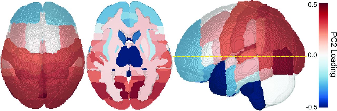
  • FIG 3.
    • Download figure
    • Open in new tab
    • Download powerpoint
    FIG 3.

    Loading values for PC2 from the PCA. Each cortical region from the Harvard Oxford brain atlas is colored according to its PC2 loading value. Red and blue loadings indicate opposite patterns of loading (red indicates positive correlation, blue indicates a negative correlation). The dashed yellow line on the lateral projection (right) indicates the plane of the axial section (center).

  • FIG 4.
    • Download figure
    • Open in new tab
    • Download powerpoint
    FIG 4.

    ROC analysis of different diffusion-weighted MR imaging–based variables for predicting outcome in patients with post–cardiac arrest coma. OHCA indicates out of hospital cardiac arrest.

Tables

  • Figures
  • Subject demographics stratified by outcome group

    VariableFollows Commands (n = 17)Does Not Follow Commands (n = 64)P Value
    Age (mean) (yr)55 (SD, 16)57 (SD, 15).741
    Female sex4/17 (24%)18/64 (28%).701
    Race and ethnicitya.757
     Asian4 (21%)6 (9%)
     Black3 (16%)14 (22%)
     Hispanic2 (11%)6 (9%)
     Hawaiian0 (0%)2 (3%)
     Non-Hispanic white2 (11%)6 (9%)
     Unknown8 (42%)30 (47%)
    Time from ROSC to MR imaging (mean) (hr)120 (SD, 26)115 (SD, 27).496
    Shockable rhythm6/17 (35%)12/64 (19%).145
    Out-of-hospital cardiac arrest14/17 (82%)49/64 (77%).610
    Time to ROSC (mean)19 (SD, 14)24 (SD, 14).182
    • ↵a Patient race and ethnicity do not add to 100% as some patients reported more than one race and ethnicity combination.

PreviousNext
Back to top

In this issue

American Journal of Neuroradiology: 44 (3)
American Journal of Neuroradiology
Vol. 44, Issue 3
1 Mar 2023
  • Table of Contents
  • Index by author
  • Complete Issue (PDF)
Advertisement
Print
Download PDF
Email Article

Thank you for your interest in spreading the word on American Journal of Neuroradiology.

NOTE: We only request your email address so that the person you are recommending the page to knows that you wanted them to see it, and that it is not junk mail. We do not capture any email address.

Enter multiple addresses on separate lines or separate them with commas.
Parieto-Occipital Injury on Diffusion MRI Correlates with Poor Neurologic Outcome following Cardiac Arrest
(Your Name) has sent you a message from American Journal of Neuroradiology
(Your Name) thought you would like to see the American Journal of Neuroradiology web site.
CAPTCHA
This question is for testing whether or not you are a human visitor and to prevent automated spam submissions.
Cite this article
E. Calabrese, S. Gandhi, J. Shih, M. Otero, D. Randazzo, C. Hemphill, R. Huie, J.F. Talbott, E. Amorim
Parieto-Occipital Injury on Diffusion MRI Correlates with Poor Neurologic Outcome following Cardiac Arrest
American Journal of Neuroradiology Mar 2023, 44 (3) 254-260; DOI: 10.3174/ajnr.A7779

Citation Manager Formats

  • BibTeX
  • Bookends
  • EasyBib
  • EndNote (tagged)
  • EndNote 8 (xml)
  • Medlars
  • Mendeley
  • Papers
  • RefWorks Tagged
  • Ref Manager
  • RIS
  • Zotero
0 Responses
Respond to this article
Share
Bookmark this article
DWI and Neurologic Outcome Post-Arrest
E. Calabrese, S. Gandhi, J. Shih, M. Otero, D. Randazzo, C. Hemphill, R. Huie, J.F. Talbott, E. Amorim
American Journal of Neuroradiology Mar 2023, 44 (3) 254-260; DOI: 10.3174/ajnr.A7779
del.icio.us logo Twitter logo Facebook logo Mendeley logo
  • Tweet Widget
  • Facebook Like
  • Google Plus One
Purchase

Jump to section

  • Article
    • Abstract
    • ABBREVIATIONS:
    • MATERIALS AND METHODS
    • RESULTS
    • DISCUSSION
    • CONCLUSIONS
    • Footnotes
    • References
  • Figures & Data
  • Info & Metrics
  • Responses
  • References
  • PDF

Related Articles

  • No related articles found.
  • PubMed
  • Google Scholar

Cited By...

  • No citing articles found.
  • Crossref (4)
  • Google Scholar

This article has been cited by the following articles in journals that are participating in Crossref Cited-by Linking.

  • Correlating Quantitative MRI-based Apparent Diffusion Coefficient Metrics with 24-month Neurodevelopmental Outcomes in Neonates from the HEAL Trial
    Evan Calabrese, Yvonne Wu, Aaron Wolfe Scheffler, Jessica L. Wisnowski, Robert C. McKinstry, Amit Mathur, Hannah C. Glass, Bryan A. Comstock, Patrick J. Heagerty, Shivani Gillon, Sandra E. Juul, Christopher P. Hess, Yi Li
    Radiology 2023 308 3
  • Brain-derived extracellular vesicles as serologic markers of brain injury following cardiac arrest: A pilot feasibility study
    Hanfei Shen, Daria Zaitseva, Zijian Yang, Liam Forsythe, Sarah Joergensen, Alea I. Zone, Joana Shehu, Sarah Maghraoui, Anahita Ghorbani, Antonio Davila, David Issadore, Benjamin S. Abella
    Resuscitation 2023 191
  • Combination strategy for prognostication in patients undergoing post-resuscitation care after cardiac arrest
    Jung Soo Park, Eun Young Kim, Yeonho You, Jin Hong Min, Wonjoon Jeong, Hong Joon Ahn, Yong Nam In, In Ho Lee, Jae Moon Kim, Changshin Kang
    Scientific Reports 2023 13 1
  • Early Burst Suppression Similarity Association with Structural Brain Injury Severity on MRI After Cardiac Arrest
    Shawn Shivdat, Tiange Zhan, Alessandro De Palma, Wei-Long Zheng, Parimala Krishnamurthy, Ezhil Paneerselvam, Samuel Snider, Matthew Bevers, Una-May O’Reilly, Jong Woo Lee, M. Brandon Westover, Edilberto Amorim
    Neurocritical Care 2025 42 1

More in this TOC Section

  • Diagnostic Neuroradiology of Monoclonal Antibodies
  • Clinical Outcomes After Chiari I Decompression
  • Segmentation of Brain Metastases with BLAST
Show more ADULT BRAIN

Similar Articles

Advertisement

Indexed Content

  • Current Issue
  • Accepted Manuscripts
  • Article Preview
  • Past Issues
  • Editorials
  • Editor's Choice
  • Fellows' Journal Club
  • Letters to the Editor
  • Video Articles

Cases

  • Case Collection
  • Archive - Case of the Week
  • Archive - Case of the Month
  • Archive - Classic Case

More from AJNR

  • Trainee Corner
  • Imaging Protocols
  • MRI Safety Corner
  • Book Reviews

Multimedia

  • AJNR Podcasts
  • AJNR Scantastics

Resources

  • Turnaround Time
  • Submit a Manuscript
  • Submit a Video Article
  • Submit an eLetter to the Editor/Response
  • Manuscript Submission Guidelines
  • Statistical Tips
  • Fast Publishing of Accepted Manuscripts
  • Graphical Abstract Preparation
  • Imaging Protocol Submission
  • Evidence-Based Medicine Level Guide
  • Publishing Checklists
  • Author Policies
  • Become a Reviewer/Academy of Reviewers
  • News and Updates

About Us

  • About AJNR
  • Editorial Board
  • Editorial Board Alumni
  • Alerts
  • Permissions
  • Not an AJNR Subscriber? Join Now
  • Advertise with Us
  • Librarian Resources
  • Feedback
  • Terms and Conditions
  • AJNR Editorial Board Alumni

American Society of Neuroradiology

  • Not an ASNR Member? Join Now

© 2025 by the American Society of Neuroradiology All rights, including for text and data mining, AI training, and similar technologies, are reserved.
Print ISSN: 0195-6108 Online ISSN: 1936-959X

Powered by HighWire