Advertisement

Main menu

  • Home
  • Content
    • Current Issue
    • Accepted Manuscripts
    • Article Preview
    • Past Issue Archive
    • Video Articles
    • AJNR Case Collection
    • Case of the Week Archive
    • Case of the Month Archive
    • Classic Case Archive
  • Special Collections
    • AJNR Awards
    • Low-Field MRI
    • Alzheimer Disease
    • ASNR Foundation Special Collection
    • Photon-Counting CT
    • View All
  • Multimedia
    • AJNR Podcasts
    • AJNR SCANtastic
    • Trainee Corner
    • MRI Safety Corner
    • Imaging Protocols
  • For Authors
    • Submit a Manuscript
    • Submit a Video Article
    • Submit an eLetter to the Editor/Response
    • Manuscript Submission Guidelines
    • Statistical Tips
    • Fast Publishing of Accepted Manuscripts
    • Graphical Abstract Preparation
    • Imaging Protocol Submission
    • Author Policies
  • About Us
    • About AJNR
    • Editorial Board
    • Editorial Board Alumni
  • More
    • Become a Reviewer/Academy of Reviewers
    • Subscribers
    • Permissions
    • Alerts
    • Feedback
    • Advertisers
    • ASNR Home

User menu

  • Alerts
  • Log in

Search

  • Advanced search
American Journal of Neuroradiology

American Journal of Neuroradiology

ASHNR American Society of Functional Neuroradiology ASHNR American Society of Pediatric Neuroradiology ASSR
  • Alerts
  • Log in

Advanced Search

  • Home
  • Content
    • Current Issue
    • Accepted Manuscripts
    • Article Preview
    • Past Issue Archive
    • Video Articles
    • AJNR Case Collection
    • Case of the Week Archive
    • Case of the Month Archive
    • Classic Case Archive
  • Special Collections
    • AJNR Awards
    • Low-Field MRI
    • Alzheimer Disease
    • ASNR Foundation Special Collection
    • Photon-Counting CT
    • View All
  • Multimedia
    • AJNR Podcasts
    • AJNR SCANtastic
    • Trainee Corner
    • MRI Safety Corner
    • Imaging Protocols
  • For Authors
    • Submit a Manuscript
    • Submit a Video Article
    • Submit an eLetter to the Editor/Response
    • Manuscript Submission Guidelines
    • Statistical Tips
    • Fast Publishing of Accepted Manuscripts
    • Graphical Abstract Preparation
    • Imaging Protocol Submission
    • Author Policies
  • About Us
    • About AJNR
    • Editorial Board
    • Editorial Board Alumni
  • More
    • Become a Reviewer/Academy of Reviewers
    • Subscribers
    • Permissions
    • Alerts
    • Feedback
    • Advertisers
    • ASNR Home
  • Follow AJNR on Twitter
  • Visit AJNR on Facebook
  • Follow AJNR on Instagram
  • Join AJNR on LinkedIn
  • RSS Feeds

AJNR Awards, New Junior Editors, and more. Read the latest AJNR updates

Research ArticleAdult Brain
Open Access

Usefulness of a Rim-Enhancing Pattern on the Contrast-Enhanced 3D-FLAIR Sequence and MRI Characteristics for Distinguishing Meningioma and Malignant Dural-Based Tumor

T. Panyaping, M. Punpichet, P. Tunlayadechanont and O. Tritanon
American Journal of Neuroradiology March 2023, 44 (3) 247-253; DOI: https://doi.org/10.3174/ajnr.A7780
T. Panyaping
aFrom the Department of Diagnostic and Therapeutic Radiology, Faculty of Medicine, Ramathibodi Hospital, Mahidol University, Bangkok, Thialand
  • Find this author on Google Scholar
  • Find this author on PubMed
  • Search for this author on this site
  • ORCID record for T. Panyaping
M. Punpichet
aFrom the Department of Diagnostic and Therapeutic Radiology, Faculty of Medicine, Ramathibodi Hospital, Mahidol University, Bangkok, Thialand
  • Find this author on Google Scholar
  • Find this author on PubMed
  • Search for this author on this site
  • ORCID record for M. Punpichet
P. Tunlayadechanont
aFrom the Department of Diagnostic and Therapeutic Radiology, Faculty of Medicine, Ramathibodi Hospital, Mahidol University, Bangkok, Thialand
  • Find this author on Google Scholar
  • Find this author on PubMed
  • Search for this author on this site
  • ORCID record for P. Tunlayadechanont
O. Tritanon
aFrom the Department of Diagnostic and Therapeutic Radiology, Faculty of Medicine, Ramathibodi Hospital, Mahidol University, Bangkok, Thialand
  • Find this author on Google Scholar
  • Find this author on PubMed
  • Search for this author on this site
  • ORCID record for O. Tritanon
  • Article
  • Figures & Data
  • Supplemental
  • Info & Metrics
  • Responses
  • References
  • PDF
Loading

Article Figures & Data

Figures

  • Tables
  • FIG 1.
    • Download figure
    • Open in new tab
    • Download powerpoint
    FIG 1.

    Four different rim-enhancement patterns at the tumor-brain interface on the CE-FLAIR sequence (arrows). A, Complete rim enhancement (CE-FLAIR rim sign). B, Rim enhancement of ≥50% but <100%. C, Rim enhancement of <50%. D, No visible rim enhancement. The pathologic results of A, B, and C are meningioma, and D is plasmacytoma.

  • FIG 2.
    • Download figure
    • Open in new tab
    • Download powerpoint
    FIG 2.

    CE-FLAIR rim sign in meningiomas at the cerebellomedullary cistern (A) and parafalcine region (B).

  • FIG 3.
    • Download figure
    • Open in new tab
    • Download powerpoint
    FIG 3.

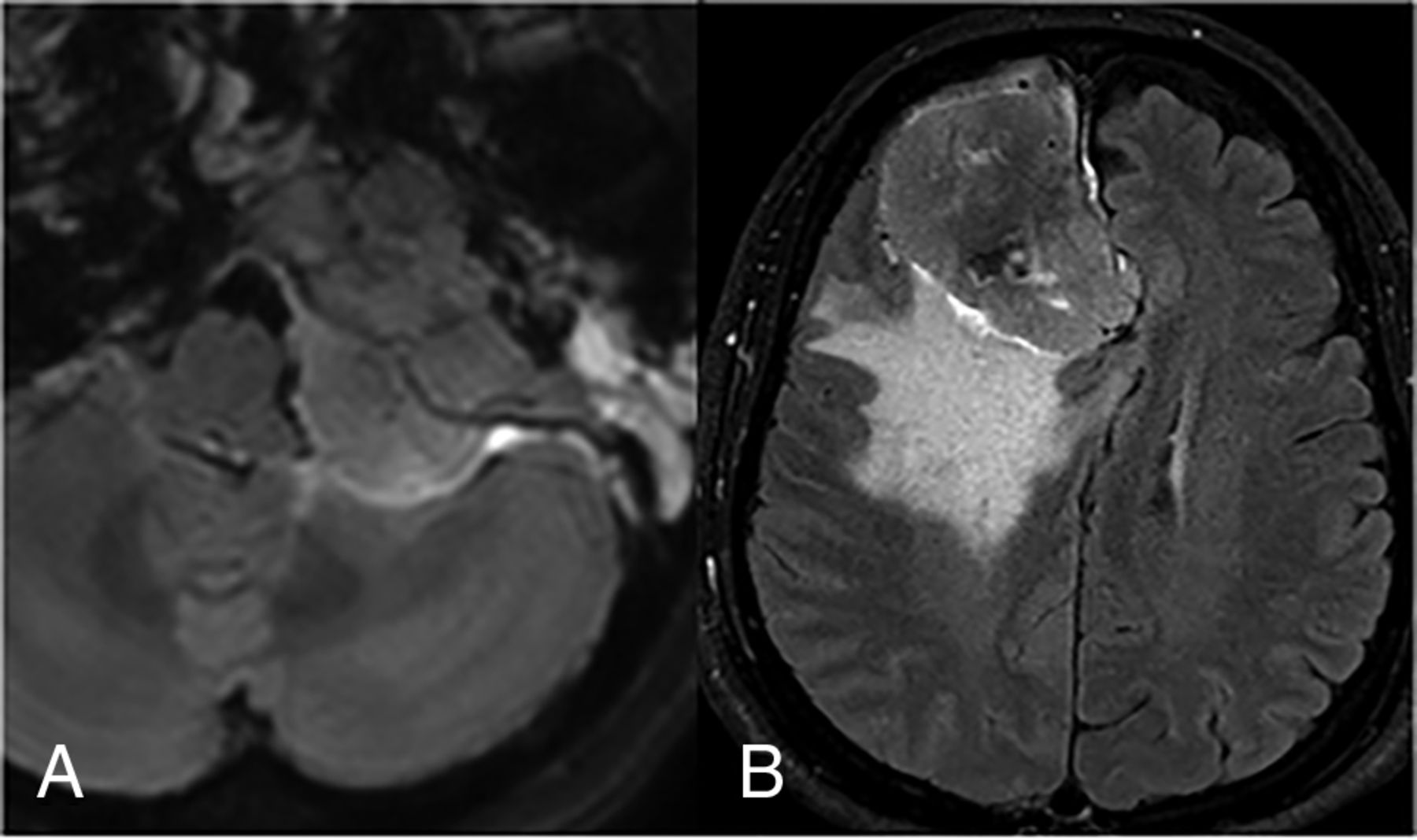
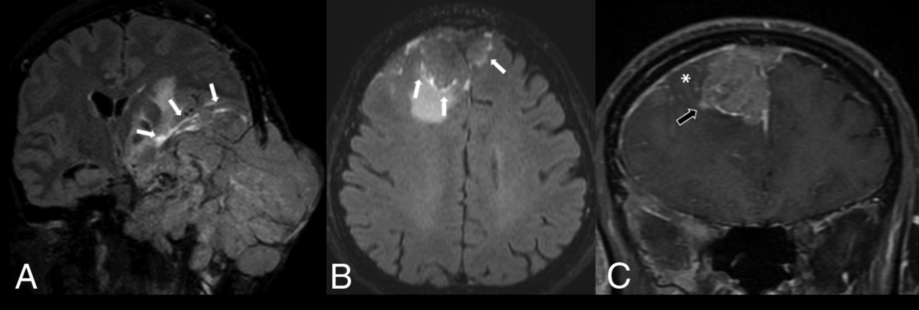
    A, Anaplastic meningioma (WHO grade III). CE-FLAIR (A) sequence demonstrates a large extra-axial mass with cortical breakthrough involving the left middle skull base and left temporal skull with the CE-FLAIR rim sign (white arrows). Malignant soft-tissue tumor was the favored diagnosis in the initial report. The pathologic result is anaplastic meningioma (WHO grade III). A malignant dural-based mass on CE-FLAIR (B) and CE-T1WI fat suppression (C) sequences shows an extra-axial heterogeneously enhancing mass at the bilateral frontal convexities that had invaded the anterior-superior sagittal sinus and demonstrates the CE-FLAIR rim sign (white arrows), accompanied by focal leptomeningeal enhancement (asterisk) and adjacent brain parenchymal invasion (black arrow). Meningioma was the favored diagnosis in the initial report. The pathologic result was metastatic mucoepidermoid carcinoma.

  • FIG 4.
    • Download figure
    • Open in new tab
    • Download powerpoint
    FIG 4.

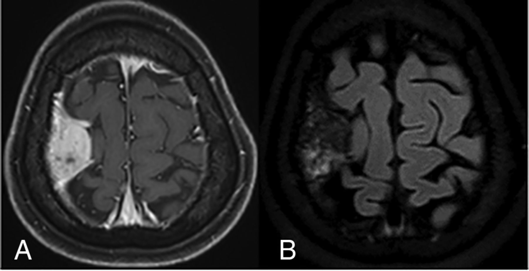
    A, CE-T1WI fat suppression sequence demonstrates an extra-axial mass at the right frontoparietal convexity with a dural tail sign that resembles a meningioma. B, CE-FLAIR sequence. No rim enhancement on the tumor-brain interface is observed. Meningioma was the favored diagnosis in the initial report. The pathologic result was osteosarcoma.

Tables

  • Figures
    • View popup
    Table 1:

    Patient demographics and pathologic findings

    VariablesMeningiomas (n = 102, 76.7%)Malignant Dural-Based Tumors (n = 31, 23.3%)
    Age (mean) (yr)51.96 (SD, 10.69)50.03 (SD, 20.81)
    Sex
     Female85 (83.3%)15 (48.4%)
     Male17 (16.7%)16 (51.6%)
    Size (mean) (cm)4.37 (SD, 1.91)4.5 (SD, 2.2)
    WHO grade
     I72 (70.6%)–
     II22 (21.6%)–
     III8 (7.8%)–
    Location
     Convexity24 (23.5%)19 (61.2%)
     Sphenoid wing20 (19.6%)3 (9.6%)
     Petroclival18 (17.6%)1 (3.3%)
     Parafalcine13 (12.7%)–
     Cavernous sinus9 (8.8%)3 (9.6%)
     Cerebellopontine angle6 (5.8%)1 (3.3%)
     Suprasellar9 (8.8%)–
     Foramen magnum2 (1.9%)–
     Olfactory groove1 (0.9%)–
     Orbit–3 (9.6%)
     Prepontine–1 (3.3%)
    • Note:—The en dash (–) indicates none.

    • View popup
    Table 2:

    Characteristics of malignant dural-based tumors

    PathologyNo. of Cases (n = 31)
    Metastasis18 (58%)
     Adenoid cystic carcinoma5
     Lung (non-small cell)3
     Breast (invasive ductal carcinoma)2
     Squamous cell carcinoma at scalp2
     Urachal carcinoma1
     Thyroid (follicular carcinoma)1
     Mucoepidermoid carcinoma1
     Nasopharynx (SCCA)1
     Base of tongue (SCCA)1
     Colon (adenocarcinoma)1
    Plasmacytoma/multiple myeloma6 (19.3%)
    Ewing sarcoma2 (6.4%)
    Lymphoma (non-Hodgkin)2 (6.4%)
    Osteosarcoma1 (3.4%)
    Spindle cell carcinoma1 (3.4%)
    Atypical teratoid/rhabdoid tumor1 (3.4%)
    • Note:—SCCA indicates squamous cell carcinoma.

    • View popup
    Table 3:

    Characteristic MR imaging findings for predicting meningiomas

    VariablesSensitivitySpecificityPPVNPVAccuracy
    Dural tail sign98.0%19.4%80.0%75.0%79.7%
    Marrow edema79.2%33.3%92.7%13.0%75.2%
    Hyperostosis74.5%100.0%100.0%54.4%80.5%
    Complete rim enhancement on CE-FLAIR89.2%93.5%97.8%72.5%90.2%
    Homogeneous enhancement on T1WI73.5%71.0%89.3%44.9%72.9%
    • View popup
    Table 4:

    Characteristic MR imaging findings for predicting malignant dural-based tumors

    VariablesSensitivitySpecificityPPVNPVAccuracy
    Lack of dural tail19.4%98.0%75.0%80.0%79.7%
    Cortical breakthrough67.7%95.1%80.8%90.7%88.7%
    Leptomeningeal enhancement32.3%100.0%100.0%82.9%84.2%
    Heterogeneous enhancement on T1WI71.0%73.5%44.9%89.3%72.9%
    Hypointense signal on T2WI53.3%86.2%66.7%78.1%75.0%
PreviousNext
Back to top

In this issue

American Journal of Neuroradiology: 44 (3)
American Journal of Neuroradiology
Vol. 44, Issue 3
1 Mar 2023
  • Table of Contents
  • Index by author
  • Complete Issue (PDF)
Advertisement
Print
Download PDF
Email Article

Thank you for your interest in spreading the word on American Journal of Neuroradiology.

NOTE: We only request your email address so that the person you are recommending the page to knows that you wanted them to see it, and that it is not junk mail. We do not capture any email address.

Enter multiple addresses on separate lines or separate them with commas.
Usefulness of a Rim-Enhancing Pattern on the Contrast-Enhanced 3D-FLAIR Sequence and MRI Characteristics for Distinguishing Meningioma and Malignant Dural-Based Tumor
(Your Name) has sent you a message from American Journal of Neuroradiology
(Your Name) thought you would like to see the American Journal of Neuroradiology web site.
CAPTCHA
This question is for testing whether or not you are a human visitor and to prevent automated spam submissions.
Cite this article
T. Panyaping, M. Punpichet, P. Tunlayadechanont, O. Tritanon
Usefulness of a Rim-Enhancing Pattern on the Contrast-Enhanced 3D-FLAIR Sequence and MRI Characteristics for Distinguishing Meningioma and Malignant Dural-Based Tumor
American Journal of Neuroradiology Mar 2023, 44 (3) 247-253; DOI: 10.3174/ajnr.A7780

Citation Manager Formats

  • BibTeX
  • Bookends
  • EasyBib
  • EndNote (tagged)
  • EndNote 8 (xml)
  • Medlars
  • Mendeley
  • Papers
  • RefWorks Tagged
  • Ref Manager
  • RIS
  • Zotero
0 Responses
Respond to this article
Share
Bookmark this article
Rim-Enhancing Pattern to Distinguish Tumors
T. Panyaping, M. Punpichet, P. Tunlayadechanont, O. Tritanon
American Journal of Neuroradiology Mar 2023, 44 (3) 247-253; DOI: 10.3174/ajnr.A7780
del.icio.us logo Twitter logo Facebook logo Mendeley logo
  • Tweet Widget
  • Facebook Like
  • Google Plus One
Purchase

Jump to section

  • Article
    • Abstract
    • ABBREVIATIONS:
    • MATERIALS AND METHODS
    • RESULTS
    • DISCUSSION
    • CONCLUSIONS
    • Footnotes
    • References
  • Figures & Data
  • Supplemental
  • Info & Metrics
  • Responses
  • References
  • PDF

Related Articles

  • PubMed
  • Google Scholar

Cited By...

  • The "Outline Sign": Thin Hyperenhancing Perimeter as an MR Imaging Feature of Meningioma. A Useful Tool in the Temporal Bone Region for Differentiating Meningiomas from Schwannomas and Paragangliomas
  • Peritumoral Signal on Postcontrast FLAIR Images: Description and Proposed Biomechanism in Vestibular Schwannomas
  • Crossref (5)
  • Google Scholar

This article has been cited by the following articles in journals that are participating in Crossref Cited-by Linking.

  • Peritumoral Signal on Postcontrast FLAIR Images: Description and Proposed Biomechanism in Vestibular Schwannomas
    John C. Benson, Matt L. Carlson, John I. Lane
    American Journal of Neuroradiology 2023 44 10
  • Meningiomas
    Lorenzo Ugga, Teresa Perillo, Serena Capasso, Davide Negroni, Renato Cuocolo
    2024
  • Radiopaedia.org
    Frank Gaillard, Liz Silverstone, Tariq Walizai
    2008
  • Real-Time Detection of Meningiomas by Image Segmentation: A Very Deep Transfer Learning Convolutional Neural Network Approach
    Debasmita Das, Chayna Sarkar, Biswadeep Das
    Tomography 2025 11 5
  • The “Outline Sign”: Thin Hyperenhancing Perimeter as an MR Imaging Feature of Meningioma. A Useful Tool in the Temporal Bone Region for Differentiating Meningiomas from Schwannomas and Paragangliomas
    Anil K. Vasireddi, Katherine L. Reinshagen, Donghoon Shin, Laura V. Romo, Amy F. Juliano
    American Journal of Neuroradiology 2025 46 2

More in this TOC Section

  • Diagnostic Neuroradiology of Monoclonal Antibodies
  • Clinical Outcomes After Chiari I Decompression
  • Segmentation of Brain Metastases with BLAST
Show more ADULT BRAIN

Similar Articles

Advertisement

Indexed Content

  • Current Issue
  • Accepted Manuscripts
  • Article Preview
  • Past Issues
  • Editorials
  • Editor's Choice
  • Fellows' Journal Club
  • Letters to the Editor
  • Video Articles

Cases

  • Case Collection
  • Archive - Case of the Week
  • Archive - Case of the Month
  • Archive - Classic Case

More from AJNR

  • Trainee Corner
  • Imaging Protocols
  • MRI Safety Corner
  • Book Reviews

Multimedia

  • AJNR Podcasts
  • AJNR Scantastics

Resources

  • Turnaround Time
  • Submit a Manuscript
  • Submit a Video Article
  • Submit an eLetter to the Editor/Response
  • Manuscript Submission Guidelines
  • Statistical Tips
  • Fast Publishing of Accepted Manuscripts
  • Graphical Abstract Preparation
  • Imaging Protocol Submission
  • Evidence-Based Medicine Level Guide
  • Publishing Checklists
  • Author Policies
  • Become a Reviewer/Academy of Reviewers
  • News and Updates

About Us

  • About AJNR
  • Editorial Board
  • Editorial Board Alumni
  • Alerts
  • Permissions
  • Not an AJNR Subscriber? Join Now
  • Advertise with Us
  • Librarian Resources
  • Feedback
  • Terms and Conditions
  • AJNR Editorial Board Alumni

American Society of Neuroradiology

  • Not an ASNR Member? Join Now

© 2025 by the American Society of Neuroradiology All rights, including for text and data mining, AI training, and similar technologies, are reserved.
Print ISSN: 0195-6108 Online ISSN: 1936-959X

Powered by HighWire