Advertisement

Main menu

  • Home
  • Content
    • Current Issue
    • Accepted Manuscripts
    • Article Preview
    • Past Issue Archive
    • Video Articles
    • AJNR Case Collection
    • Case of the Week Archive
    • Case of the Month Archive
    • Classic Case Archive
  • Special Collections
    • AJNR Awards
    • Low-Field MRI
    • Alzheimer Disease
    • ASNR Foundation Special Collection
    • Photon-Counting CT
    • View All
  • Multimedia
    • AJNR Podcasts
    • AJNR SCANtastic
    • Trainee Corner
    • MRI Safety Corner
    • Imaging Protocols
  • For Authors
    • Submit a Manuscript
    • Submit a Video Article
    • Submit an eLetter to the Editor/Response
    • Manuscript Submission Guidelines
    • Statistical Tips
    • Fast Publishing of Accepted Manuscripts
    • Graphical Abstract Preparation
    • Imaging Protocol Submission
    • Author Policies
  • About Us
    • About AJNR
    • Editorial Board
    • Editorial Board Alumni
  • More
    • Become a Reviewer/Academy of Reviewers
    • Subscribers
    • Permissions
    • Alerts
    • Feedback
    • Advertisers
    • ASNR Home

User menu

  • Alerts
  • Log in

Search

  • Advanced search
American Journal of Neuroradiology

American Journal of Neuroradiology

ASHNR American Society of Functional Neuroradiology ASHNR American Society of Pediatric Neuroradiology ASSR
  • Alerts
  • Log in

Advanced Search

  • Home
  • Content
    • Current Issue
    • Accepted Manuscripts
    • Article Preview
    • Past Issue Archive
    • Video Articles
    • AJNR Case Collection
    • Case of the Week Archive
    • Case of the Month Archive
    • Classic Case Archive
  • Special Collections
    • AJNR Awards
    • Low-Field MRI
    • Alzheimer Disease
    • ASNR Foundation Special Collection
    • Photon-Counting CT
    • View All
  • Multimedia
    • AJNR Podcasts
    • AJNR SCANtastic
    • Trainee Corner
    • MRI Safety Corner
    • Imaging Protocols
  • For Authors
    • Submit a Manuscript
    • Submit a Video Article
    • Submit an eLetter to the Editor/Response
    • Manuscript Submission Guidelines
    • Statistical Tips
    • Fast Publishing of Accepted Manuscripts
    • Graphical Abstract Preparation
    • Imaging Protocol Submission
    • Author Policies
  • About Us
    • About AJNR
    • Editorial Board
    • Editorial Board Alumni
  • More
    • Become a Reviewer/Academy of Reviewers
    • Subscribers
    • Permissions
    • Alerts
    • Feedback
    • Advertisers
    • ASNR Home
  • Follow AJNR on Twitter
  • Visit AJNR on Facebook
  • Follow AJNR on Instagram
  • Join AJNR on LinkedIn
  • RSS Feeds

AJNR Awards, New Junior Editors, and more. Read the latest AJNR updates

Research ArticlePediatric Neuroimaging

Fetal MR Imaging Anatomy of the Transverse Temporal Gyrus (Heschl Gyrus)

Eleonora Piccirilli, Chiara Marchetti, Valentina Panara, Claudio Celentano, Francesco D'Antonio, Stefano Sensi, Andrea Righini and Massimo Caulo
American Journal of Neuroradiology November 2023, 44 (11) 1325-1331; DOI: https://doi.org/10.3174/ajnr.A8026
Eleonora Piccirilli
aFrom the Department of Neuroscience (E.P., S.S., M.C.), Imaging and Clinical Sciences, University of Chieti, Chieti, Italy
bNeuro-Oncology Unit (E.P.), Department of Imaging, Bambino Gesù Children's Hospital, Istituto di Ricovero e Cura a Carattere Scientifico, Rome, Italy
h ITAB-Institute of Advanced Biomedical Technologies, Department of Neuroscience, Imaging and Clinical Sciences (E.P., C.M., V.P., S.S., M.C.), University G. d'Annunzio of Chieti-Pescara, Chieti, Italy
  • Find this author on Google Scholar
  • Find this author on PubMed
  • Search for this author on this site
  • ORCID record for Eleonora Piccirilli
Chiara Marchetti
cRadiology Unit (C.M.), San Pio Da Pietrelcina Hospital, Vasto, Chieti, Italy
h ITAB-Institute of Advanced Biomedical Technologies, Department of Neuroscience, Imaging and Clinical Sciences (E.P., C.M., V.P., S.S., M.C.), University G. d'Annunzio of Chieti-Pescara, Chieti, Italy
  • Find this author on Google Scholar
  • Find this author on PubMed
  • Search for this author on this site
  • ORCID record for Chiara Marchetti
Valentina Panara
dDepartment of Radiology (V.P., M.C.), Santissima Annunziata Hospital, Chieti, Italy
h ITAB-Institute of Advanced Biomedical Technologies, Department of Neuroscience, Imaging and Clinical Sciences (E.P., C.M., V.P., S.S., M.C.), University G. d'Annunzio of Chieti-Pescara, Chieti, Italy
  • Find this author on Google Scholar
  • Find this author on PubMed
  • Search for this author on this site
  • ORCID record for Valentina Panara
Claudio Celentano
eObstetrics and Gynaecology Unit (C.C.), Santo Spirito Hospital, Pescara, Italy
  • Find this author on Google Scholar
  • Find this author on PubMed
  • Search for this author on this site
  • ORCID record for Claudio Celentano
Francesco D'Antonio
fCenter for Fetal Care and High-Risk Pregnancy (F.D.), Department of Obstetrics and Gynecology, University of Chieti, Chieti, Italy
  • Find this author on Google Scholar
  • Find this author on PubMed
  • Search for this author on this site
Stefano Sensi
aFrom the Department of Neuroscience (E.P., S.S., M.C.), Imaging and Clinical Sciences, University of Chieti, Chieti, Italy
h ITAB-Institute of Advanced Biomedical Technologies, Department of Neuroscience, Imaging and Clinical Sciences (E.P., C.M., V.P., S.S., M.C.), University G. d'Annunzio of Chieti-Pescara, Chieti, Italy
  • Find this author on Google Scholar
  • Find this author on PubMed
  • Search for this author on this site
Andrea Righini
gNeuroradiology Unit (A.R.), Pediatric Radiology Department, Vittore Buzzi Children's Hospital, Milan, Italy.
  • Find this author on Google Scholar
  • Find this author on PubMed
  • Search for this author on this site
  • ORCID record for Andrea Righini
Massimo Caulo
aFrom the Department of Neuroscience (E.P., S.S., M.C.), Imaging and Clinical Sciences, University of Chieti, Chieti, Italy
dDepartment of Radiology (V.P., M.C.), Santissima Annunziata Hospital, Chieti, Italy
h ITAB-Institute of Advanced Biomedical Technologies, Department of Neuroscience, Imaging and Clinical Sciences (E.P., C.M., V.P., S.S., M.C.), University G. d'Annunzio of Chieti-Pescara, Chieti, Italy
  • Find this author on Google Scholar
  • Find this author on PubMed
  • Search for this author on this site
  • ORCID record for Massimo Caulo
  • Article
  • Figures & Data
  • Info & Metrics
  • Responses
  • References
  • PDF
Loading

Article Figures & Data

Figures

  • Tables
  • FIG 1.
    • Download figure
    • Open in new tab
    • Download powerpoint
    FIG 1.

    Representative images of adult (upper row) and fetal (lower row, 28 weeks' GA) HGs on the 3 orthogonal planes. Adult landmarks were used to correctly identify the HG on the fetal brains (asterisk).
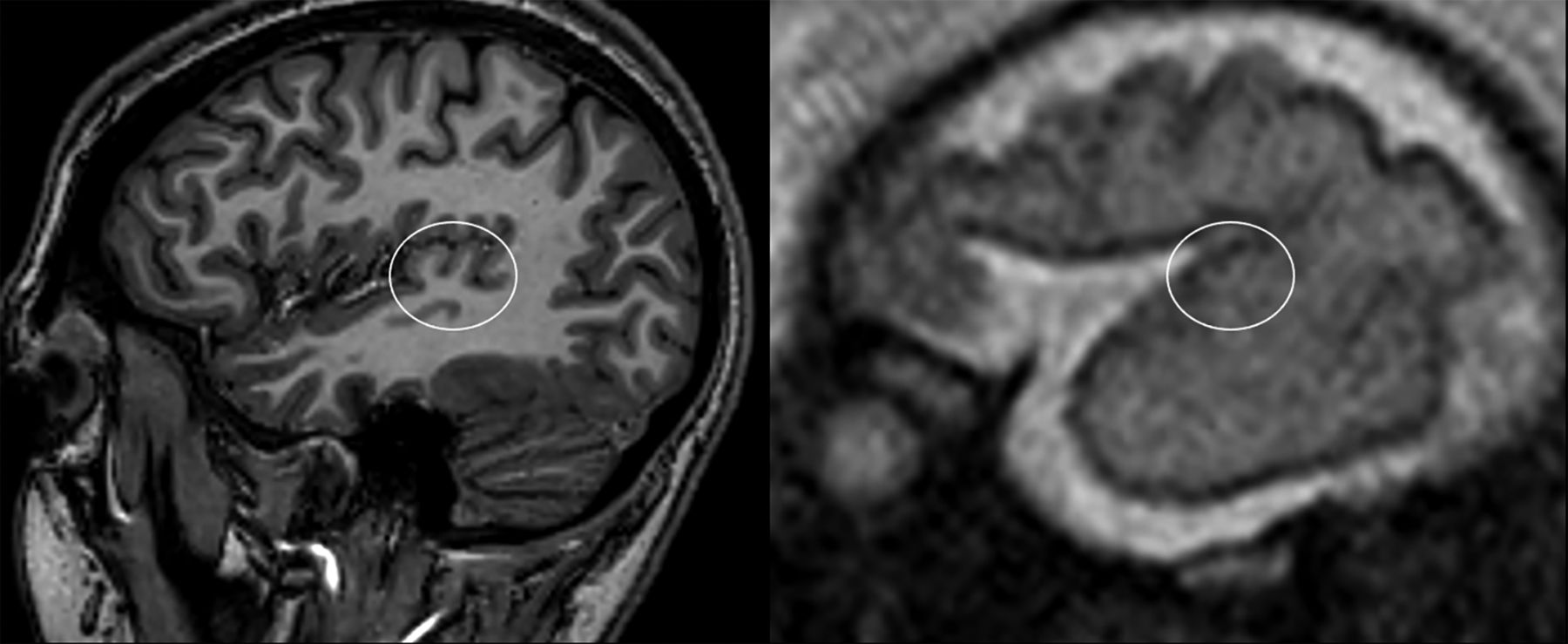
  • FIG 2.
    • Download figure
    • Open in new tab
    • Download powerpoint
    FIG 2.

    The SI of the HG in representative adult (left) and fetal (right, 28 weeks' GA) brains. The characteristic adult heart-shaped configuration in the sagittal plane was used for SI identification on fetal scans.
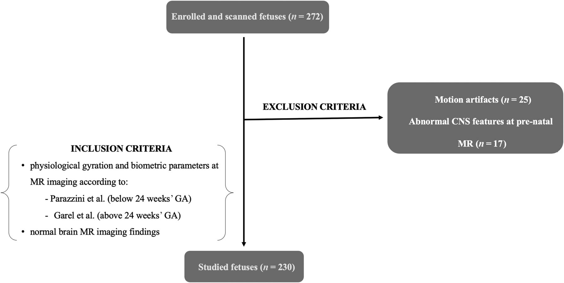
  • FIG 3.
    • Download figure
    • Open in new tab
    • Download powerpoint
    FIG 3.

    Flow chart detailing inclusion and exclusion criteria.
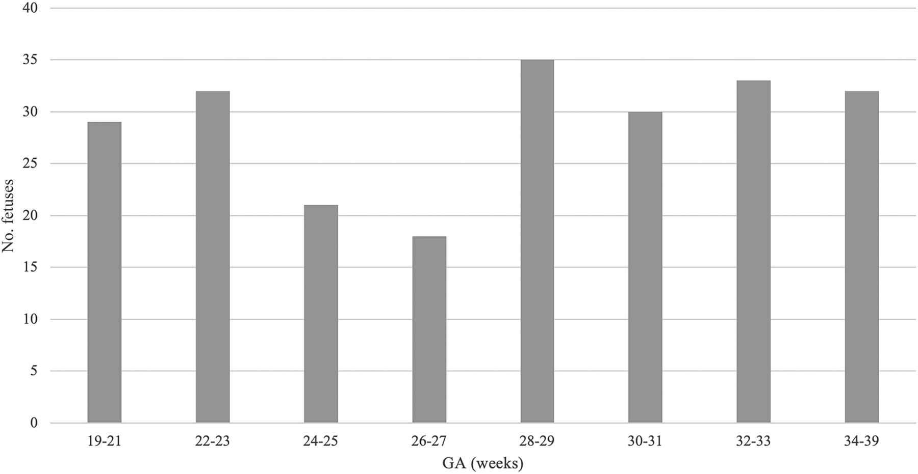
  • FIG 4.
    • Download figure
    • Open in new tab
    • Download powerpoint
    FIG 4.

    Distribution of the number of fetuses as a function of GA.

  • FIG 5.
    • Download figure
    • Open in new tab
    • Download powerpoint
    FIG 5.

    Detection of the HG on sagittal (black line), coronal (gray line), and axial (light gray line) T2-weighted SSh MR images with increasing GA (in weeks).
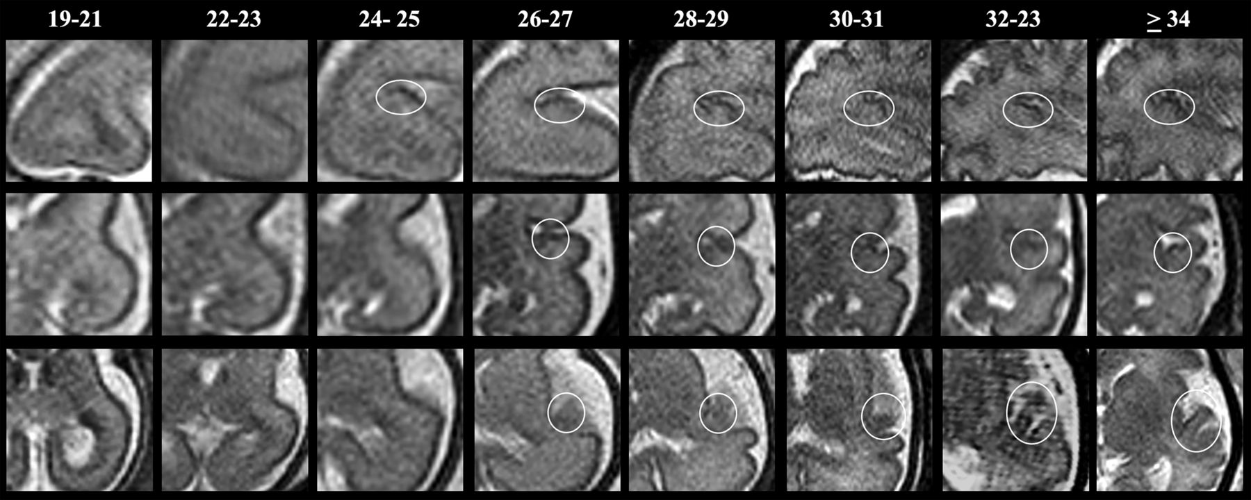
  • FIG 6.
    • Download figure
    • Open in new tab
    • Download powerpoint
    FIG 6.

    SSh T2WI sagittal (upper row), coronal (middle row), and axial (lower row) MR images illustrating changes in HG morphology with increasing GA.

Tables

  • Figures
  • Frequencies of HG appearance in the three orthogonal planes according to GA groups

    GA (week)Sagittal Plane (%)Coronal Plane (%)Axial Plane (%)
    19–21000
    22–23000
    24–25679.50
    26–279488.867
    28–29100100100
    30–31100100100
    32–33100100100
    34–39100100100
PreviousNext
Back to top

In this issue

American Journal of Neuroradiology: 44 (11)
American Journal of Neuroradiology
Vol. 44, Issue 11
1 Nov 2023
  • Table of Contents
  • Index by author
  • Complete Issue (PDF)
Advertisement
Print
Download PDF
Email Article

Thank you for your interest in spreading the word on American Journal of Neuroradiology.

NOTE: We only request your email address so that the person you are recommending the page to knows that you wanted them to see it, and that it is not junk mail. We do not capture any email address.

Enter multiple addresses on separate lines or separate them with commas.
Fetal MR Imaging Anatomy of the Transverse Temporal Gyrus (Heschl Gyrus)
(Your Name) has sent you a message from American Journal of Neuroradiology
(Your Name) thought you would like to see the American Journal of Neuroradiology web site.
CAPTCHA
This question is for testing whether or not you are a human visitor and to prevent automated spam submissions.
Cite this article
Eleonora Piccirilli, Chiara Marchetti, Valentina Panara, Claudio Celentano, Francesco D'Antonio, Stefano Sensi, Andrea Righini, Massimo Caulo
Fetal MR Imaging Anatomy of the Transverse Temporal Gyrus (Heschl Gyrus)
American Journal of Neuroradiology Nov 2023, 44 (11) 1325-1331; DOI: 10.3174/ajnr.A8026

Citation Manager Formats

  • BibTeX
  • Bookends
  • EasyBib
  • EndNote (tagged)
  • EndNote 8 (xml)
  • Medlars
  • Mendeley
  • Papers
  • RefWorks Tagged
  • Ref Manager
  • RIS
  • Zotero
0 Responses
Respond to this article
Share
Bookmark this article
Fetal MR Imaging of Heschl Gyrus Anatomy
Eleonora Piccirilli, Chiara Marchetti, Valentina Panara, Claudio Celentano, Francesco D'Antonio, Stefano Sensi, Andrea Righini, Massimo Caulo
American Journal of Neuroradiology Nov 2023, 44 (11) 1325-1331; DOI: 10.3174/ajnr.A8026
del.icio.us logo Twitter logo Facebook logo Mendeley logo
  • Tweet Widget
  • Facebook Like
  • Google Plus One
Purchase

Jump to section

  • Article
    • Abstract
    • ABBREVIATIONS:
    • MATERIALS AND MATERIALS
    • RESULTS
    • DISCUSSION
    • CONCLUSIONS
    • Footnotes
    • References
  • Figures & Data
  • Info & Metrics
  • Responses
  • References
  • PDF

Related Articles

  • No related articles found.
  • PubMed
  • Google Scholar

Cited By...

  • No citing articles found.
  • Crossref (2)
  • Google Scholar

This article has been cited by the following articles in journals that are participating in Crossref Cited-by Linking.

  • Improving Image Quality and Decreasing SAR With High Dielectric Constant Pads in 3 T Fetal MRI
    Zhengyang Zhu, Xunwen Xue, Tang Tang, Chao Luo, Ye Li, Jing Chen, Biyun Xu, Zengping Lin, Xin Zhang, Zhengge Wang, Jun Chen, Jiaming Lu, Wen Zhang, Xin Li, Qian Chen, Zhuoru Jiang, Junxia Wang, Qing Hu, Sven Haller, Ming Li, Chenchen Yan, Bing Zhang
    Journal of Magnetic Resonance Imaging 2025 61 6
  • Tracking microstructural adaptations in primary visual areas: A diffusion kurtosis imaging study on visuomotor learning
    Fahad Alharshan, Abdulrahman Aloufi, Fiona Rowe, Georg Meyer
    NeuroImage 2025 310

More in this TOC Section

  • fetal brain development of 10 weeks gestation
  • CHARGE fetal MRI clival cleft
  • Neuroimaging Delineation and Progression of SLSMD
Show more Pediatric Neuroimaging

Similar Articles

Advertisement

Indexed Content

  • Current Issue
  • Accepted Manuscripts
  • Article Preview
  • Past Issues
  • Editorials
  • Editor's Choice
  • Fellows' Journal Club
  • Letters to the Editor
  • Video Articles

Cases

  • Case Collection
  • Archive - Case of the Week
  • Archive - Case of the Month
  • Archive - Classic Case

More from AJNR

  • Trainee Corner
  • Imaging Protocols
  • MRI Safety Corner

Multimedia

  • AJNR Podcasts
  • AJNR Scantastics

Resources

  • Turnaround Time
  • Submit a Manuscript
  • Submit a Video Article
  • Submit an eLetter to the Editor/Response
  • Manuscript Submission Guidelines
  • Statistical Tips
  • Fast Publishing of Accepted Manuscripts
  • Graphical Abstract Preparation
  • Imaging Protocol Submission
  • Evidence-Based Medicine Level Guide
  • Publishing Checklists
  • Author Policies
  • Become a Reviewer/Academy of Reviewers
  • News and Updates

About Us

  • About AJNR
  • Editorial Board
  • Editorial Board Alumni
  • Alerts
  • Permissions
  • Not an AJNR Subscriber? Join Now
  • Advertise with Us
  • Librarian Resources
  • Feedback
  • Terms and Conditions
  • AJNR Editorial Board Alumni

American Society of Neuroradiology

  • Not an ASNR Member? Join Now

© 2025 by the American Society of Neuroradiology All rights, including for text and data mining, AI training, and similar technologies, are reserved.
Print ISSN: 0195-6108 Online ISSN: 1936-959X

Powered by HighWire