
Article Figures & Data
Figures
- FIG 1.
The architecture of the U-Net model for landmark detection. The U-Net converts a sagittal fetal brain image into gray-scale masks in which the vertex represents the locations of different landmark points. Up-conv indicates up-sampling operation; conv, convolutional layer; ReLU, Rectified Linear Activation.
- FIG 2.
The cut surface of a 3D rotationally symmetric Gaussian distribution function with the radius (R), and the top represents the landmark point position.
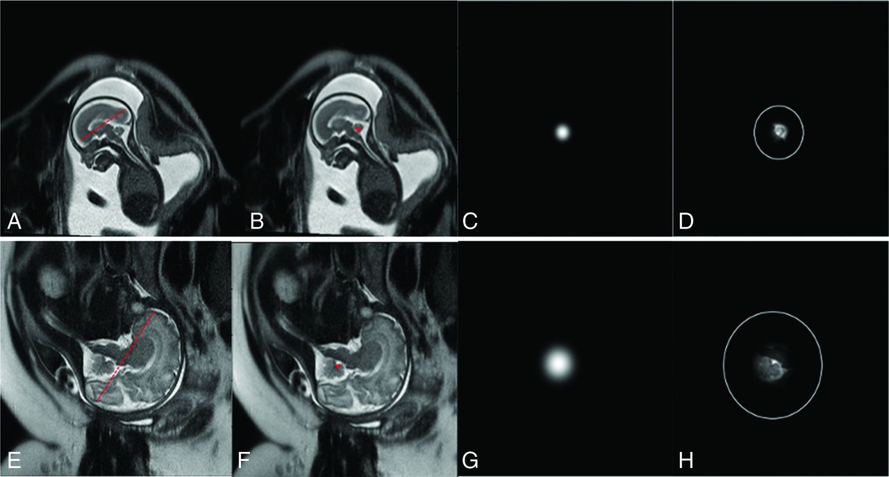
- FIG 3.
Fronto-occipital radius of the fetal brain determines the radius of Gaussian distribution function in a patient at GA week 20 (A–D) and another at GA week 33 (E–H). The left 2 columns are MR images with a fronto-occipital radius (A and E) and annotated landmark points on the vermis (B and F). The third column (C and G) shows the image mask with the Gaussian distribution used in model training. The fourth column shows the image area surrounding the landmark (D and H) determined by the Gaussian distribution function, with an added white circle indicating the radius range.
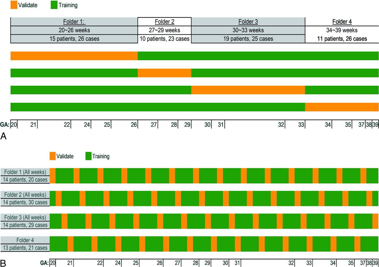
- FIG 4.
Two 4-fold cross-validation methods for DL-model training and testing. Method 1 divided the data sets by sorting the ranges of GA weeks (A). Method 2 divided the data sets randomly with mixed GA weeks (B).
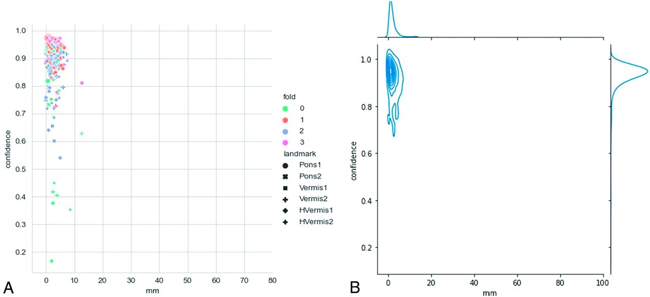
- FIG 5.
The scatterplot (A) and contour line plot (B) of the prediction error distribution with associated confidence scores in the sorted GA week validation method. The x-axis represents the distance (millimeters) between the predicted landmark and the ground truth, and the y-axis represents the confidence score of the prediction.
- FIG 6.
The scatterplot (A) and contour line plot (B) of the distribution of prediction error with associated confidence scores in the randomly mixed GA week validation method. The x-axis represents the distance (millimeters) between the predicted landmark and the ground truth, and the y-axis represents the confidence score of the prediction.
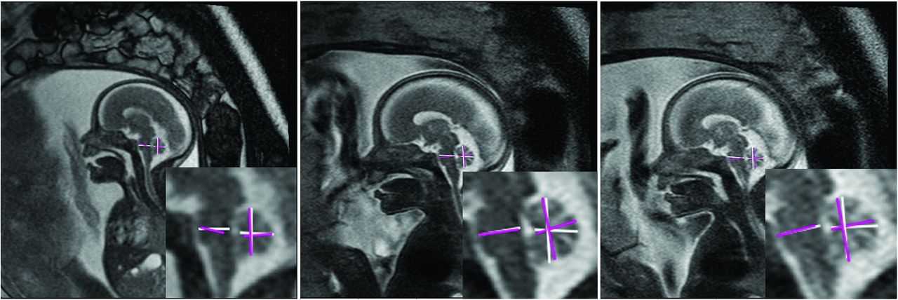
- FIG 7.
Representative image examples of model-predicted landmark locations with biometric measurements. (The white line is manual annotations by radiologists, and the purple line is DL-model predicted measurements). Three patients (A, 22 weeks; B, 22 weeks; C, 27 weeks) with accurate model-predicted landmarks compared with radiologists.
- FIG 8.
An interactive tool integrating DL-model-based prediction of landmarks. It helps the radiologist quickly locate, confirm, or adjust the landmarks on the automatically selected image section. After the landmark locations are confirmed, the distance between 2 related landmarks on a particular brain structure is calculated. The white line is the “Pons” AP diameter, and red crossed lines are “Vermis” AP diameter and height.


Tables
Compared manual landmark localization conducted by a radiologist and an expert pediatric neuroradiologist
Pons1 Pons2 Vermis1 Vermis2 HVermis1 HVermis2 Mean (mm) 1.41 0.79 0.42 1.87 1.28 1.51 SD (mm) 1.12 0.76 0.59 1.81 1.38 1.68 Note:—Pons1 indicates Anterior landmark of Pons; Pons2, Posterior landmark of Pons; Vermis1, Anterior landmark of Vermis; Vermis2, Posterior landmark of Vermis; Hvermis1, Superior landmark of Vermis (height of vermis); Hvermis2, Inferior landmark of Vermis (height of vermis).


Jump to section
Related Articles
Cited By...
- No citing articles found.