
Article Figures & Data
Figures
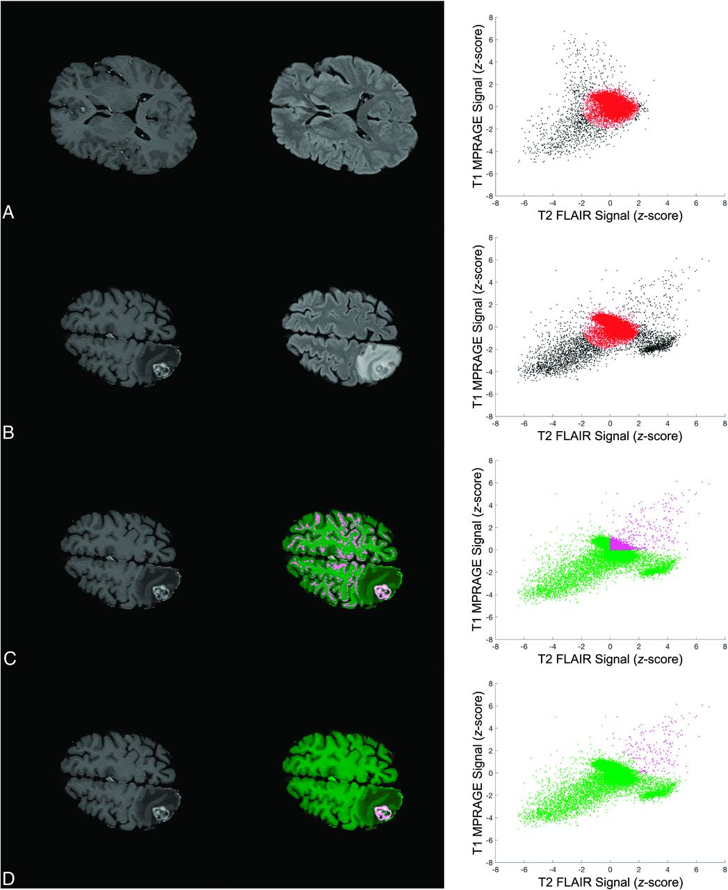
- FIG 1.
Key steps in defining the BLAST parameter space and performing segmentations with BLAST. Images and segmentations are shown in the left and middle columns, and the corresponding parameter space is shown in the right column. Each point in the scatterplot represents a voxel in the image. A, The statistics of the background layer (gray and white matter) are first defined using K-means clustering on a normal section of the brain. The origin of the parameter space is defined by the centroid of the background layer cluster and the axes expressed as z score relative to the background layer cluster SD. B, An ellipsoid approximating the background layer is applied to all slices (red ellipse). Voxels corresponding to enhancing brain tumor, edema, and CSF are found in the upper right, lower right, and lower left corner of parameter space, respectively. C, Thresholds in both T1 MPRAGE and T2 FLAIR are applied to exclude nontumoral voxels to perform segmentations. In the sample shown, thresholds in both parameters are set to the mean of the background layer (z score = 0). D, Voxels falling within the background layer ellipsoid are subtracted to further exclude the background layer. The segmentation is selected to save a mask of the 3D-connected enhancing BM voxels. An algorithm to fill in necrotic areas is applied for the final segmentation (not shown).
- FIG 2.
Sample contours derived from BLAST segmentations for parenchymal and dural metastases. The first column shows ground truth segmentations for 5 metastases (red). Corresponding contours created with BLAST are shown in the second column (green) for the operator with the best DSC. The mean DSCs for A, B, and C are 0.90, 0.83, 0.80, respectively. Two small metastases measuring 7 and 8 mm are shown in D with DSCs of 0.67 and 0.82, respectively.
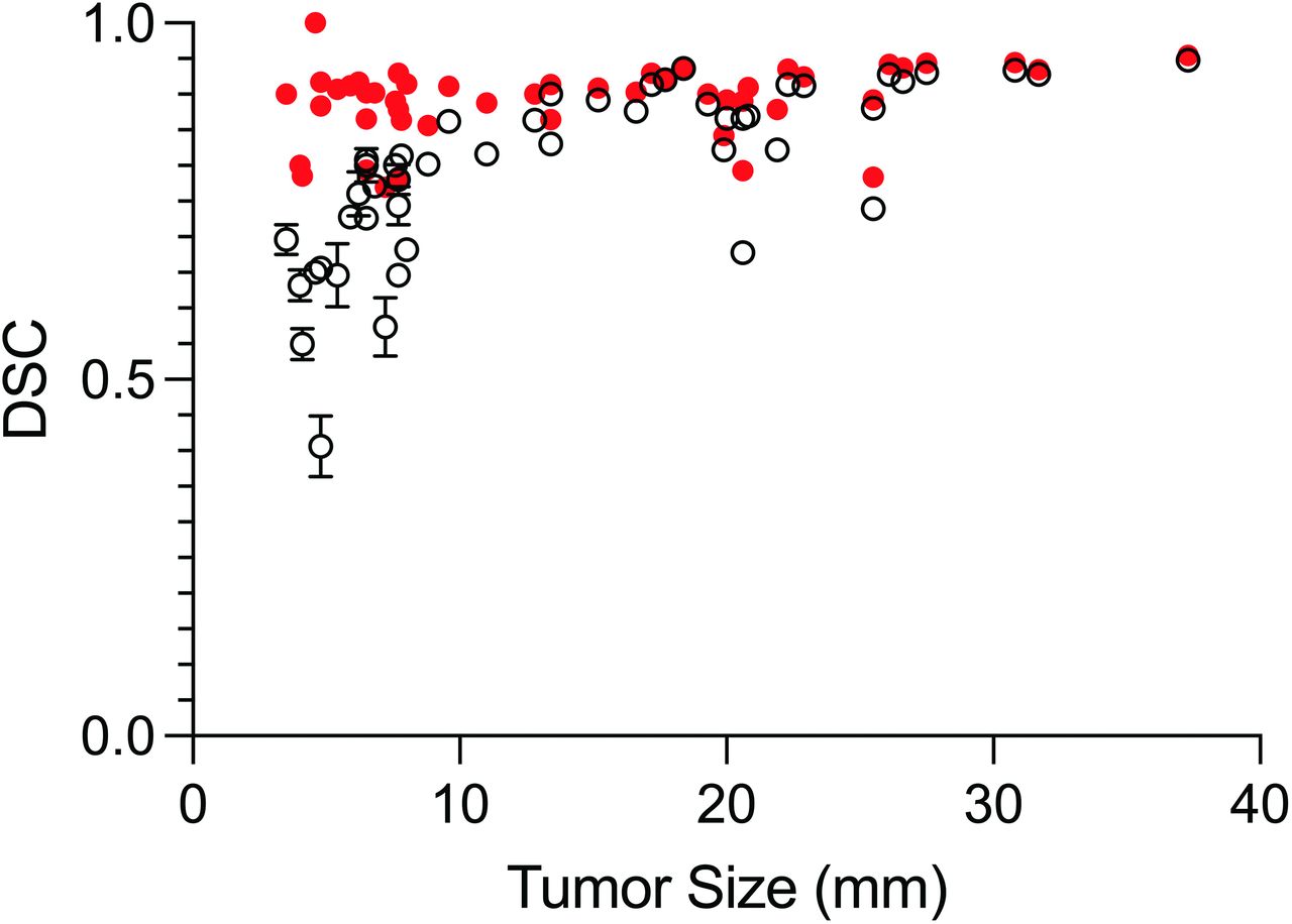
- FIG 3.
BLAST segmentation performance measured by DSC (mean) as a function of metastasis size for all operators (black open circles). Segmentation performance for BLAST generated by automatically iterating through combinations of thresholds with fixed background layer subtraction (red closed circles) outperforms operator-generated segmentations for small (<10 mm) metastases (P < .001).
- FIG 4.
Comparison of tumor volumes measured by operators with BLAST versus ground truth. Linear regression of the data (dashed lines indicate 95% CI) shows an excellent fit (R2 = 0.9951). The measured volumes closely approximate the ground truth volumes, with a slope close to unity (1.05; 95% CI, 1.04–1.06).


Tables
Patient demographics and tumor characteristics
Parameter Demographics No. of patients 19 Average age (yr) 65.7 (SD, 14) No. women 9 (47.3%) Primary cancer type Lung NSCLC 9 SCLC 1 Breast 2 Melanoma 2 Esophagus 1 Gastric 1 Pancreas 1 Vagina 1 Nasopharynx 1 Metastasis information Total No. Parenchymal 38 Dural 10 Median No./patient 2 (IQR, 1–3.5) Median size (mm) 21.8 (IQR, 6.7–20.7) Median volume (cm3) 0.70 (IQR, 0.10–2.1) Note:—NSCLC indicates non-small cell lung cancer; SCLC, small cell lung cancer.


