
Article Figures & Data
Figures
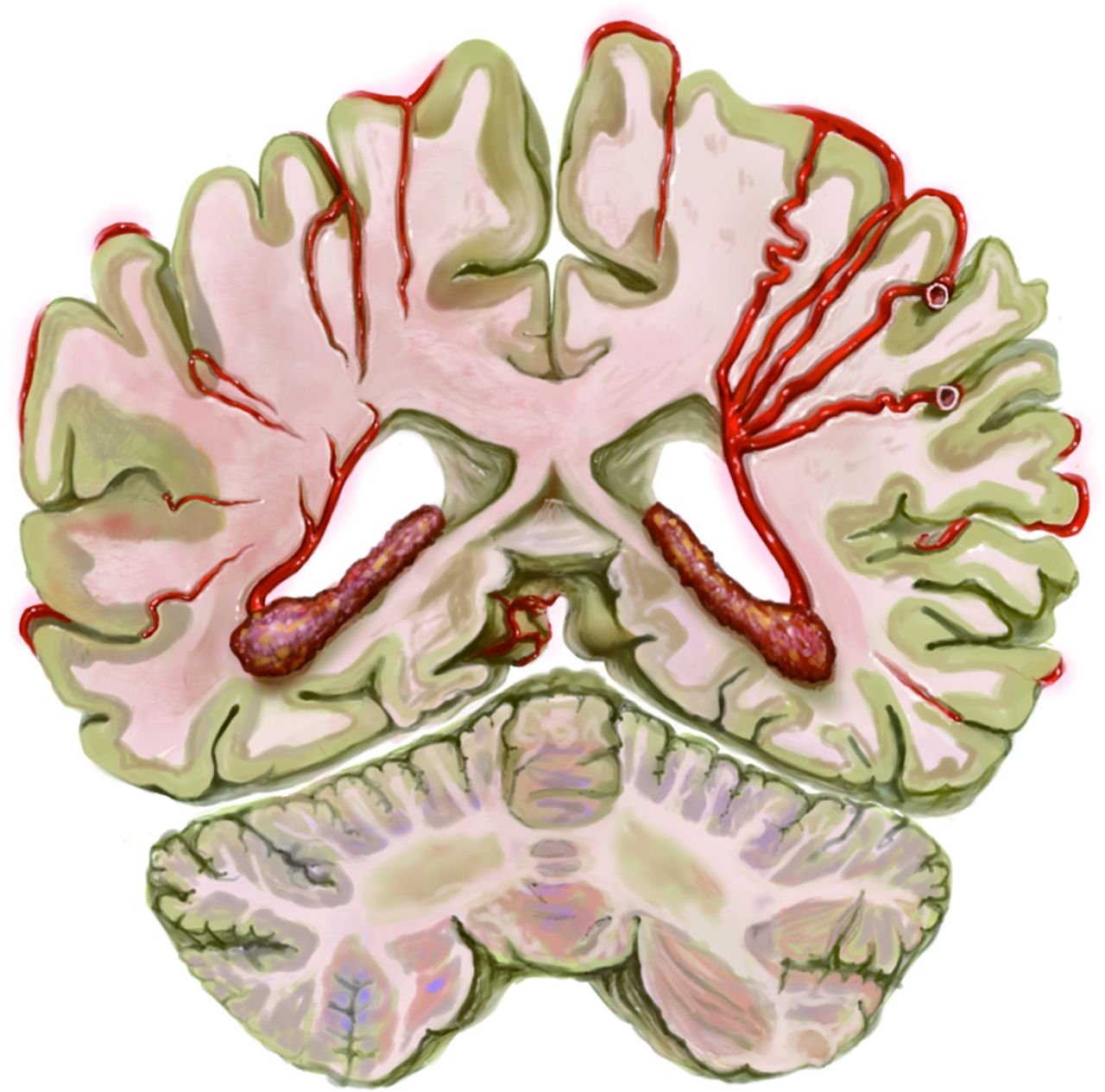
- FIG 1.
Schematic illustration showing choroidal anastomosis (left hemisphere) in the coronal plane.
- FIG 2.
Quantitative measurement of revascularization areas. A, Original image of the postoperative external carotid angiography in the capillary phase. B, Images imported into the software (ImageJ, Version 1.52a). The revascularization area (RA) posterior to the CS (blue line) is 188805/747497 = 25.3%.
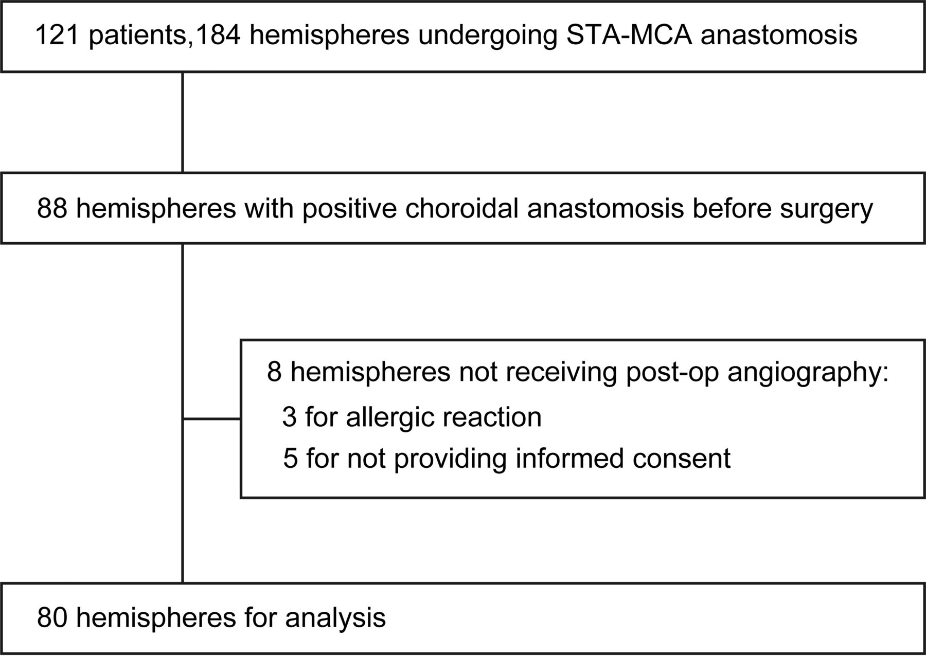
- FIG 3.
Flow chart for patient inclusion.
- FIG 4.
Comparison of each revascularization area between hemispheres exhibiting reduction of choroidal anastomosis and those exhibiting no reduction. The dotted line indicates the cutoff value (10.7%).
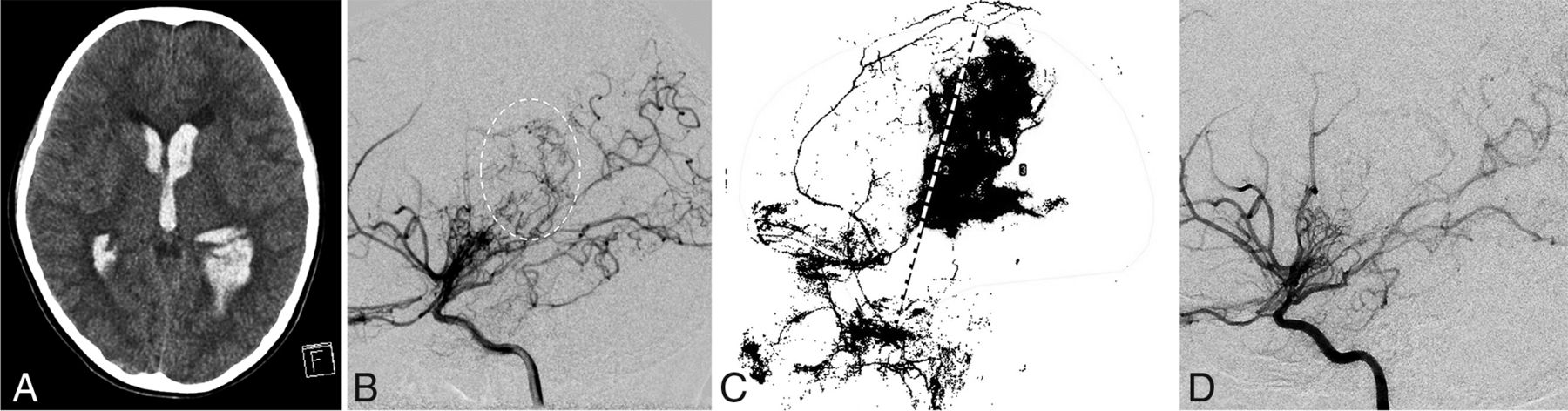
- FIG 5.
Case 1 (a 9-year-old girl). A, CT at onset shows intraventricular hemorrhage. B, Preoperative left internal carotid angiography shows choroidal anastomosis (dotted circle), which corresponds to the hemorrhage site. C, Quantitative measurement of the revascularization area in the left hemisphere. The revascularization areas anterior and posterior to the CS are 3.4% and 16.1%, respectively. The dotted line indicates the CS. D, Postoperative left internal carotid angiography shows reduction of choroidal anastomosis.
- FIG 6.
Case 2. The patient had initially manifested seizure at 43 years of age and had undergone direct bypass. A, Quantitative measurement of the revascularization area in the left hemisphere. The revascularization areas anterior and posterior to the CS were 12.3% and 1.5%, respectively. The dotted line indicates the CS. B, Postoperative left ICA shows persistence of choroidal anastomosis (dotted circle). C, CT obtained at the onset of de novo intracranial hemorrhage, which occurred 13 years after the operation.


Tables
Total Reduction of Choroidal Anastomosis P Value Yes No No. of hemispheres 80 68 12 NA Revascularization area (mean) (%)a Posterior to CS 13.6 (SD, 7.7) 15.2 (SD, 7.1) 4.2 (SD, 3.4) <.001 Anterior to CS 12.6 (SD, 7.0) 12.5 (SD, 6.7) 13.2 (SD, 8.9) .74 Median age (yr) (IQR) 12 (8–36) 11 (8–33) 19.5 (5.25–47.5) .57 Female (%) 47 (58.8) 43 (63.2) 4 (33.3) .05 Hemorrhagic presentation (%) 17 (21.3) 12 (17.7) 5 (41.7) .06 Median Suzuki stage (IQR) 3 (3–3) 3 (3–3) 3 (2–3) .33 PCA involvement (%) 10 (12.5) 10 (14.7) 0 .34 SPECT stage 2 MCA territory (%) 37 (46.3) 32 (47.1) 5 (41.7) .73 Posterior MCA territory (%) 27 (33.8) 25 (36.8) 2 (16.7) .17 Addition of EMS (%) 21 (26.3) 16 (23.5) 5 (41.7) .19 Note:—EMS indicates encephalomyosynangiosis; IQR, interquartile range; NA, not applicable.
↵a Percentage relative to whole supratentorial area.


Jump to section
Related Articles
Cited By...
- No citing articles found.