Advertisement

Main menu

  • Home
  • Content
    • Current Issue
    • Accepted Manuscripts
    • Article Preview
    • Past Issue Archive
    • Video Articles
    • AJNR Case Collection
    • Case of the Week Archive
    • Case of the Month Archive
    • Classic Case Archive
  • Special Collections
    • AJNR Awards
    • Low-Field MRI
    • Alzheimer Disease
    • ASNR Foundation Special Collection
    • Photon-Counting CT
    • View All
  • Multimedia
    • AJNR Podcasts
    • AJNR SCANtastic
    • Trainee Corner
    • MRI Safety Corner
    • Imaging Protocols
  • For Authors
    • Submit a Manuscript
    • Submit a Video Article
    • Submit an eLetter to the Editor/Response
    • Manuscript Submission Guidelines
    • Statistical Tips
    • Fast Publishing of Accepted Manuscripts
    • Graphical Abstract Preparation
    • Imaging Protocol Submission
    • Author Policies
  • About Us
    • About AJNR
    • Editorial Board
    • Editorial Board Alumni
  • More
    • Become a Reviewer/Academy of Reviewers
    • Subscribers
    • Permissions
    • Alerts
    • Feedback
    • Advertisers
    • ASNR Home

User menu

  • Alerts
  • Log in

Search

  • Advanced search
American Journal of Neuroradiology

American Journal of Neuroradiology

ASHNR American Society of Functional Neuroradiology ASHNR American Society of Pediatric Neuroradiology ASSR
  • Alerts
  • Log in

Advanced Search

  • Home
  • Content
    • Current Issue
    • Accepted Manuscripts
    • Article Preview
    • Past Issue Archive
    • Video Articles
    • AJNR Case Collection
    • Case of the Week Archive
    • Case of the Month Archive
    • Classic Case Archive
  • Special Collections
    • AJNR Awards
    • Low-Field MRI
    • Alzheimer Disease
    • ASNR Foundation Special Collection
    • Photon-Counting CT
    • View All
  • Multimedia
    • AJNR Podcasts
    • AJNR SCANtastic
    • Trainee Corner
    • MRI Safety Corner
    • Imaging Protocols
  • For Authors
    • Submit a Manuscript
    • Submit a Video Article
    • Submit an eLetter to the Editor/Response
    • Manuscript Submission Guidelines
    • Statistical Tips
    • Fast Publishing of Accepted Manuscripts
    • Graphical Abstract Preparation
    • Imaging Protocol Submission
    • Author Policies
  • About Us
    • About AJNR
    • Editorial Board
    • Editorial Board Alumni
  • More
    • Become a Reviewer/Academy of Reviewers
    • Subscribers
    • Permissions
    • Alerts
    • Feedback
    • Advertisers
    • ASNR Home
  • Follow AJNR on Twitter
  • Visit AJNR on Facebook
  • Follow AJNR on Instagram
  • Join AJNR on LinkedIn
  • RSS Feeds

AJNR Awards, New Junior Editors, and more. Read the latest AJNR updates

Research ArticleAdult Brain
Open Access

Detection of Early Ischemic Changes with Virtual Noncontrast Dual-Energy CT in Acute Ischemic Stroke: A Noninferiority Analysis

F. Kauw, V.Y. Ding, J.W. Dankbaar, F. van Ommen, G. Zhu, D.B. Boothroyd, D.N. Wolman, L. Molvin, H.W.A.M. de Jong, L.J. Kappelle, B.K. Velthuis, J.J. Heit and M. Wintermark
American Journal of Neuroradiology September 2022, 43 (9) 1259-1264; DOI: https://doi.org/10.3174/ajnr.A7600
F. Kauw
aFrom the Departments of Radiology (F.K., F.v.O., G.Z., D.N.W., L.M., J.J.H., M.W.)
cDepartments of Radiology (F.K., J.W.D., F.v.O., H.W.A.M.d.J., B.K.V.)
dNeurology (F.K., L.J.K.), University Medical Center Utrecht, Utrecht University, Utrecht, the Netherlands
  • Find this author on Google Scholar
  • Find this author on PubMed
  • Search for this author on this site
  • ORCID record for F. Kauw
V.Y. Ding
bMedicine (V.Y.D., D.B.B.), Stanford University, Stanford, California
  • Find this author on Google Scholar
  • Find this author on PubMed
  • Search for this author on this site
  • ORCID record for V.Y. Ding
J.W. Dankbaar
cDepartments of Radiology (F.K., J.W.D., F.v.O., H.W.A.M.d.J., B.K.V.)
  • Find this author on Google Scholar
  • Find this author on PubMed
  • Search for this author on this site
  • ORCID record for J.W. Dankbaar
F. van Ommen
aFrom the Departments of Radiology (F.K., F.v.O., G.Z., D.N.W., L.M., J.J.H., M.W.)
cDepartments of Radiology (F.K., J.W.D., F.v.O., H.W.A.M.d.J., B.K.V.)
  • Find this author on Google Scholar
  • Find this author on PubMed
  • Search for this author on this site
  • ORCID record for F. van Ommen
G. Zhu
aFrom the Departments of Radiology (F.K., F.v.O., G.Z., D.N.W., L.M., J.J.H., M.W.)
  • Find this author on Google Scholar
  • Find this author on PubMed
  • Search for this author on this site
  • ORCID record for G. Zhu
D.B. Boothroyd
bMedicine (V.Y.D., D.B.B.), Stanford University, Stanford, California
  • Find this author on Google Scholar
  • Find this author on PubMed
  • Search for this author on this site
  • ORCID record for D.B. Boothroyd
D.N. Wolman
aFrom the Departments of Radiology (F.K., F.v.O., G.Z., D.N.W., L.M., J.J.H., M.W.)
  • Find this author on Google Scholar
  • Find this author on PubMed
  • Search for this author on this site
  • ORCID record for D.N. Wolman
L. Molvin
aFrom the Departments of Radiology (F.K., F.v.O., G.Z., D.N.W., L.M., J.J.H., M.W.)
  • Find this author on Google Scholar
  • Find this author on PubMed
  • Search for this author on this site
  • ORCID record for L. Molvin
H.W.A.M. de Jong
cDepartments of Radiology (F.K., J.W.D., F.v.O., H.W.A.M.d.J., B.K.V.)
  • Find this author on Google Scholar
  • Find this author on PubMed
  • Search for this author on this site
  • ORCID record for H.W.A.M. de Jong
L.J. Kappelle
dNeurology (F.K., L.J.K.), University Medical Center Utrecht, Utrecht University, Utrecht, the Netherlands
  • Find this author on Google Scholar
  • Find this author on PubMed
  • Search for this author on this site
  • ORCID record for L.J. Kappelle
B.K. Velthuis
cDepartments of Radiology (F.K., J.W.D., F.v.O., H.W.A.M.d.J., B.K.V.)
  • Find this author on Google Scholar
  • Find this author on PubMed
  • Search for this author on this site
  • ORCID record for B.K. Velthuis
J.J. Heit
aFrom the Departments of Radiology (F.K., F.v.O., G.Z., D.N.W., L.M., J.J.H., M.W.)
  • Find this author on Google Scholar
  • Find this author on PubMed
  • Search for this author on this site
  • ORCID record for J.J. Heit
M. Wintermark
aFrom the Departments of Radiology (F.K., F.v.O., G.Z., D.N.W., L.M., J.J.H., M.W.)
  • Find this author on Google Scholar
  • Find this author on PubMed
  • Search for this author on this site
  • ORCID record for M. Wintermark
  • Article
  • Figures & Data
  • Info & Metrics
  • Responses
  • References
  • PDF
Loading

Abstract

BACKGROUND AND PURPOSE: Dual-energy virtual NCCT has the potential to replace conventional NCCT to detect early ischemic changes in acute ischemic stroke. In this study, we evaluated whether virtual NCCT is noninferior compared with standard linearly blended NCCT, a surrogate of conventional NCCT, regarding the detection of early ischemic changes with ASPECTS.

MATERIALS AND METHODS: Adult patients who presented with suspected acute ischemic stroke and who underwent dual-energy NCCT and CTA and brain MR imaging within 48 hours were included. Standard linearly blended images were reconstructed to match a conventional NCCT. Virtual NCCT images were reconstructed from CTA. ASPECTS was evaluated on conventional NCCT, virtual NCCT, and DWI, which served as the reference standard. Agreement between CT assessments and the reference standard was evaluated with the Lin concordance correlation coefficient. Noninferiority was assessed with bootstrapped estimates of the differences in ASPECTS between conventional and virtual NCCT with 95% CIs.

RESULTS: Of the 193 included patients, 100 patients (52%) had ischemia on DWI. Compared with the reference standard, the ASPECTS concordance correlation coefficient for conventional and virtual NCCT was 0.23 (95% CI, 0.15–0.32) and 0.44 (95% CI, 0.33–0.53), respectively. The difference in the concordance correlation coefficient between virtual and conventional NCCT was 0.20 (95% CI, 0.01–0.39) and did not cross the prespecified noninferiority margin of −0.10.

CONCLUSIONS: Dual-energy virtual NCCT is noninferior compared with conventional NCCT for the detection of early ischemic changes with ASPECTS.

ABBREVIATION:

CCC
concordance correlation coefficient

CT of the head is the most commonly used technique in the evaluation of stroke.1 Initial NCCT is the first-line study in the evaluation of suspected stroke to exclude intracranial hemorrhage or an alternative explanation for the patient’s symptoms. While early ischemic changes can be detected with NCCT, sensitivity is limited relative to DWI, which remains the reference standard for the detection of cerebral ischemia. However, MR imaging has limited availability.2 Early ischemic changes can be evaluated with ASPECTS on CT and are prognostic for functional outcome after IV thrombolysis and endovascular thrombectomy.3-5 ASPECTS is used for thrombectomy decision-making, because the large thrombectomy trials excluded patients with a low ASPECTS.6,7

Dual-energy CT enables acquisition of CT images at 2 different tube voltages instead of one and has several applications in neurointervention and neuroradiology.8 For instance, iodine contrast can be separated from blood components in patients with large-vessel occlusion who underwent endovascular thrombectomy.9,10 Similarly, it is also possible to subtract iodine maps from the CTA images, resulting in virtual NCCT images.

It is unknown whether virtual NCCT is noninferior to conventional NCCT for the detection of early ischemic changes. If virtual NCCT is noninferior compared with conventional NCCT, then the latter may be omitted from the stroke imaging protocol, reducing the radiation exposure to the patient. We performed a noninferiority analysis comparing virtual NCCT with standard linearly blended NCCT, a surrogate of conventional NCCT, for the detection of early ischemic changes with ASPECTS in patients with acute ischemic stroke.

MATERIALS AND METHODS

Patient Selection

Adult (18 years of age or older) patients who presented with suspected acute ischemic stroke and who underwent dual-energy CT (Somatom Force; Siemens) between February 2018 and February 2019 were selected for this study. The inclusion criteria were the availability of dual-energy NCCT and CTA scans and brain MR imaging within 48 hours after CT. Imaging was routinely performed as part of our standard-of-care stroke protocol. This study was approved by the Stanford University Institutional Review Board and complied with the Health Insurance Portability and Accountability Act. The need for informed consent was waived.

Image Acquisition and Preparation

NCCT and CTA were acquired helically using tube voltages of 80 and 140 kV (peak) with a tin filter and tube currents of 640 and 320 mAs, respectively. The images were acquired in dual-energy mode. The conventional NCCT was a standard linearly blended reconstruction to match a conventional 120-kVp NCCT. The virtual NCCT was reconstructed from CTA. The dual-energy data sets were routinely postprocessed using available dedicated software (syngo; Siemens) that was provided by the vendor to reconstruct virtual NCCT images. Section thicknesses of conventional and virtual NCCT were 3 mm.

DWI parameters were the following: TR = 6000 ms, TE = 78.2 ms, b-values = 0 and 1000, flip angle = 90°, and section thickness = 5 mm.

Interobserver Study

CT data were saved and presented in a random order to 2 experienced neuroradiologists (M.W. and J.W.D.) in the form of videos with fixed window settings (window width = 50 HU and window level = 40 HU). The reviewers evaluated the presence of early ischemic changes (yes or no) and rated ASPECTS, which is a prognostic score and allows quantification of the extensiveness of anterior circulation ischemic stroke.3 ASPECTS ranges from 0 to 10, with 0 indicating involvement of all the ASPECTS areas and 10 indicating involvement of none of the ASPECTS areas. One point is subtracted for every involved area. The areas include the caudate, internal capsule, lentiform nucleus, insular ribbon, anterior MCA cortex (M1), MCA cortex lateral to the insular ribbon (M2), posterior MCA cortex (M3), anterior cortex immediately rostral to M1 (M4), lateral cortex immediately rostral to M3 (M5), and posterior cortex immediately rostral to M3 (M6). Two separate scoring rounds were organized per reviewer, and the presentations contained a mix of virtual and conventional NCCT images. In case of disagreement, a third reviewer (G.Z.) was consulted to reach adjudicated assessments.

The DWIs were anonymized and presented to 1 neurointerventional radiologist with 7 years of experience (J.J.H.) who was blinded to the clinical and other imaging data. The reviewer was able to adjust the window settings and evaluated the presence of ischemia with ASPECTS, which can also be rated on DWI.11

We assessed interobserver agreement of early ischemic changes and ASPECTS, as well as the agreement between adjudicated CT assessments and MR imaging assessments.

Statistical Analysis

We reported frequencies and percentages for categoric characteristics and medians with first (Q1) and third (Q3) quartiles for continuous characteristics. These included symptom laterality (left/right/both/unknown), image assessability (yes/no), as well as MR imaging–assessed early ischemic change (presence/absence)—overall and for each of the 10 brain regions (M1–M6, caudate, lentiform nucleus, internal capsule, and insular cortex). Proportions of the involved brain regions were visualized as barplots.

The agreement between the 2 reviewers was quantified by calculating the Cohen κ, with disagreements weighted according to their squared distance from perfect agreement.12 The level of agreement was interpreted on the basis of the following intervals for κ: poor, <0.20; fair, 0.21–0.40; moderate, 0.41–0.60; good, 0.61–0.80; and very good, 0.81–1.00. Confidence intervals for κ statistics were based on 5000 bootstrap resamples.

Measures of sensitivity and specificity were calculated for virtual and conventional NCCT. We used a global test to compare the sensitivity and specificity.13

For feasibility, we concluded that the maximum study size was 200 patients. Our a priori power calculation determined that this would provide 77% power to show noninferiority, given a noninferiority margin of −0.10. Concordance between adjudicated CT assessments and the MR imaging criterion standard was quantified using the Lin concordance correlation coefficient (CCC),14,15 which is also a measure of accuracy against the criterion standard. Bootstrapped estimates were obtained of the differences between virtual and conventional NCCT with 2-sided 95% CIs. Noninferiority of virtual NCCT to conventional NCCT was prespecified as the lower boundary greater than −0.10 for the 95% CI of their difference in the CCC. In case the 95% CI of the difference in the CCC was greater than 0, superiority could be inferred.

All analyses were performed in the R statistical computing framework, Version 4.0 (http://www.r-project.org/), and the CCC was estimated using the epiR R package.

RESULTS

Imaging assessment was not possible in 3 of the 196 included patients. As a result, 193 patients remained for the final analysis (Table 1). In 100 patients (52%), ischemia was detected with MR imaging in different ASPECTS regions.

View this table:
  • View inline
  • View popup
Table 1:

Baseline characteristics

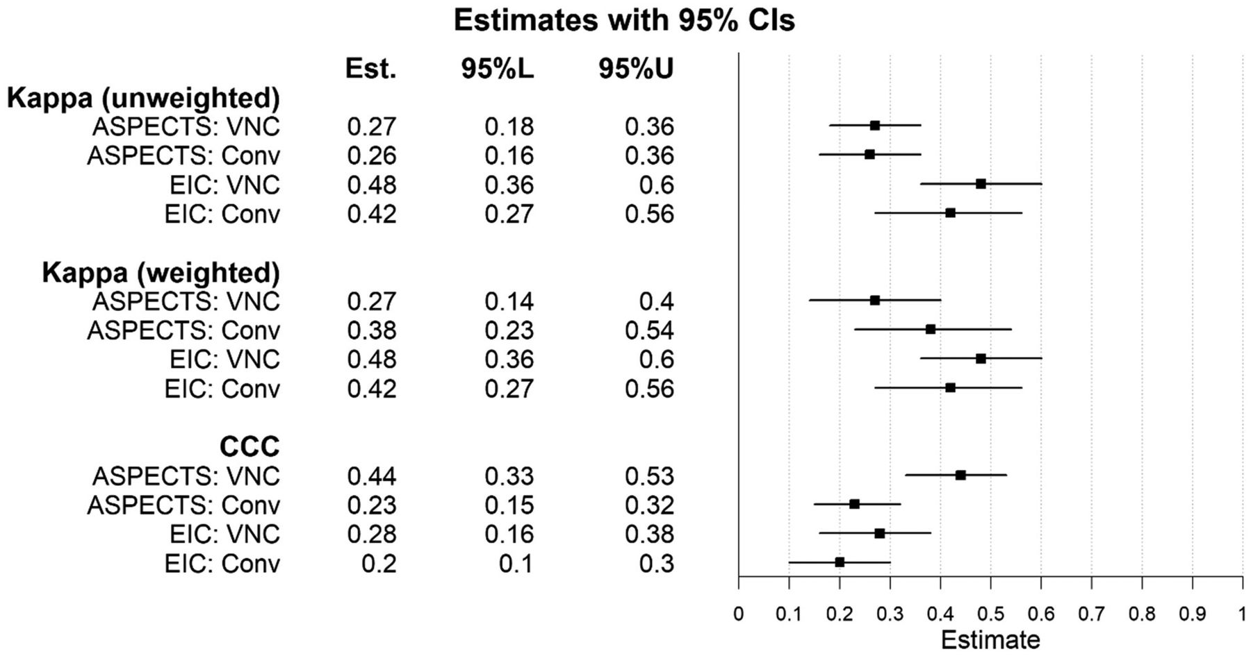
Some differences were observed between the observations of reviewers 1 and 2 (Fig 1). Interobserver agreement regarding the presence of early ischemic changes was moderate for conventional (weighted κ = 0.42; 95% CI, 0.27–0.56) and virtual NCCT (weighted κ = 0.48; 95% CI, 0.36–0.6) (Fig 2). Interobserver agreement regarding ASPECTS was fair for conventional NCCT (weighted κ = 0.38; 95% CI, 0.23–0.54) and virtual NCCT (weighted κ = 0.27; 95% CI, 0.14–0.4).

FIG 1.
  • Download figure
  • Open in new tab
  • Download powerpoint
FIG 1.

The proportion of CT scans showing early ischemic changes by reviewer and technique. VNC indicates virtual noncontrast; EIC, early ischemic changes; Cau, caudate nucleus; LNuc, lentiform nucleus; ICap, internal capsule; ICor, insular cortex.

FIG 2.
  • Download figure
  • Open in new tab
  • Download powerpoint
FIG 2.

Estimates of interobserver agreement between CT and the reference standard and accuracy with 95% confidence intervals. VNC indicates virtual noncontrast; EIC, early ischemic changes; Conv, conventional; 95%L, lower bound of the 95% confidence interval; 95%U, upper bound of the 95% confidence interval.

The sensitivity of virtual and conventional NCCT for the detection of early ischemic changes was 0.37 (95% CI, 0.28–0.47) and 0.27 (95% CI, 0.19–0.37), respectively (P = .078). Specificity was 0.91 (95% CI, 0.83–0.96) and 0.94 (95% CI, 0.86–0.98), respectively (Table 2). The global test finding was not significant (P = .111).

View this table:
  • View inline
  • View popup
Table 2:

Adjudicated-versus–criterion standard assessments

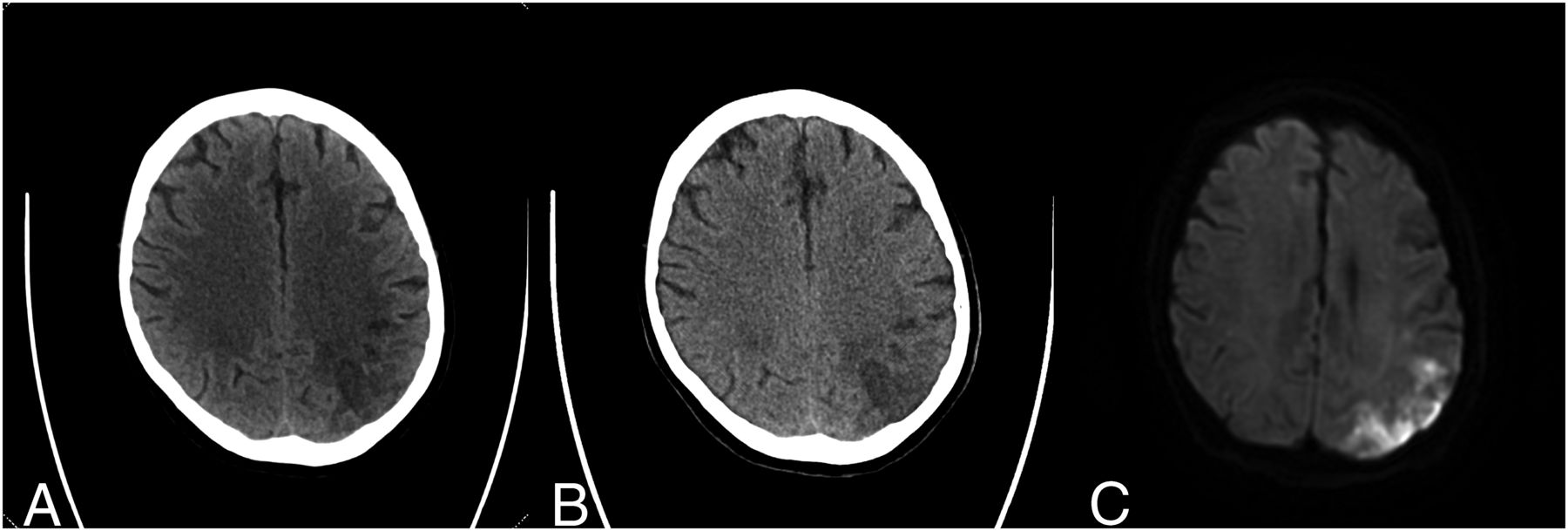
The forest plots (Fig 2) for the CCC showed overlapping intervals for early ischemic changes and nonoverlapping 95% CIs for ASPECTS. The difference in the CCC between virtual and conventional NCCT was 0.201 (95% CI, 0.014–0.389) regarding ASPECTS and 0.077 (95% CI, −0.041–0.191) regarding early ischemic changes. Neither 95% CI for the difference in the CCC crossed the prespecified noninferiority margin of −0.10, thereby establishing noninferiority of virtual NCCT compared with conventional NCCT with respect to both ASPECTS and early ischemic changes. Furthermore, virtual NCCT was demonstrated to be superior to conventional NCCT with respect to ASPECTS. Sample acquisitions of conventional NCCT, virtual NCCT, and DWI MR imaging are shown in Fig 3.

FIG 3.
  • Download figure
  • Open in new tab
  • Download powerpoint
FIG 3.

A sample case of a 66-year-old male patient who presented with right-sided weakness and numbness (NIHSS 7). His medical history included diabetes mellitus, hypertension, hyperlipidemia, prior smoking, and prior ischemic stroke, for which the patient used dual-antiplatelet therapy. The time between last seen well and CT was 14 hours. The time between CT and DWI was 12 hours. Conventional NCCT (A) and virtual NCCT (B) show early ischemic changes in the left parietal lobe (ASPECTS region M6). In the same region, DWI (C) shows hyperintensity, indicating diffusion restriction compatible with ischemia.

DISCUSSION

This study shows that detection of early ischemic changes with dual-energy virtual NCCT in patients with suspected stroke is noninferior compared with standard linearly blended NCCT. Furthermore, virtual NCCT was demonstrated to be superior to conventional NCCT with respect to ASPECTS.

To the best of our knowledge, no studies have evaluated early ischemic changes on virtual NCCT images reconstructed from CTA. Studies in fields other than neuroradiology have shown contradictory results, and the reliability of the virtual NCCT maps seems to be influenced by the clinical indication.16,17 Excluding intracranial hemorrhage is an important application of NCCT in the acute stroke setting and can also be done with dual-energy virtual NCCT images.18-20 Dual-energy CT can also be used to distinguish intracranial hemorrhage and extravasation of iodine contrast after endovascular treatment of stroke.21-23 In addition, 1 study showed high sensitivity and specificity for detecting the hyperdense artery sign on dual-energy virtual NCCT.24 Previous studies showed that the detection of the ischemic core with dual-energy virtual NCCT images derived from NCCT was superior compared with conventional NCCT.22,25,26 It is difficult to compare these studies with our study because the reconstructions were acquired from different CT images (NCCT versus CTA). However, all studies indicated that dual-energy virtual NCCT has the potential to replace conventional NCCT with respect to the detection of early ischemic changes.

The observed interobserver variability with regard to the detection of early ischemic changes and ASPECTS was moderate and fair, respectively, and similar for virtual NCCT and conventional NCCT in this study. ASPECTS is known to have relatively poor intraobserver and interobserver agreement, as was the case in this study.27 Recently, this finding was supported by a study that demonstrated a relatively poor interobserver agreement in a large group of 100 readers.28 The agreement, based on the CCC, between the dual-energy virtual NCCT assessments and MR imaging assessments regarding the presence of early ischemic changes was slightly better than the agreement between conventional NCCT assessments and MR imaging assessments. With regard to ASPECTS, the CCC showed fair agreement between both CT modalities and MR imaging, which is in line with findings in a previous study.29

The applications of dual-energy CT are becoming increasingly apparent. If dual-energy virtual NCCT of the brain can replace conventional NCCT, this would result in less radiation exposure for the patient. Whether dual-energy virtual NCCT saves time in the acute stroke work-up needs to be elucidated because dual-energy reconstructions also take time. Automated dual-energy reconstructions do not add extra time to the imaging protocol, unless manual processing is required. The disadvantages of replacing conventional NCCT with virtual NCCT include the necessity of acquiring CTA, turning on the dual-energy mode, and the requirements of added storage and bandwidth. Besides replacing conventional NCCT, future applications of virtual NCCT may include improvement of the conventional NCCT in case of severe movement artifacts and reduction of the radiation dose in a nonacute setting.

Most dual-energy CT scanners, using a dual source with a dual detector or a monosource with rapid kilovolt (peak) switching, require that the dual-energy setting be switched on beforehand to acquire dual-energy data.30 Other detector-based spectral CT scanners have a single source and a single dual-layered detector that is always switched on. In the acute stroke imaging work-up, NCCT needs to be acquired first to exclude intracranial hemorrhage. Because the dual-energy virtual NCCT images need to be reconstructed from the CTA, which is often only performed if an NCCT has ruled out an intracranial hemorrhage, it is difficult to omit the conventional NCCT in clinical practice. On the other hand, even in patients who have intracranial hemorrhage, CTA is often performed to look for cerebral aneurysms, cerebral arteriovenous malformations, or other vascular malformations as the cause of the bleed. Taken together, the role of dual-energy virtual NCCT seems dependent on the indication and on whether CTA is performed regardless of the findings on NCCT.

Several strengths can be noted in this study. To prevent observation bias, the observers who assessed the CT were blinded to clinical information and MR images; vice versa, the observer who assessed the MR images was blinded to the CT images. Another strength was the power calculation, which helped to achieve a reliable noninferiority analysis.

A limitation of this study was the potential risk of selection bias because patients were selected who underwent dual-energy CT and diffusion-weighted MR imaging. However, besides the imaging criteria, we used wide inclusion criteria (adult patients with a suspicion of acute ischemic stroke). Nevertheless, the results of this study need to be validated, preferably in a large prospective study. Due to missing data, extensive patient characteristics were not reported in this study. Although we did not use conventional single-source NCCT in this study, standard linearly blended images closely resemble conventional NCCT.31 Another limitation is the time interval between the acquired CT scan and the DWI. Although the time window was within 48 hours, this theoretically could have resulted in larger or additional infarcts on MR imaging compared with CT. This possibility may also partially explain the fair agreement between CT and MR imaging in this study. In addition, MR imaging is more sensitive for detecting ischemia; therefore, ASPECTS may differ between CT and MR imaging assessments, which are acquired simultaneously because small infarcts may be visible on MR imaging but not on CT. In this study, the dual-energy CT scanner and the software of 1 vendor were used, limiting the ability to generalize these findings. Future studies should include a broader range of CT vendors and software to increase generalizability.

CONCLUSIONS

Dual-energy virtual NCCT is noninferior compared with NCCT for the detection of early ischemic changes with ASPECTS.

Footnotes

  • This research has been made possible by the Dutch Heart Foundation, the Netherlands Organisation for Scientific Research, and the Domain Applied and Engineering Sciences, as part of their joint strategic research program: “Earlier Recognition of Cardiovascular Diseases” (grant No. 14732).

  • Disclosure forms provided by the authors are available with the full text and PDF of this article at www.ajnr.org.

Indicates open access to non-subscribers at www.ajnr.org

References

  1. 1.↵
    1. Wintermark M,
    2. Luby M,
    3. Bornstein NM, et al
    . International survey of acute stroke imaging used to make revascularization treatment decisions. Int J Stroke 2015;10:759–62 doi:10.1111/ijs.12491 pmid:25833105
    CrossRefPubMed
  2. 2.↵
    1. Wardlaw JM,
    2. Mielke O
    . Early signs of brain infarction at CT: observer reliability and outcome after thrombolytic treatment—systematic review. Radiology 2005;235:444–53 doi:10.1148/radiol.2352040262 pmid:15858087
    CrossRefPubMed
  3. 3.↵
    1. Barber PA,
    2. Demchuk AM,
    3. Zhang J
    , et al. Validity and reliability of a quantitative computed tomography score in predicting outcome of hyperacute stroke before thrombolytic therapy: ASPECTS Study Group—Alberta Stroke Programme. Lancet 2000;355:1670–74 doi:10.1016/S0140-673(00)02237-6 pmid:10905241
    CrossRefPubMed
  4. 4.
    1. Jovin TG,
    2. Chamorro A,
    3. Cobo E, et al
    ; REVASCAT Trial Investigators. Thrombectomy within 8 hours after symptom onset in ischemic stroke. N Engl J Med 2015;372:2296–2306 doi:10.1056/NEJMoa1503780 pmid:25882510
    CrossRefPubMed
  5. 5.↵
    1. Goyal M,
    2. Demchuk AM,
    3. Menon BK, et al
    . Randomized assessment of rapid endovascular treatment of ischemic stroke. N Engl J Med 2015;372:1019–30 doi:10.1056/NEJMoa1414905 pmid:25671798
    CrossRefPubMed
  6. 6.↵
    1. Goyal M,
    2. Menon BK,
    3. van Zwam WH, et al
    ; HERMES Collaborators. Endovascular thrombectomy after large-vessel ischaemic stroke: a meta-analysis of individual patient data from five randomised trials. Lancet 2016;387:1723–31 doi:10.1016/S0140-6736(16)00163-X pmid:26898852
    CrossRefPubMed
  7. 7.↵
    1. Jovin TG,
    2. Nogueira RG,
    3. Lansberg MG, et al
    . Thrombectomy for anterior circulation stroke beyond 6 h from time last known well (AURORA): a systematic review and individual patient data meta-analysis. Lancet 2021;399:249–58 doi:10.1016/S0140-6736(21)01341-6 pmid:34774198
    CrossRefPubMed
  8. 8.↵
    1. Wolman DN,
    2. Patel BP,
    3. Wintermark M, et al
    . Dual-energy computed tomography applications in neurointervention. J Comput Assist Tomogr 2018;42:31–38 doi:10.1097/RCT.0000000000000779 pmid:30052616
    CrossRefPubMed
  9. 9.↵
    1. Almqvist H,
    2. Holmin S,
    3. Mazya MV
    . Dual energy CT after stroke thrombectomy alters assessment of hemorrhagic complications. Neurology 2019;93:e1068–75 doi:10.1212/WNL.0000000000008093 pmid:31409735
    Abstract/FREE Full Text
  10. 10.↵
    1. Liu K,
    2. Jiang L,
    3. Ruan J, et al
    . The role of dual energy CT in evaluating hemorrhagic complications at different stages after thrombectomy. Front Neurol 2020;11:583411 doi:10.3389/fneur.2020.583411 pmid:33117268
    CrossRefPubMed
  11. 11.↵
    1. Barber PA,
    2. Hill MD,
    3. Eliasziw M, et al
    . Imaging of the brain in acute ischaemic stroke: comparison of computed tomography and magnetic resonance diffusion-weighted imaging. J Neurol Neurosurg Psychiatry 2005;76:1528–33 doi:10.1136/jnnp.2004.059261 pmid:16227545
    Abstract/FREE Full Text
  12. 12.↵
    1. Cohen J
    . Weighted kappa: nominal scale agreement with provision for scaled disagreement or partial credit. Psychol Bull 1968;70:213–20 doi:10.1037/h0026256 pmid:19673146
    CrossRefPubMed
  13. 13.↵
    1. Roldán-Nofuentes JA
    . Compbdt: an R program to compare two binary diagnostic tests subject to a paired design. BMC Med Res Methodol 2020;20:143 doi:10.1186/s12874-020-00988-y pmid:32503431
    CrossRefPubMed
  14. 14.↵
    1. Lin LI
    . A concordance correlation coefficient to evaluate reproducibility. Biometrics 1989;45:255–68 pmid:2720055
    CrossRefPubMed
  15. 15.↵
    1. Morgan CJ,
    2. Aban I
    . Methods for evaluating the agreement between diagnostic tests. J Nucl Cardiol 2016;23:511–13 doi:10.1007/s12350-015-0175-7 pmid:25995184
    CrossRefPubMed
  16. 16.↵
    1. Lehti L,
    2. Söderberg M,
    3. Höglund P, et al
    . Reliability of virtual non-contrast computed tomography angiography: comparing it with the real deal. Acta Radiol Open 2018;7:2058460118790115 doi:10.1177/2058460118790115 pmid:30181911
    CrossRefPubMed
  17. 17.↵
    1. Noid G,
    2. Schott D,
    3. Paulson E, et al
    . Technical note: using virtual noncontrast images from dual-energy CT to eliminate the need of precontrast CT for x-ray radiation treatment planning of abdominal tumors. Med Phys 2021;48:1365–71 doi:10.1002/mp.14702 pmid:33386614
    CrossRefPubMed
  18. 18.↵
    1. Ferda J,
    2. Novák M,
    3. Mírka H, et al
    . The assessment of intracranial bleeding with virtual unenhanced imaging by means of dual-energy CT angiography. Eur Radiol 2009;19:2518–22 doi:10.1007/s00330-009-1495-2 pmid:19585123
    CrossRefPubMed
  19. 19.
    1. Jiang XY,
    2. Zhang SH,
    3. Xie QZ, et al
    . Evaluation of virtual noncontrast images obtained from dual-energy CTA for diagnosing subarachnoid hemorrhage. AJNR Am J Neuroradiol 2015;36:855–60 doi:10.3174/ajnr.A4223 pmid:25614477
    Abstract/FREE Full Text
  20. 20.↵
    1. Bonatti M,
    2. Lombardo F,
    3. Zamboni GA, et al
    . Dual-energy CT of the brain: comparison between DECT angiography-derived virtual unenhanced images and true unenhanced images in the detection of intracranial haemorrhage. Eur Radiol 2017;27:2690–97 doi:10.1007/s00330-016-4658-y pmid:27882426
    CrossRefPubMed
  21. 21.↵
    1. Tijssen MP,
    2. Hofman PA,
    3. Stadler AA, et al
    . The role of dual energy CT in differentiating between brain haemorrhage and contrast medium after mechanical revascularisation in acute ischaemic stroke. Eur Radiol 2014;24:834–40 doi:10.1007/s00330-013-3073-x pmid:24258277
    CrossRefPubMed
  22. 22.↵
    1. Gariani J,
    2. Cuvinciuc V,
    3. Courvoisier D, et al
    . Diagnosis of acute ischemia using dual energy CT after mechanical thrombectomy. J Neurointerv Surg 2016;8:996–1000 doi:10.1136/neurintsurg-2015-011988 pmid:26534867
    Abstract/FREE Full Text
  23. 23.↵
    1. Ebashi R,
    2. Ogata A,
    3. Nishihara M, et al
    . Significance of simulated conventional images on dual energy CT after endovascular treatment for ischemic stroke. J Neurointerv Surg 2019;11:898–902 doi:10.1136/neurintsurg-2018-014486 pmid:30670626
    Abstract/FREE Full Text
  24. 24.↵
    1. Winklhofer S,
    2. Vittoria De Martini I,
    3. Nern C, et al
    . Dual-energy computed tomography in stroke imaging: technical and clinical considerations of virtual noncontrast images for detection of the hyperdense artery sign. J Comput Assist Tomogr 2017;41:843–48 doi:10.1097/RCT.0000000000000638 pmid:28708725
    CrossRefPubMed
  25. 25.↵
    1. Mohammed MF,
    2. Marais O,
    3. Min A, et al
    . Unenhanced dual-energy computed tomography: visualization of brain edema. Invest Radiol 2018;53:63–69 doi:10.1097/RLI.0000000000000413 pmid:28915161
    CrossRefPubMed
  26. 26.↵
    1. Wolman DN,
    2. van Ommen F,
    3. Tong E, et al
    . Non-contrast dual-energy CT virtual ischemia maps accurately estimate ischemic core size in large-vessel occlusive stroke. Sci Rep 2021;11:6745 doi:10.1038/s41598-021-85143-3 pmid:33762589
    CrossRefPubMed
  27. 27.↵
    1. Gupta AC,
    2. Schaefer PW,
    3. Chaudhry ZA, et al
    . Interobserver reliability of baseline noncontrast CT Alberta Stroke Program Early CT Score for intra-arterial stroke treatment selection. AJNR Am J Neuroradiol 2012;33:1046–49 doi:10.3174/ajnr.A2942 pmid:22322602
    Abstract/FREE Full Text
  28. 28.↵
    1. van Horn N,
    2. Kniep H,
    3. Broocks G, et al
    . ASPECTS interobserver agreement of 100 investigators from the TENSION study. Clin Neuroradiol 2021;31:1093–1100 doi:10.1007/s00062-020-00988-x pmid:33502563
    CrossRefPubMed
  29. 29.↵
    1. Lansberg MG,
    2. Albers GW,
    3. Beaulieu C, et al
    . Comparison of diffusion-weighted MRI and CT in acute stroke. Neurology 2000;54:1557–61 doi:10.1212/wnl.54.8.1557 pmid:10762493
    Abstract/FREE Full Text
  30. 30.↵
    1. McCollough CH,
    2. Leng S,
    3. Yu L, et al
    . Dual- and multi-energy CT: principles, technical approaches, and clinical applications. Radiology 2015;276:637–53 doi:10.1148/radiol.2015142631 pmid:26302388
    CrossRefPubMed
  31. 31.↵
    1. Yu L,
    2. Primak AN,
    3. Liu X, et al
    . Image quality optimization and evaluation of linearly mixed images in dual-source, dual-energy CT. Med Phys 2009;36:1019–24 doi:10.1118/1.3077921 pmid:19378762
    CrossRefPubMed
  • Received January 21, 2022.
  • Accepted after revision June 17, 2022.
  • © 2022 by American Journal of Neuroradiology
PreviousNext
Back to top

In this issue

American Journal of Neuroradiology: 43 (9)
American Journal of Neuroradiology
Vol. 43, Issue 9
1 Sep 2022
  • Table of Contents
  • Index by author
  • Complete Issue (PDF)
Advertisement
Print
Download PDF
Email Article

Thank you for your interest in spreading the word on American Journal of Neuroradiology.

NOTE: We only request your email address so that the person you are recommending the page to knows that you wanted them to see it, and that it is not junk mail. We do not capture any email address.

Enter multiple addresses on separate lines or separate them with commas.
Detection of Early Ischemic Changes with Virtual Noncontrast Dual-Energy CT in Acute Ischemic Stroke: A Noninferiority Analysis
(Your Name) has sent you a message from American Journal of Neuroradiology
(Your Name) thought you would like to see the American Journal of Neuroradiology web site.
CAPTCHA
This question is for testing whether or not you are a human visitor and to prevent automated spam submissions.
Cite this article
F. Kauw, V.Y. Ding, J.W. Dankbaar, F. van Ommen, G. Zhu, D.B. Boothroyd, D.N. Wolman, L. Molvin, H.W.A.M. de Jong, L.J. Kappelle, B.K. Velthuis, J.J. Heit, M. Wintermark
Detection of Early Ischemic Changes with Virtual Noncontrast Dual-Energy CT in Acute Ischemic Stroke: A Noninferiority Analysis
American Journal of Neuroradiology Sep 2022, 43 (9) 1259-1264; DOI: 10.3174/ajnr.A7600

Citation Manager Formats

  • BibTeX
  • Bookends
  • EasyBib
  • EndNote (tagged)
  • EndNote 8 (xml)
  • Medlars
  • Mendeley
  • Papers
  • RefWorks Tagged
  • Ref Manager
  • RIS
  • Zotero
0 Responses
Respond to this article
Share
Bookmark this article
Early Ischemic Changes with Dual-Energy CT
F. Kauw, V.Y. Ding, J.W. Dankbaar, F. van Ommen, G. Zhu, D.B. Boothroyd, D.N. Wolman, L. Molvin, H.W.A.M. de Jong, L.J. Kappelle, B.K. Velthuis, J.J. Heit, M. Wintermark
American Journal of Neuroradiology Sep 2022, 43 (9) 1259-1264; DOI: 10.3174/ajnr.A7600
del.icio.us logo Twitter logo Facebook logo Mendeley logo
  • Tweet Widget
  • Facebook Like
  • Google Plus One
Purchase

Jump to section

  • Article
    • Abstract
    • ABBREVIATION:
    • MATERIALS AND METHODS
    • RESULTS
    • DISCUSSION
    • CONCLUSIONS
    • Footnotes
    • References
  • Figures & Data
  • Info & Metrics
  • Responses
  • References
  • PDF

Related Articles

  • No related articles found.
  • PubMed
  • Google Scholar

Cited By...

  • No citing articles found.
  • Crossref (6)
  • Google Scholar

This article has been cited by the following articles in journals that are participating in Crossref Cited-by Linking.

  • Perioperative stroke
    Jonathon P. Fanning, Bruce C. V. Campbell, Richard Bulbulia, Rebecca F. Gottesman, Sang-Bae Ko, Thomas F. Floyd, Steven R. Messé
    Nature Reviews Disease Primers 2024 10 1
  • Dual-Energy CT in Acute Stroke: Could Non-Contrast CT Be Replaced by Virtual Non-Contrast CT? A Feasibility Study
    Guillaume Herpe, Alexandra Platon, Pierre-Alexandre Poletti, Karl O. Lövblad, Paolo Machi, Minerva Becker, Michel Muster, Thomas Perneger, Rémy Guillevin
    Journal of Clinical Medicine 2024 13 13
  • Neuroradiology Applications of Dual and Multi-energy Computed Tomography
    Natthawut Jarunnarumol, Shahmir Kamalian, Michael H. Lev, Rajiv Gupta
    Radiologic Clinics of North America 2023 61 6
  • A Dual-Stream Deep Learning Architecture With Adaptive Random Vector Functional Link for Multi-Center Ischemic Stroke Classification
    Mahesh Anil Inamdar, Anjan Gudigar, U. Raghavendra, Massimo Salvi, Raja Rizal Azman Bin Raja Aman, Nadia Fareeda Muhammad Gowdh, Izzah Amirah Binti Mohd Ahir, Mohd Salahuddin Bin Kamaruddin, Khairul Azmi Abdul Kadir, Filippo Molinari, Ajay Hegde, Girish R. Menon, U. Rajendra Acharya
    IEEE Access 2025 13
  • Advances in Imaging of Neurovascular Emergencies on Computer Tomography CT
    Ahmad Alghamdi, Muhammad Ejaz Ahmed
    Current Radiology Reports 2023 12 1
  • Iodine-Based Dual-Energy Computed Tomography After Mechanical Thrombectomy Predicts Secondary Neurologic Decline from Cerebral Edema After Severe Stroke
    William Zimmerman, Melissa Pergakis, Ghasan Ahmad, Nicholas A. Morris, Jamie Podell, Wan-Tsu Chang, Melissa Motta, Hegang Chen, Gaurav Jindal, Uttam Bodanapally, J. Marc Simard, Neeraj Badjatia, Gunjan Y. Parikh
    Neurocritical Care 2024

More in this TOC Section

  • Diagnostic Neuroradiology of Monoclonal Antibodies
  • Clinical Outcomes After Chiari I Decompression
  • Segmentation of Brain Metastases with BLAST
Show more ADULT BRAIN

Similar Articles

Advertisement

Indexed Content

  • Current Issue
  • Accepted Manuscripts
  • Article Preview
  • Past Issues
  • Editorials
  • Editor's Choice
  • Fellows' Journal Club
  • Letters to the Editor
  • Video Articles

Cases

  • Case Collection
  • Archive - Case of the Week
  • Archive - Case of the Month
  • Archive - Classic Case

More from AJNR

  • Trainee Corner
  • Imaging Protocols
  • MRI Safety Corner

Multimedia

  • AJNR Podcasts
  • AJNR Scantastics

Resources

  • Turnaround Time
  • Submit a Manuscript
  • Submit a Video Article
  • Submit an eLetter to the Editor/Response
  • Manuscript Submission Guidelines
  • Statistical Tips
  • Fast Publishing of Accepted Manuscripts
  • Graphical Abstract Preparation
  • Imaging Protocol Submission
  • Evidence-Based Medicine Level Guide
  • Publishing Checklists
  • Author Policies
  • Become a Reviewer/Academy of Reviewers
  • News and Updates

About Us

  • About AJNR
  • Editorial Board
  • Editorial Board Alumni
  • Alerts
  • Permissions
  • Not an AJNR Subscriber? Join Now
  • Advertise with Us
  • Librarian Resources
  • Feedback
  • Terms and Conditions
  • AJNR Editorial Board Alumni

American Society of Neuroradiology

  • Not an ASNR Member? Join Now

© 2025 by the American Society of Neuroradiology All rights, including for text and data mining, AI training, and similar technologies, are reserved.
Print ISSN: 0195-6108 Online ISSN: 1936-959X

Powered by HighWire