Advertisement

Main menu

  • Home
  • Content
    • Current Issue
    • Accepted Manuscripts
    • Article Preview
    • Past Issue Archive
    • Video Articles
    • AJNR Case Collection
    • Case of the Week Archive
    • Case of the Month Archive
    • Classic Case Archive
  • Special Collections
    • AJNR Awards
    • Low-Field MRI
    • Alzheimer Disease
    • ASNR Foundation Special Collection
    • Photon-Counting CT
    • View All
  • Multimedia
    • AJNR Podcasts
    • AJNR SCANtastic
    • Trainee Corner
    • MRI Safety Corner
    • Imaging Protocols
  • For Authors
    • Submit a Manuscript
    • Submit a Video Article
    • Submit an eLetter to the Editor/Response
    • Manuscript Submission Guidelines
    • Statistical Tips
    • Fast Publishing of Accepted Manuscripts
    • Graphical Abstract Preparation
    • Imaging Protocol Submission
    • Author Policies
  • About Us
    • About AJNR
    • Editorial Board
    • Editorial Board Alumni
  • More
    • Become a Reviewer/Academy of Reviewers
    • Subscribers
    • Permissions
    • Alerts
    • Feedback
    • Advertisers
    • ASNR Home

User menu

  • Alerts
  • Log in

Search

  • Advanced search
American Journal of Neuroradiology

American Journal of Neuroradiology

ASHNR American Society of Functional Neuroradiology ASHNR American Society of Pediatric Neuroradiology ASSR
  • Alerts
  • Log in

Advanced Search

  • Home
  • Content
    • Current Issue
    • Accepted Manuscripts
    • Article Preview
    • Past Issue Archive
    • Video Articles
    • AJNR Case Collection
    • Case of the Week Archive
    • Case of the Month Archive
    • Classic Case Archive
  • Special Collections
    • AJNR Awards
    • Low-Field MRI
    • Alzheimer Disease
    • ASNR Foundation Special Collection
    • Photon-Counting CT
    • View All
  • Multimedia
    • AJNR Podcasts
    • AJNR SCANtastic
    • Trainee Corner
    • MRI Safety Corner
    • Imaging Protocols
  • For Authors
    • Submit a Manuscript
    • Submit a Video Article
    • Submit an eLetter to the Editor/Response
    • Manuscript Submission Guidelines
    • Statistical Tips
    • Fast Publishing of Accepted Manuscripts
    • Graphical Abstract Preparation
    • Imaging Protocol Submission
    • Author Policies
  • About Us
    • About AJNR
    • Editorial Board
    • Editorial Board Alumni
  • More
    • Become a Reviewer/Academy of Reviewers
    • Subscribers
    • Permissions
    • Alerts
    • Feedback
    • Advertisers
    • ASNR Home
  • Follow AJNR on Twitter
  • Visit AJNR on Facebook
  • Follow AJNR on Instagram
  • Join AJNR on LinkedIn
  • RSS Feeds

AJNR Awards, New Junior Editors, and more. Read the latest AJNR updates

Research ArticlePediatrics

Evaluation of the Prevalence of Punctate White Matter Lesions in a Healthy Volunteer Neonatal Population

S. Kular, H. Holmes, A. Hart, P. Griffiths and D. Connolly
American Journal of Neuroradiology August 2022, 43 (8) 1210-1213; DOI: https://doi.org/10.3174/ajnr.A7578
S. Kular
aFrom the Departments of Neuroradiology (S.K., H.H., P.G., D.C.)
  • Find this author on Google Scholar
  • Find this author on PubMed
  • Search for this author on this site
  • ORCID record for S. Kular
H. Holmes
aFrom the Departments of Neuroradiology (S.K., H.H., P.G., D.C.)
  • Find this author on Google Scholar
  • Find this author on PubMed
  • Search for this author on this site
  • ORCID record for H. Holmes
A. Hart
bNeurology (A.H.), Sheffield Children’s Hospital, Sheffield, UK
  • Find this author on Google Scholar
  • Find this author on PubMed
  • Search for this author on this site
  • ORCID record for A. Hart
P. Griffiths
aFrom the Departments of Neuroradiology (S.K., H.H., P.G., D.C.)
  • Find this author on Google Scholar
  • Find this author on PubMed
  • Search for this author on this site
  • ORCID record for P. Griffiths
D. Connolly
aFrom the Departments of Neuroradiology (S.K., H.H., P.G., D.C.)
  • Find this author on Google Scholar
  • Find this author on PubMed
  • Search for this author on this site
  • ORCID record for D. Connolly
  • Article
  • Figures & Data
  • Info & Metrics
  • Responses
  • References
  • PDF
Loading

Abstract

SUMMARY: Hypoxic-ischemic injury is the most common cause of neonatal encephalopathy. T1-weighted punctate white matter lesions have been described in hypoxic-ischemic injury. We have reviewed a healthy volunteer neonatal population to assess the prevalence of punctate white matter lesions in neonates with no clinical signs of hypoxic-ischemic injury. Fifty-two subjects were scanned on a neonatal-specific 3T MR imaging scanner. Twelve patients were excluded due to the lack of T1-weighted imaging, leaving a total of 40 patients (35 term, 5 preterm) assessed in the study. One had a solitary T1-punctate white matter lesion. We concluded that solitary punctate white matter lesions have a low prevalence in healthy neonates.

ABBREVIATIONS:

HII
hypoxic-ischemic injury
PWML
punctate white matter lesion
SDH
subdural hematoma

Neonatal hypoxic-ischemic injury (HII) is the most common cause of neonatal encephalopathy, ahead of infection, metabolic disorder, and trauma.1,2 HII can occur in preterm or term infants.1 On MR imaging, HII with brain injury can manifest as signal abnormality within the posterior putamen, ventral-posterolateral thalamic injury, central sulcus cortical injury (acute profound asphyxia), or watershed injury (chronic partial asphyxia).3 Punctate white matter lesions (PWMLs) are hyperintense on T1-weighted and iso- to hypointense on T2-weighted imaging. PWMLs have been described in association with hypoxic-ischemic injury.4 PWMLs can be multifactorial, with a combination of gliotic scarring, hemorrhage, and mineralization contributing to their MR imaging appearance; however, it is often not possible to ascertain which of these is the causative factor in each case. A histologic study has, however, found that approximately 25% of PWMLs are secondary to hemorrhage.5

SWI may help differentiate hemorrhagic PWMLs from nonhemorrhagic PWMLs, with the latter attributed to early scarring or mineralization.6 Hemorrhagic PWMLs are also associated with greater proportions of infantile intraventricular hemorrhage, with nonhemorrhagic PWMLs being associated more with ischemia-related injury.7 In either etiology, the general consensus is that PWMLs are derived from pathologic processes occurring via hemorrhagic or ischemic pathways. Thus, these lesions, one would assume, should not be present in a healthy neonate who has not been subjected to any known pathologic insult.

In our study, we reviewed MR images of a healthy volunteer neonate population obtained on a specific neonatal MR imaging scanner to assess the prevalence of PWMLs in neonates without clinical signs of HII.

MATERIALS AND METHODS

A 3T neonatal MR imaging scanner (GE Healthcare) was installed in the neonatal unit of the local obstetric department.

Ethics approval was governed by the Medicines and Healthcare products Regulatory Agency of the UK. Conditions of use granted under the Medicines and Healthcare products Regulatory Agency were that the scanner was not to be used for primary diagnostic purpose and the manufacturer sponsored the study. Enrollment for up to 60 neonatal subjects was granted. Two groups were scanned. Group 1 consisted of healthy term neonates (≥37 weeks’ gestational age) whose parents agreed to MR imaging before discharge. Group 2 involved premature neonates about to be discharged at term-corrected age, with no other clinical complications.

All subjects had 2 scheduled sonography examinations throughout pregnancy as per local guidelines, at approximately 12 and 19 weeks for the first and second scans, respectively. No subjects were found to have screening anomalies on fetal sonography.

All parents provided informed, written consent and completed an MR imaging safety form for the neonate and themselves because 1 parent could accompany their child during scanning.

Healthy volunteer subjects were imaged as part of the Firefly 3T MR imaging project evaluation.8 The scans were performed within 1 hour of feeding. The 3T magnet strength was deemed safe for neonatal scanning.9 Subjects’ vital observations encompassing body temperature, heart rate, and oxygen saturation were monitored throughout scanning. Scanning time was limited to 1 hour inclusive of the time for room acclimatization, monitoring, and subject positioning.

Initially, T2-weighted images were acquired in a trial format and optimized for contrast, resolution, and scan time before using them for both ultrafast single-shot FSE and T2-FSE imaging. The optimized T2-sequence parameters were subsequently used for all cases with long-TE single-voxel proton spectroscopy, T1-volume/spin-echo, DWI, and MR arteriography added as tolerated.

Inclusion criteria were patients with T2-weighted imaging and either or both standard T1-weighted spin-echo and T1-volumetric sequences. Excluded cases were those without T1-weighted imaging because they were unable to be adequately assessed for PWML.

Acquired images were reviewed by 2 consultant pediatric neuroradiologists. Further reviews were conducted by 2 post-board examination Royal College of Radiologists (FRCR, UK) neuroradiology fellows.

RESULTS

Fifty-two subjects were recruited into the study in total; however, 12 had no T1-weighted imaging; therefore, 40 cases were evaluated for evidence of PWML (35 in group 1, five in group 2).

One subject had a pre-existing bradycardia, and while having a normal sinus rhythm before scanning, the subject developed bradycardia during scanning. This was, however, not deemed to be attributed to MR imaging, and the neonate did not require further follow-up on discharge. No other subject demonstrated abnormal vital signs before, during, or immediately after MR imaging.

One hundred percent of imaging studies (40/40) were deemed evaluable, either completely or partially. Twenty-five percent (10/40) of studies were deemed partially evaluable, predominantly secondary to motion artifacts. The partially evaluable studies were, however, still thought to be accurate enough to identify notable abnormal findings on T1-weighted imaging.

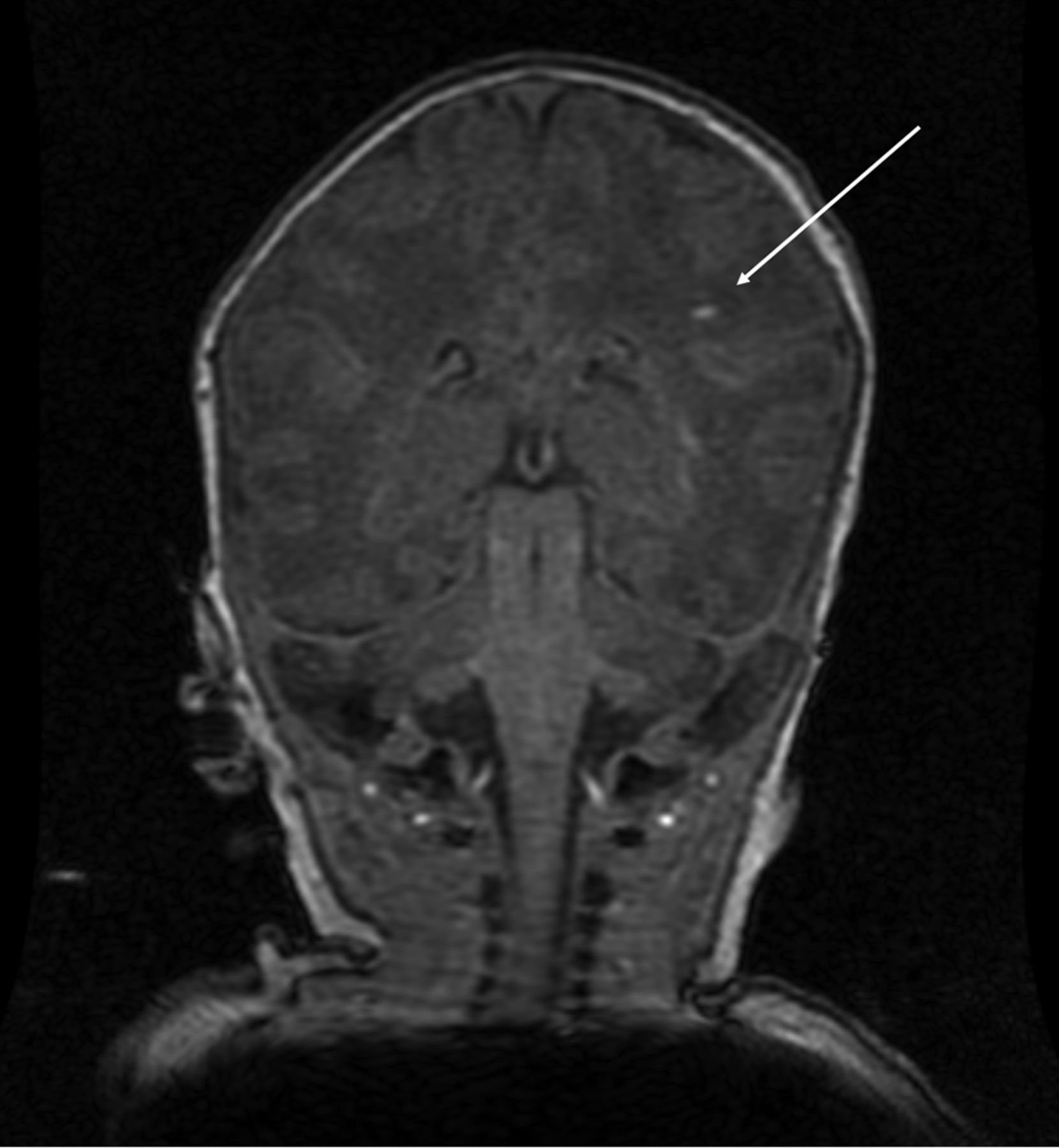
Of the 40 subjects, 1 subject in group 1 (2.5%) had evidence of a 2-mm solitary PWML, which was hyperintense on T1-weighted imaging (Fig 1). It was not possible to further distinguish this PWML as hemorrhagic or nonhemorrhagic (scarring/mineralization) due to absence of SWI. In this case, no pre- or perinatal injury was known to have occurred, nor was there any evidence of neonatal encephalopathy. The finding was, therefore, of unknown etiology but highly unlikely to be from perinatal asphyxia. The long-term follow-up of this PWML is not known.

FIG 1.
  • Download figure
  • Open in new tab
  • Download powerpoint
FIG 1.

A solitary T1-weighted PWML was demonstrated in the left corona radiata of 1 study patient (white arrow).

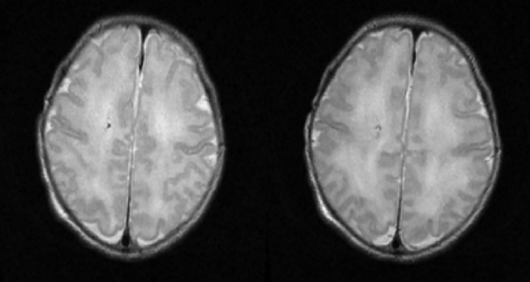
One group 2 subject (2.5%) had a finding of a right frontal developmental venous anomaly (Fig 2) with small-volume intraventricular hemorrhage; however, clinical review was unremarkable, and the subject was developing normally on 2-month assessment.

FIG 2.
  • Download figure
  • Open in new tab
  • Download powerpoint
FIG 2.

One subject demonstrated a right frontal developmental venous anomaly.

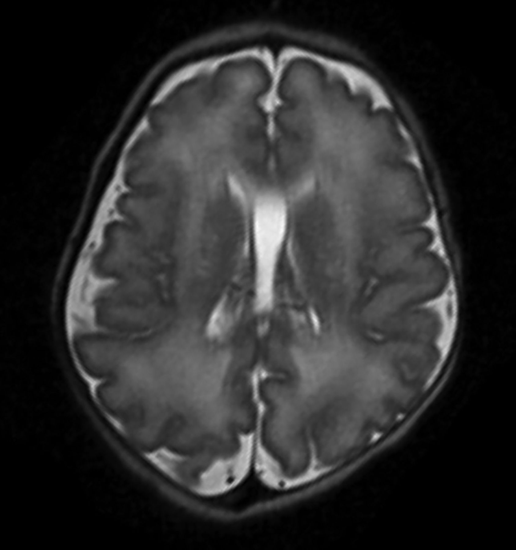
One group 1 subject (2.5%) had a cavum vergae, a normal anatomic variant with no clinical consequences (Fig 3). One group 1 subject (2.5%) also had a maturing left subdural hematoma (SDH) (Fig 4).

FIG 3.
  • Download figure
  • Open in new tab
  • Download powerpoint
FIG 3.

An incidental normal variant of a cavum vergae was seen in 1 study subject.

FIG 4.
  • Download figure
  • Open in new tab
  • Download powerpoint
FIG 4.

A moderate-sized left-convexity SDH was observed in 1 otherwise clinically well patient on T1-weighted imaging. This can be a common finding postbirth, seen in up to 38% of the population, spontaneously self-resolving in most cases.

DISCUSSION

In our population, there was an overwhelmingly large proportion of subjects presenting with no evidence of PWMLs (97.5%). In the single patient presenting with a tiny, solitary PWML, no discernible signs of physiologic impairment were seen on early medical assessment of the infant. Recent studies performed at approximately 18 months postpartum have demonstrated a similar, albeit slightly higher incidence of incidental PWMLs at approximately 12%; however, this is still consistent with a substantial minority within the population.10 The slightly higher incidence in a delayed-scan cohort could potentially be secondary to a greater number of lesions being visible from gliosis or scarring, which may not necessarily have been immediately visible in our postpartum scan cohort; however, with time, these lesions may become more apparent.

The same proportion of subjects that presented with a PWML also presented with an incidental normal variant (cavum vergae) and a left-sided SDH. SDH is a common finding, seen in up to 38% of births.11

A limitation of our study was that some studies were affected by motion artifacts; therefore, not all studies were of an equivalent reading standard. Nevertheless, all studies were deemed by participating radiologists to be of sufficient quality to provide conclusive PWML identification/exclusion. Furthermore, there was a significant difference in subject numbers within the 2 groups (35 versus 5); and while the overall number of 40 subjects is low, given the limitations of the population, this is the largest cohort of healthy neonates in the literature to be scanned postpartum for PWMLs.

Our findings conclude that PWMLs have very low prevalence in healthy neonates, and the presence of 1 or 2 PWMLs in otherwise healthy patients is not likely to be of clinical relevance. Furthermore, PWMLs in these cases may not necessarily be secondary to hemorrhage or scarring, but from physiologic mineralization. In contrast, when several T1 PWMLs (approximating ≥6) are present, this is more likely related to perinatal asphyxia, and closer follow-up is recommended.3

CONCLUSIONS

We recommend that in the event of neonates demonstrating 1 or 2 PWMLs with no other clinical or radiologic abnormality, no routine follow-up imaging is indicated. Further long-term follow-up studies to investigate the chronic relevance of solitary PWMLs in otherwise healthy neonates are required.

Footnotes

  • Disclosure forms provided by the authors are available with the full text and PDF of this article at www.ajnr.org.

References

  1. 1.↵
    1. Cowan F,
    2. Rutherford M,
    3. Groenendaal F, et al
    . Origin and timing of brain lesions in term infants with neonatal encephalopathy. Lancet 2003;361:736–42 doi:10.1016/S0140-6736(03)12658-X pmid:12620738
    CrossRefPubMed
  2. 2.↵
    1. Wagenaar N,
    2. Chau V,
    3. Groenendaal F, et al
    . Clinical risk factors for punctate white matter lesions on early magnetic resonance imaging in preterm newborns. J Pediatr 2017;182:34–40.e1 doi:10.1016/j.jpeds.2016.11.073 pmid:28063691
    CrossRefPubMed
  3. 3.↵
    1. Hayman M,
    2. van Wezel-Meijler G,
    3. van Straaten H, et al
    . Punctate white-matter lesions in the full-term newborn: underlying aetiology and outcome. Eur J Paediatr Neurol 2019;23:280–87 doi:10.1016/j.ejpn.2019.01.005 pmid:30744982
    CrossRefPubMed
  4. 4.↵
    1. Martinez-Biarge M,
    2. Groenendaal F,
    3. Kersbergen KJ, et al
    . MRI based preterm white matter injury classification: the importance of sequential imaging in determining severity of injury. PLoS One 2016;11:e0156245 doi:10.1371/journal.pone.0156245 pmid:27257863
    CrossRefPubMed
  5. 5.↵
    1. Paneth N,
    2. Rudelli R,
    3. Monte W, et al
    . White matter necrosis in very low birth weight infants: neuropathologic and ultrasonographic findings in infants surviving six days or longer. J Pediatr 1990;116:975–84 doi:10.1016/S0022-3476(05)80664-X pmid:2189978
    CrossRefPubMed
  6. 6.↵
    1. Childs AM,
    2. Cornette L,
    3. Ramenghi LA, et al
    . Magnetic resonance and cranial ultrasound characteristics of periventricular white matter abnormalities in newborn infants. Clin Radiol 2001;56:647–55 doi:10.1053/crad.2001.0754 pmid:11467866
    CrossRefPubMed
  7. 7.↵
    1. Niwa T,
    2. de Vries LS,
    3. Benders MJ, et al
    . Punctate white matter lesions in infants: new insights using susceptibility-weighted imaging. Neuroradiology 2011;53:669–79 doi:10.1007/s00234-011-0872-0 pmid:21553013
    CrossRefPubMed
  8. 8.↵
    1. Griffiths PD,
    2. Jarvis D,
    3. Armstrong L, et al
    . Initial experience of an investigational 3T MR scanner designed for use on neonatal wards. Eur Radiol 2018;28:4438–46 doi:10.1007/s00330-018-5357-7 pmid:29713778
    CrossRefPubMed
  9. 9.↵
    International Electrotechnical Commission. Medical electrical equipment, Part 2-33: Particular requirements for the safety of MR equipment for medical diagnosis IEC 60601-2-33 ed 2.2 (Geneva, 2008) with amendments 1 and 2. https://standards.globalspec.com/std/1099061/iec-60601-2-33. Accessed October 9, 2021
  10. 10.↵
    1. Carney O,
    2. Hughes E,
    3. Tusor N, et al
    . Incidental findings on brain MR imaging of asymptomatic term neonates in the Developing Human Connectome Project. EClinicalMedicine 2021;38:100984 doi:10.1016/j.eclinm.2021.100984 pmid:34355154
    CrossRefPubMed
  11. 11.↵
    1. Nikam RM,
    2. Kandula VV,
    3. Yue X, et al
    . Birth-related subdural hemorrhage: prevalence and imaging morphology. Pediatr Radiol 2021;51:939–46 doi:10.1007/s00247-021-05060-5 pmid:33830289
    CrossRefPubMed
  • Received March 17, 2022.
  • Accepted after revision May 24, 2022.
  • © 2022 by American Journal of Neuroradiology
PreviousNext
Back to top

In this issue

American Journal of Neuroradiology: 43 (8)
American Journal of Neuroradiology
Vol. 43, Issue 8
1 Aug 2022
  • Table of Contents
  • Index by author
  • Complete Issue (PDF)
Advertisement
Print
Download PDF
Email Article

Thank you for your interest in spreading the word on American Journal of Neuroradiology.

NOTE: We only request your email address so that the person you are recommending the page to knows that you wanted them to see it, and that it is not junk mail. We do not capture any email address.

Enter multiple addresses on separate lines or separate them with commas.
Evaluation of the Prevalence of Punctate White Matter Lesions in a Healthy Volunteer Neonatal Population
(Your Name) has sent you a message from American Journal of Neuroradiology
(Your Name) thought you would like to see the American Journal of Neuroradiology web site.
CAPTCHA
This question is for testing whether or not you are a human visitor and to prevent automated spam submissions.
Cite this article
S. Kular, H. Holmes, A. Hart, P. Griffiths, D. Connolly
Evaluation of the Prevalence of Punctate White Matter Lesions in a Healthy Volunteer Neonatal Population
American Journal of Neuroradiology Aug 2022, 43 (8) 1210-1213; DOI: 10.3174/ajnr.A7578

Citation Manager Formats

  • BibTeX
  • Bookends
  • EasyBib
  • EndNote (tagged)
  • EndNote 8 (xml)
  • Medlars
  • Mendeley
  • Papers
  • RefWorks Tagged
  • Ref Manager
  • RIS
  • Zotero
0 Responses
Respond to this article
Share
Bookmark this article
Prevalence of White Matter Lesions in Neonates
S. Kular, H. Holmes, A. Hart, P. Griffiths, D. Connolly
American Journal of Neuroradiology Aug 2022, 43 (8) 1210-1213; DOI: 10.3174/ajnr.A7578
del.icio.us logo Twitter logo Facebook logo Mendeley logo
  • Tweet Widget
  • Facebook Like
  • Google Plus One
Purchase

Jump to section

  • Article
    • Abstract
    • ABBREVIATIONS:
    • MATERIALS AND METHODS
    • RESULTS
    • DISCUSSION
    • CONCLUSIONS
    • Footnotes
    • References
  • Figures & Data
  • Info & Metrics
  • Responses
  • References
  • PDF

Related Articles

  • No related articles found.
  • PubMed
  • Google Scholar

Cited By...

  • No citing articles found.
  • Crossref (1)
  • Google Scholar

This article has been cited by the following articles in journals that are participating in Crossref Cited-by Linking.

  • Prenatal Opioid Exposure is Associated with Punctate White Matter Lesions in Term Newborns
    Stephanie L. Merhar, Carla M. Bann, Nicole Mack, Jamie E. Newman, Catherine Limperopoulos, Namasivayam Ambalavanan, Jonathan M. Davis, Sara B. DeMauro, Scott Lorch, Deanne E. Wilson-Costello, Myriam Peralta-Carcelan, Lindsay M. Parlberg, Brenda B. Poindexter, Kushal Kapse, Beth Kline-Fath, Jonathan Murnick
    The Journal of Pediatrics 2025

More in this TOC Section

  • Comparison of Image Quality and Radiation Dose in Pediatric Temporal Bone CT Using Photon-Counting Detector CT and Energy-Integrating Detector CT
  • SyMRI & MR Fingerprinting in Brainstem Myelination
  • Dual-Layer Detector CT for PEDS Image Quality
Show more Pediatrics

Similar Articles

Advertisement

Indexed Content

  • Current Issue
  • Accepted Manuscripts
  • Article Preview
  • Past Issues
  • Editorials
  • Editor's Choice
  • Fellows' Journal Club
  • Letters to the Editor
  • Video Articles

Cases

  • Case Collection
  • Archive - Case of the Week
  • Archive - Case of the Month
  • Archive - Classic Case

More from AJNR

  • Trainee Corner
  • Imaging Protocols
  • MRI Safety Corner

Multimedia

  • AJNR Podcasts
  • AJNR Scantastics

Resources

  • Turnaround Time
  • Submit a Manuscript
  • Submit a Video Article
  • Submit an eLetter to the Editor/Response
  • Manuscript Submission Guidelines
  • Statistical Tips
  • Fast Publishing of Accepted Manuscripts
  • Graphical Abstract Preparation
  • Imaging Protocol Submission
  • Evidence-Based Medicine Level Guide
  • Publishing Checklists
  • Author Policies
  • Become a Reviewer/Academy of Reviewers
  • News and Updates

About Us

  • About AJNR
  • Editorial Board
  • Editorial Board Alumni
  • Alerts
  • Permissions
  • Not an AJNR Subscriber? Join Now
  • Advertise with Us
  • Librarian Resources
  • Feedback
  • Terms and Conditions
  • AJNR Editorial Board Alumni

American Society of Neuroradiology

  • Not an ASNR Member? Join Now

© 2025 by the American Society of Neuroradiology All rights, including for text and data mining, AI training, and similar technologies, are reserved.
Print ISSN: 0195-6108 Online ISSN: 1936-959X

Powered by HighWire