Advertisement

Main menu

  • Home
  • Content
    • Current Issue
    • Accepted Manuscripts
    • Article Preview
    • Past Issue Archive
    • Video Articles
    • AJNR Case Collection
    • Case of the Week Archive
    • Case of the Month Archive
    • Classic Case Archive
  • Special Collections
    • AJNR Awards
    • Low-Field MRI
    • Alzheimer Disease
    • ASNR Foundation Special Collection
    • Photon-Counting CT
    • View All
  • Multimedia
    • AJNR Podcasts
    • AJNR SCANtastic
    • Trainee Corner
    • MRI Safety Corner
    • Imaging Protocols
  • For Authors
    • Submit a Manuscript
    • Submit a Video Article
    • Submit an eLetter to the Editor/Response
    • Manuscript Submission Guidelines
    • Statistical Tips
    • Fast Publishing of Accepted Manuscripts
    • Graphical Abstract Preparation
    • Imaging Protocol Submission
    • Author Policies
  • About Us
    • About AJNR
    • Editorial Board
    • Editorial Board Alumni
  • More
    • Become a Reviewer/Academy of Reviewers
    • Subscribers
    • Permissions
    • Alerts
    • Feedback
    • Advertisers
    • ASNR Home

User menu

  • Alerts
  • Log in

Search

  • Advanced search
American Journal of Neuroradiology

American Journal of Neuroradiology

ASHNR American Society of Functional Neuroradiology ASHNR American Society of Pediatric Neuroradiology ASSR
  • Alerts
  • Log in

Advanced Search

  • Home
  • Content
    • Current Issue
    • Accepted Manuscripts
    • Article Preview
    • Past Issue Archive
    • Video Articles
    • AJNR Case Collection
    • Case of the Week Archive
    • Case of the Month Archive
    • Classic Case Archive
  • Special Collections
    • AJNR Awards
    • Low-Field MRI
    • Alzheimer Disease
    • ASNR Foundation Special Collection
    • Photon-Counting CT
    • View All
  • Multimedia
    • AJNR Podcasts
    • AJNR SCANtastic
    • Trainee Corner
    • MRI Safety Corner
    • Imaging Protocols
  • For Authors
    • Submit a Manuscript
    • Submit a Video Article
    • Submit an eLetter to the Editor/Response
    • Manuscript Submission Guidelines
    • Statistical Tips
    • Fast Publishing of Accepted Manuscripts
    • Graphical Abstract Preparation
    • Imaging Protocol Submission
    • Author Policies
  • About Us
    • About AJNR
    • Editorial Board
    • Editorial Board Alumni
  • More
    • Become a Reviewer/Academy of Reviewers
    • Subscribers
    • Permissions
    • Alerts
    • Feedback
    • Advertisers
    • ASNR Home
  • Follow AJNR on Twitter
  • Visit AJNR on Facebook
  • Follow AJNR on Instagram
  • Join AJNR on LinkedIn
  • RSS Feeds

AJNR Awards, New Junior Editors, and more. Read the latest AJNR updates

Research ArticlePediatrics
Open Access

MR Imaging Detection of CNS Lesions in Tuberous Sclerosis Complex: The Usefulness of T1WI with Chemical Shift Selective Images

H. Fujii, N. Sato, Y. Kimura, M. Mizutani, M. Kusama, N. Sumitomo, E. Chiba, Y. Shigemoto, M. Takao, Y. Takayama, M. Iwasaki, E. Nakagawa and H. Mori
American Journal of Neuroradiology August 2022, 43 (8) 1202-1209; DOI: https://doi.org/10.3174/ajnr.A7573
H. Fujii
aFrom the Departments of Radiology (H.F., N.Sato, Y.K., M.K., E.C., Y.S.)
eDepartment of Radiology (H.F., H.M.), Jichi Medical University, School of Medicine, Shimotsuke, Tochigi, Japan
  • Find this author on Google Scholar
  • Find this author on PubMed
  • Search for this author on this site
  • ORCID record for H. Fujii
N. Sato
aFrom the Departments of Radiology (H.F., N.Sato, Y.K., M.K., E.C., Y.S.)
  • Find this author on Google Scholar
  • Find this author on PubMed
  • Search for this author on this site
  • ORCID record for N. Sato
Y. Kimura
aFrom the Departments of Radiology (H.F., N.Sato, Y.K., M.K., E.C., Y.S.)
  • Find this author on Google Scholar
  • Find this author on PubMed
  • Search for this author on this site
  • ORCID record for Y. Kimura
M. Mizutani
bPathology and Laboratory Medicine (M.M., M.T.)
  • Find this author on Google Scholar
  • Find this author on PubMed
  • Search for this author on this site
  • ORCID record for M. Mizutani
M. Kusama
aFrom the Departments of Radiology (H.F., N.Sato, Y.K., M.K., E.C., Y.S.)
  • Find this author on Google Scholar
  • Find this author on PubMed
  • Search for this author on this site
  • ORCID record for M. Kusama
N. Sumitomo
cChild Neurology (N.Sumitomo, E.N.)
  • Find this author on Google Scholar
  • Find this author on PubMed
  • Search for this author on this site
  • ORCID record for N. Sumitomo
E. Chiba
aFrom the Departments of Radiology (H.F., N.Sato, Y.K., M.K., E.C., Y.S.)
  • Find this author on Google Scholar
  • Find this author on PubMed
  • Search for this author on this site
  • ORCID record for E. Chiba
Y. Shigemoto
aFrom the Departments of Radiology (H.F., N.Sato, Y.K., M.K., E.C., Y.S.)
  • Find this author on Google Scholar
  • Find this author on PubMed
  • Search for this author on this site
  • ORCID record for Y. Shigemoto
M. Takao
bPathology and Laboratory Medicine (M.M., M.T.)
  • Find this author on Google Scholar
  • Find this author on PubMed
  • Search for this author on this site
  • ORCID record for M. Takao
Y. Takayama
dNeurosurgery (Y.T., M.I.), National Center Hospital, National Center of Neurology and Psychiatry, Kodaira, Tokyo, Japan
  • Find this author on Google Scholar
  • Find this author on PubMed
  • Search for this author on this site
  • ORCID record for Y. Takayama
M. Iwasaki
dNeurosurgery (Y.T., M.I.), National Center Hospital, National Center of Neurology and Psychiatry, Kodaira, Tokyo, Japan
  • Find this author on Google Scholar
  • Find this author on PubMed
  • Search for this author on this site
  • ORCID record for M. Iwasaki
E. Nakagawa
cChild Neurology (N.Sumitomo, E.N.)
  • Find this author on Google Scholar
  • Find this author on PubMed
  • Search for this author on this site
  • ORCID record for E. Nakagawa
H. Mori
eDepartment of Radiology (H.F., H.M.), Jichi Medical University, School of Medicine, Shimotsuke, Tochigi, Japan
  • Find this author on Google Scholar
  • Find this author on PubMed
  • Search for this author on this site
  • ORCID record for H. Mori
  • Article
  • Figures & Data
  • Info & Metrics
  • Responses
  • References
  • PDF
Loading

Article Figures & Data

Figures

  • Tables
  • FIG 1.
    • Download figure
    • Open in new tab
    • Download powerpoint
    FIG 1.

    Study flow chart. NCNP indicates National Center of Neurology and Psychiatry.
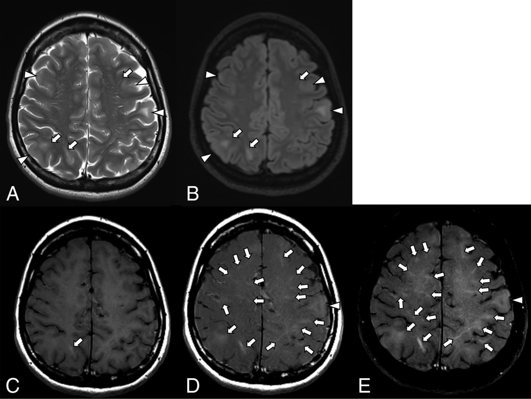
  • FIG 2.
    • Download figure
    • Open in new tab
    • Download powerpoint
    FIG 2.

    Patient 19. A 26-month-old boy. Axial T2WI (A), FLAIR (B), T1WI (C), T1WI-MTC (D), and T1WI-CHESS (E). T2WI and FLAIR images demonstrate swelling of the cortex and subcortical hyperintensity, plus a linear hyperintensity extending to the ventricle, representing cortical tubers and RMLs, respectively (A and B, arrowheads and arrows, respectively). On T1WI, several RMLs can be seen as hyperintensity (C, arrows). The RMLs are more apparent on T1WI-MTC and especially on T1WI-CHESS (D and E, arrows).

  • FIG 3.
    • Download figure
    • Open in new tab
    • Download powerpoint
    FIG 3.

    Patient 4. A 29-year-old woman. Axial T2WI (A), FLAIR (B), T1WI (C), T1WI-MTC (D), and T1WI-CHESS (E). T2WI and FLAIR images demonstrate swollen cortices, some of which are accompanied by subcortical hyperintensities, representing cortical tubers (A and B, arrowheads). Linear hyperintensities in the subcortical area extending to the ventricle are observed, representing the RMLs (A and B, arrows). On T1WI, an RML is found in the right parietal lobe (C, arrow). T1WI-TMC and T1WI-CHESS are able to detect more RMLs than T1WI, T2WI, and FLAIR (D and E, arrows). The RMLs are more apparent on T1WI-CHESS than on T1WI-MTC (D and E, arrows). Although no obvious cortical tuber is shown by T1WI, both T1WI-MTC and T1WI-CHESS detect a cortical tuber as a slight hyperintensity (D and E, arrowheads).
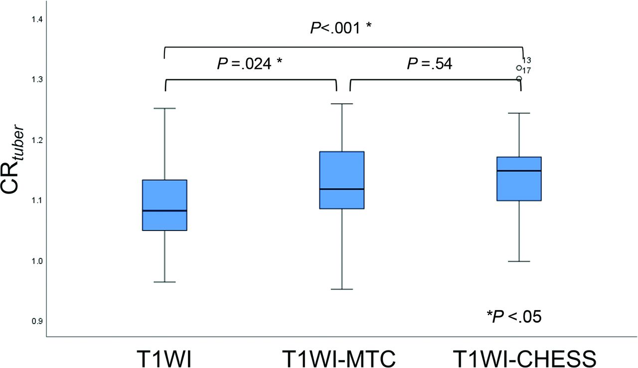
  • FIG 4.
    • Download figure
    • Open in new tab
    • Download powerpoint
    FIG 4.

    Comparison of the CRtuber among T1WI, T1WI-MTC, and T1WI-CHESS.

  • FIG 5.
    • Download figure
    • Open in new tab
    • Download powerpoint
    FIG 5.

    Comparison of the CRRML among T1WI, T1WI-MTC, and T1WI-CHESS.
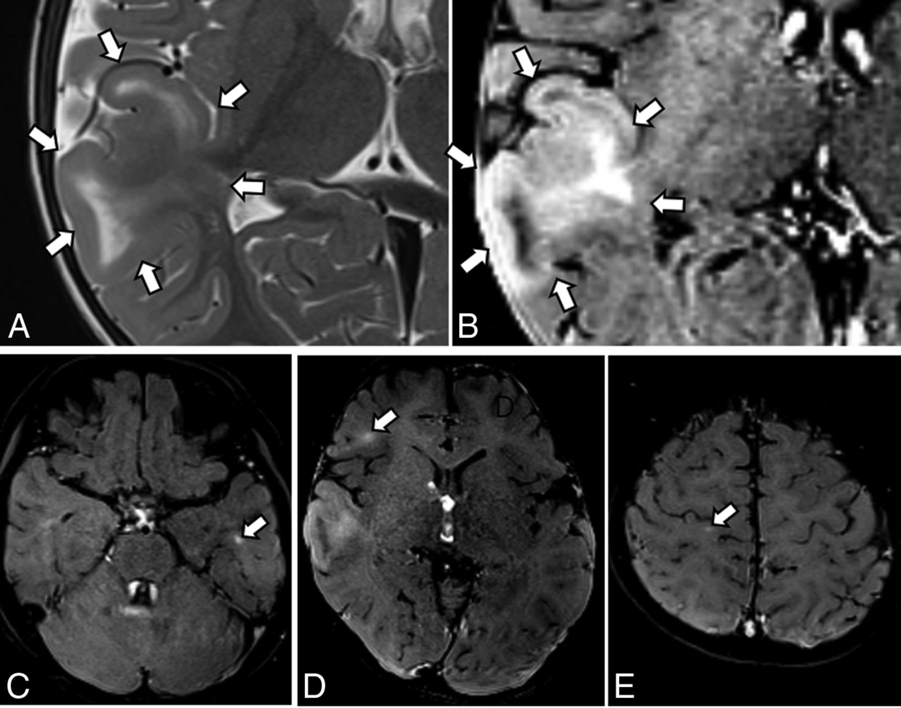
  • FIG 6.
    • Download figure
    • Open in new tab
    • Download powerpoint
    FIG 6.

    MR imaging findings of the surgical case, a 20-month-old boy with repeat epileptic seizures. A, Axial T2WI shows swelling of the cortex and subcortical hyperintensity (arrows), consistent with a cortical tuber. No other lesions are noted on T1WI, T2WI, and FLAIR (not shown); suggesting focal cortical dysplasia or a solitary cortical tuber. B, Axial T1WI-CHESS shows marked hyperintensity in the swollen cortex and subcortical white matter (arrows). C–E, Axial T1WI-CHESS shows several RMLs as linear hyperintense areas (arrows). On the basis of these findings, the patient was diagnosed with TSC.
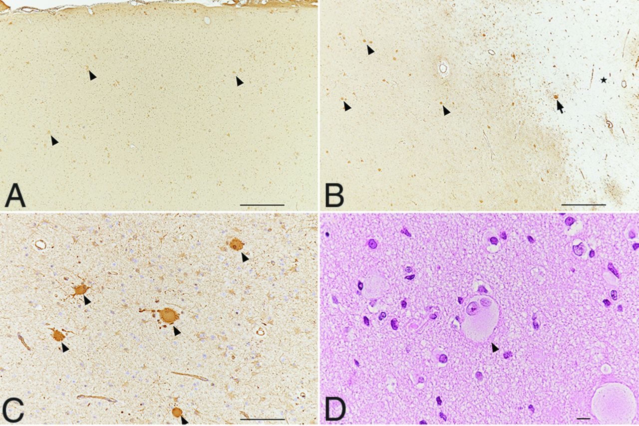
  • FIG 7.
    • Download figure
    • Open in new tab
    • Download powerpoint
    FIG 7.

    Pathologic findings of the surgical case, a 20-month-old boy with repeat epileptic seizures. A, Many balloon cells are seen in the thickened cortex (arrowheads as examples). B, In subcortical WM, many balloon cells (arrowheads as examples) are seen in the left part of the white rectangle. Rare balloon cells (arrow) are present in the right part of the rectangle (star). C and D, Many balloon cells are observed in the subcortical white matter (arrowheads) and are immunoreactive for a monoclonal antibody against vimentin (1:400, M7020; Dako). A–C, Monoclonal antivimentin antibody. D, Hematoxylin-eosin staining. Scale bars: A and B, 500 μm. C, 100 μm. D, 10 μm.

Tables

  • Figures
    • View popup
    Table 1:

    The number of CNS lesions in each sequencea

    T2WIFLAIRT1WIT1WI-MTCT1WI-CHESS
    Cortical tuber17.1 (SD, 10.8)17.5 (SD, 10.8)1.1 (SD, 2.3)10.2 (SD, 6.7)12.2 (SD, 7.7)
    RML3.56 (SD, 2.3)10.5 (SD, 8.1)1.4 (SD, 3.9)20.4 (SD, 12.1)24.8 (SD, 15.4)
    Subependymal nodule6.2 (SD, 4.3)4.5 (SD, 3.0)5.1 (SD, 3.2)6.4 (SD, 3.7)7.2 (SD, 3.9)
    • ↵a Data are mean (SD).

    • View popup
    Table 2:

    Interobserver reproducibility of the number of CNS lesions in each sequence

    T2WIFLAIRT1WIT1WI-MTCT1WI-CHESS
    Cortical tuber0.9590.9030.9840.7860.768
    RML0.6480.8660.8870.8830.799
    Subependymal nodule0.8260.6180.8220.9470.899
    • View popup
    Table 3:

    Comparison of the detection of CNS lesions among T1WI, T1WI-MTC, and T1WI-CHESS

    Multiple Comparison (P Value)
    T1WI vs. T1WI-MTCT1WI vs. T1WI-CHESST1WI-MTC vs. T1WI-CHESS
    Cortical tuber<.001a<.001a.198
    RML.001a<.001a.022a
    Subependymal nodule.007a<.001a.35
    • ↵a P  < .05.

    • View popup
    Table 4:

    Comparison of the detection of CNS lesions among T2WI, FLAIR, and T1WI-CHESS

    Multiple Comparison (P Value)
    T2WI vs FLAIRT2WI vs T1WI-CHESSFLAIR v. T1WI-CHESS
    Cortical tuber1.014a.011a
    RML.007a<.001a.014a
    Subependymal nodule.027a.085<.001a
    • ↵a P < .05.

    • View popup
    Table 5:

    Interobserver reproducibility of CRtuber and CRRML in each sequence

    T1WIT1WI-MTCT1WI-CHESS
    CRtuber0.7880.8630.797
    CRRML0.7130.8840.792
PreviousNext
Back to top

In this issue

American Journal of Neuroradiology: 43 (8)
American Journal of Neuroradiology
Vol. 43, Issue 8
1 Aug 2022
  • Table of Contents
  • Index by author
  • Complete Issue (PDF)
Advertisement
Print
Download PDF
Email Article

Thank you for your interest in spreading the word on American Journal of Neuroradiology.

NOTE: We only request your email address so that the person you are recommending the page to knows that you wanted them to see it, and that it is not junk mail. We do not capture any email address.

Enter multiple addresses on separate lines or separate them with commas.
MR Imaging Detection of CNS Lesions in Tuberous Sclerosis Complex: The Usefulness of T1WI with Chemical Shift Selective Images
(Your Name) has sent you a message from American Journal of Neuroradiology
(Your Name) thought you would like to see the American Journal of Neuroradiology web site.
CAPTCHA
This question is for testing whether or not you are a human visitor and to prevent automated spam submissions.
Cite this article
H. Fujii, N. Sato, Y. Kimura, M. Mizutani, M. Kusama, N. Sumitomo, E. Chiba, Y. Shigemoto, M. Takao, Y. Takayama, M. Iwasaki, E. Nakagawa, H. Mori
MR Imaging Detection of CNS Lesions in Tuberous Sclerosis Complex: The Usefulness of T1WI with Chemical Shift Selective Images
American Journal of Neuroradiology Aug 2022, 43 (8) 1202-1209; DOI: 10.3174/ajnr.A7573

Citation Manager Formats

  • BibTeX
  • Bookends
  • EasyBib
  • EndNote (tagged)
  • EndNote 8 (xml)
  • Medlars
  • Mendeley
  • Papers
  • RefWorks Tagged
  • Ref Manager
  • RIS
  • Zotero
0 Responses
Respond to this article
Share
Bookmark this article
MR Imaging of CNS Lesions in Tuberous Sclerosis
H. Fujii, N. Sato, Y. Kimura, M. Mizutani, M. Kusama, N. Sumitomo, E. Chiba, Y. Shigemoto, M. Takao, Y. Takayama, M. Iwasaki, E. Nakagawa, H. Mori
American Journal of Neuroradiology Aug 2022, 43 (8) 1202-1209; DOI: 10.3174/ajnr.A7573
del.icio.us logo Twitter logo Facebook logo Mendeley logo
  • Tweet Widget
  • Facebook Like
  • Google Plus One
Purchase

Jump to section

  • Article
    • Abstract
    • ABBREVIATIONS:
    • MATERIALS AND METHODS
    • RESULTS
    • DISCUSSION
    • CONCLUSIONS
    • Footnotes
    • References
  • Figures & Data
  • Info & Metrics
  • Responses
  • References
  • PDF

Related Articles

  • No related articles found.
  • PubMed
  • Google Scholar

Cited By...

  • No citing articles found.
  • Crossref (7)
  • Google Scholar

This article has been cited by the following articles in journals that are participating in Crossref Cited-by Linking.

  • MRI detection of mild malformation of cortical development with oligodendroglial hyperplasia (MOGHE) on T1WI-CHESS
    Elly Arizono, Zen-ichi Tanei, Keiya Iijima, Yukio Kimura, Yoko Shigemoto, Hiroyuki Maki, Midori Kusama, Kumiko Murayama, Masaki Iwasaki, Takashi Saito, Yuko Saito, Kazuhiro Saito, Noriko Sato
    Epilepsy & Behavior Reports 2024 26
  • Comparison of 3D Magnetization-transfer- and Spectral-presaturation-with-inversion-recovery-based Neuromelanin Imaging
    Midori Kusama, Yukio Kimura, Masami Yoneyama, Takashi Namiki, Takeshi Tamaru, Kenji Miyagi, Noriko Sato
    Magnetic Resonance in Medical Sciences 2025 24 2
  • Differences between Tuberous Sclerosis Complex Patients with and without Epilepsy: The Results of a Quantitative Diffusion Tensor Imaging Study
    Anna B. Marcinkowska, Sergiusz Jóźwiak, Agnieszka Sabisz, Agnieszka Tarasewicz, Beata Rutkowska, Alicja Dębska-Ślizień, Edyta Szurowska
    Biomedicines 2024 12 9
  • In vivo Metabolic Investigation of Oxygen, Light, and Temperature Effects on Dormancy Alleviation of Sesame (Sesamum indicum L.) Seeds
    Honghao Cai, Xiayi Ruan, Yumin Wan, Mengting Chen, Xianqin Wu, Yingqiang Cai
    Tarım Bilimleri Dergisi 2025 31 1
  • Multi-parametric diffusion spectrum imaging in tuberous sclerosis complex: Identifying cortical tubers and predicting genotypes
    Hui Sun, Zhiping Yan, Junhang Gao, Yingzhi Zheng, Yueyu Zheng, Yang Song, Yongji Liu, Zhixian Lin, Wencai Shen, Jin Fang, Hong Qu, Yingying Song, Yanzhao Diao, Sulian Su, Guihua Jiang
    European Journal of Radiology 2025 184
  • Neuroradiologic Findings Associated with Tuberous Sclerosis Complex: A Comprehensive Analysis of 24 Cases
    Mehmet Demir, Özlem Öz, Mehmet Sinan Benzer
    Harran Üniversitesi Tıp Fakültesi Dergisi 2025 22 1
  • Superior lesion detection on 3D FSE T1WI with magnetization transfer and CHESS preparation pulses in children with focal epilepsy: Preliminary results
    Xing‐Chang Wei, Zarina Assis, Martin Sherriff, Walter J. Hader, Juan Pablo Appendino
    Epileptic Disorders 2024 26 6

More in this TOC Section

  • SyMRI & MR Fingerprinting in Brainstem Myelination
  • Comparison of Image Quality and Radiation Dose in Pediatric Temporal Bone CT Using Photon-Counting Detector CT and Energy-Integrating Detector CT
  • Axenfeld-Rieger Syndrome: Neuroimaging Findings
Show more Pediatrics

Similar Articles

Advertisement

Indexed Content

  • Current Issue
  • Accepted Manuscripts
  • Article Preview
  • Past Issues
  • Editorials
  • Editor's Choice
  • Fellows' Journal Club
  • Letters to the Editor
  • Video Articles

Cases

  • Case Collection
  • Archive - Case of the Week
  • Archive - Case of the Month
  • Archive - Classic Case

More from AJNR

  • Trainee Corner
  • Imaging Protocols
  • MRI Safety Corner

Multimedia

  • AJNR Podcasts
  • AJNR Scantastics

Resources

  • Turnaround Time
  • Submit a Manuscript
  • Submit a Video Article
  • Submit an eLetter to the Editor/Response
  • Manuscript Submission Guidelines
  • Statistical Tips
  • Fast Publishing of Accepted Manuscripts
  • Graphical Abstract Preparation
  • Imaging Protocol Submission
  • Evidence-Based Medicine Level Guide
  • Publishing Checklists
  • Author Policies
  • Become a Reviewer/Academy of Reviewers
  • News and Updates

About Us

  • About AJNR
  • Editorial Board
  • Editorial Board Alumni
  • Alerts
  • Permissions
  • Not an AJNR Subscriber? Join Now
  • Advertise with Us
  • Librarian Resources
  • Feedback
  • Terms and Conditions
  • AJNR Editorial Board Alumni

American Society of Neuroradiology

  • Not an ASNR Member? Join Now

© 2025 by the American Society of Neuroradiology All rights, including for text and data mining, AI training, and similar technologies, are reserved.
Print ISSN: 0195-6108 Online ISSN: 1936-959X

Powered by HighWire