Advertisement

Main menu

  • Home
  • Content
    • Current Issue
    • Accepted Manuscripts
    • Article Preview
    • Past Issue Archive
    • Video Articles
    • AJNR Case Collection
    • Case of the Week Archive
    • Case of the Month Archive
    • Classic Case Archive
  • Special Collections
    • AJNR Awards
    • Low-Field MRI
    • Alzheimer Disease
    • ASNR Foundation Special Collection
    • Photon-Counting CT
    • View All
  • Multimedia
    • AJNR Podcasts
    • AJNR SCANtastic
    • Trainee Corner
    • MRI Safety Corner
    • Imaging Protocols
  • For Authors
    • Submit a Manuscript
    • Submit a Video Article
    • Submit an eLetter to the Editor/Response
    • Manuscript Submission Guidelines
    • Statistical Tips
    • Fast Publishing of Accepted Manuscripts
    • Graphical Abstract Preparation
    • Imaging Protocol Submission
    • Author Policies
  • About Us
    • About AJNR
    • Editorial Board
    • Editorial Board Alumni
  • More
    • Become a Reviewer/Academy of Reviewers
    • Subscribers
    • Permissions
    • Alerts
    • Feedback
    • Advertisers
    • ASNR Home

User menu

  • Alerts
  • Log in

Search

  • Advanced search
American Journal of Neuroradiology

American Journal of Neuroradiology

ASHNR American Society of Functional Neuroradiology ASHNR American Society of Pediatric Neuroradiology ASSR
  • Alerts
  • Log in

Advanced Search

  • Home
  • Content
    • Current Issue
    • Accepted Manuscripts
    • Article Preview
    • Past Issue Archive
    • Video Articles
    • AJNR Case Collection
    • Case of the Week Archive
    • Case of the Month Archive
    • Classic Case Archive
  • Special Collections
    • AJNR Awards
    • Low-Field MRI
    • Alzheimer Disease
    • ASNR Foundation Special Collection
    • Photon-Counting CT
    • View All
  • Multimedia
    • AJNR Podcasts
    • AJNR SCANtastic
    • Trainee Corner
    • MRI Safety Corner
    • Imaging Protocols
  • For Authors
    • Submit a Manuscript
    • Submit a Video Article
    • Submit an eLetter to the Editor/Response
    • Manuscript Submission Guidelines
    • Statistical Tips
    • Fast Publishing of Accepted Manuscripts
    • Graphical Abstract Preparation
    • Imaging Protocol Submission
    • Author Policies
  • About Us
    • About AJNR
    • Editorial Board
    • Editorial Board Alumni
  • More
    • Become a Reviewer/Academy of Reviewers
    • Subscribers
    • Permissions
    • Alerts
    • Feedback
    • Advertisers
    • ASNR Home
  • Follow AJNR on Twitter
  • Visit AJNR on Facebook
  • Follow AJNR on Instagram
  • Join AJNR on LinkedIn
  • RSS Feeds

AJNR Awards, New Junior Editors, and more. Read the latest AJNR updates

Research ArticleBrain Tumor Imaging

Do We Need Gadolinium-Based Contrast Agents for Routine MRI Surveillance of Unoperated Pituitary Macroadenoma?

A.A. Alali, P.B. Hanagandi and P.J. Maralani
American Journal of Neuroradiology July 2022, 43 (7) 1024-1028; DOI: https://doi.org/10.3174/ajnr.A7554
A.A. Alali
aFrom the Division of Neuroradiology (A.A.A.), King Saud bin Abdulaziz University for Health Sciences, Riyadh, Saudi Arabia
  • Find this author on Google Scholar
  • Find this author on PubMed
  • Search for this author on this site
  • ORCID record for A.A. Alali
P.B. Hanagandi
bDepartment of Medical Imaging (P.B.H.), King Abdulaziz Medical City NGHA, Riyadh, Saudi Arabia
  • Find this author on Google Scholar
  • Find this author on PubMed
  • Search for this author on this site
  • ORCID record for P.B. Hanagandi
P.J. Maralani
cDivision of Neuroradiology (P.J.M.), Department of Medical Imaging, University of Toronto, Sunnybrook Health Sciences Centre, Toronto, Ontario, Canada
  • Find this author on Google Scholar
  • Find this author on PubMed
  • Search for this author on this site
  • ORCID record for P.J. Maralani
  • Article
  • Figures & Data
  • Info & Metrics
  • Responses
  • References
  • PDF
Loading

Article Figures & Data

Figures

  • Tables
  • FIG 1.
    • Download figure
    • Open in new tab
    • Download powerpoint
    FIG 1.

    Flow chart shows patient selection.

  • FIG 2.
    • Download figure
    • Open in new tab
    • Download powerpoint
    FIG 2.

    Coronal T2WI versus T1C show the different grades of cavernous sinus invasion using the Knosp classification; grade 0, the lesion does not extend beyond the medial carotid line (red line); grade 1, the lesion extends to the medial line but does not reach the intercarotid line (blue line); grade 2, the tumor extends beyond the intercarotid line but does not extend beyond the lateral line (yellow line); grade 3, the tumor extends to the lateral line more so on the left (yellow line); and grade 4, there is complete encasement of the cavernous carotid artery.
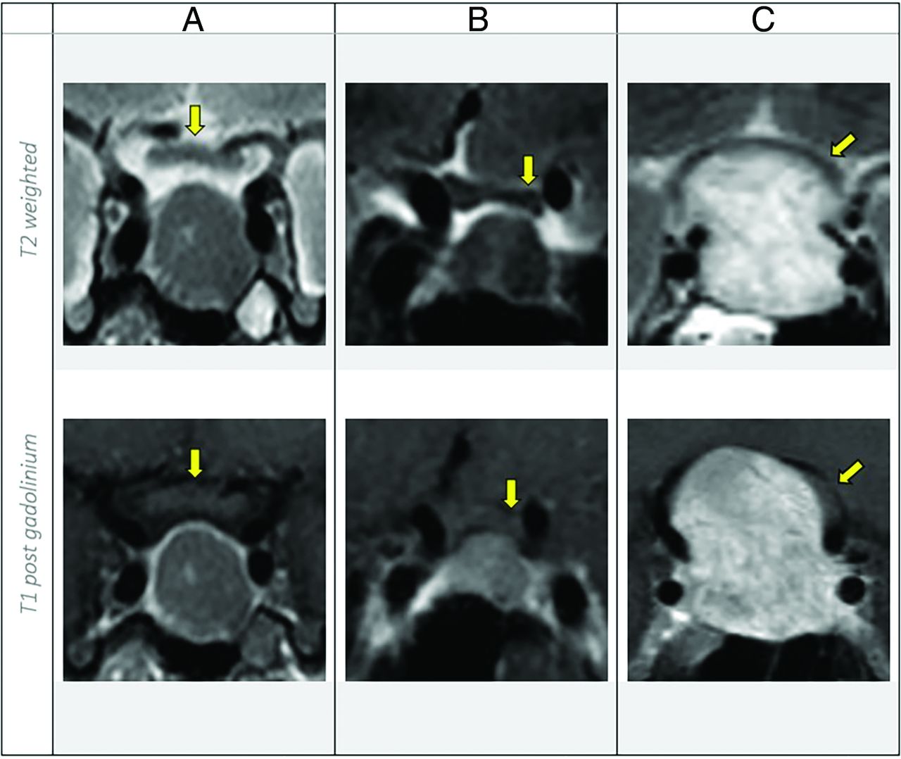
  • FIG 3.
    • Download figure
    • Open in new tab
    • Download powerpoint
    FIG 3.

    Coronal T2WI versus T1C show the optic pathway (depicted by yellow arrows) compression classification. A, There is no contact. B, The pituitary macroadenoma is abutting the left aspect of the optic chiasm without displacement. C, There are mass effect and displacement of the optic pathway.

  • FIG 4.
    • Download figure
    • Open in new tab
    • Download powerpoint
    FIG 4.

    MR imaging of the sella shows internal hemorrhage with the hematocrit level on different sequences. Sag indicates sagittal; pre gad, pregadolinium; post gad, postgadolinium.

Tables

  • Figures
    • View popup
    Table 1:

    MR imaging parameters

    ScannerCoronal SequenceTR
    (ms)
    TE
    (ms)
    Flip AngleFOV
    (mm)
    Section Thickness (mm)Voxel Size (mm)Image MatrixAcquisition Time (Min)
    PhilipsT1WI4501090°1302.5 or 30.7 × 0.7 × 2.5184 × 1843:45
    T2WI30008090°1302.5 or 30.7 × 0.7 × 2.5184 × 1852:32
    GET1WI4719111°1502.5 or 30.6 × 0.8 × 2.5256 × 1922:35
    T2WI2800103.9111°1502.5 or 30.5 × 0.8 × 2.5320 × 1923:06
    SiemensT1WI4008.6150°1402.5 or 30.8 × 0.5 × 2.5179 × 2562:50
    T2WI320096150°1402.5 or 30.8 × 0.5 × 2.5184 × 2562:25
    • View popup
    Table 2:

    Summary statistics comparing the craniocaudal dimension of pituitary macroadenoma, cavernous sinus invasion, and optic pathway compression on T2WI versus T1C

    Coronal T2WICoronal T1CAgreement
    ICC/κ
    Craniocaudal dimension
     Mean23.1 (SD, 10.7) mm23.1 (SD, 10.8) mmICC = 0.96
     Range9–55 mm8–55 mm
    Knosp Classification (No.) (%)
     016 (15.2)16 (15.2)κ = 0.95
     128 (26.7)27 (25.7)
     233 (31.4)33 (31.4)
     315 (14.3)16 (15.2)
     413 (12.4)13 (12.4)
    Optic pathway compression (No.) (%)
     No contact32 (30.5)34 (32.4)κ = 0.84
     Abutment without displacement18 (17.1)20 (19)
     Compression with displacement55 (52.4)51 (48.6)
PreviousNext
Back to top

In this issue

American Journal of Neuroradiology: 43 (7)
American Journal of Neuroradiology
Vol. 43, Issue 7
1 Jul 2022
  • Table of Contents
  • Index by author
  • Complete Issue (PDF)
Advertisement
Print
Download PDF
Email Article

Thank you for your interest in spreading the word on American Journal of Neuroradiology.

NOTE: We only request your email address so that the person you are recommending the page to knows that you wanted them to see it, and that it is not junk mail. We do not capture any email address.

Enter multiple addresses on separate lines or separate them with commas.
Do We Need Gadolinium-Based Contrast Agents for Routine MRI Surveillance of Unoperated Pituitary Macroadenoma?
(Your Name) has sent you a message from American Journal of Neuroradiology
(Your Name) thought you would like to see the American Journal of Neuroradiology web site.
CAPTCHA
This question is for testing whether or not you are a human visitor and to prevent automated spam submissions.
Cite this article
A.A. Alali, P.B. Hanagandi, P.J. Maralani
Do We Need Gadolinium-Based Contrast Agents for Routine MRI Surveillance of Unoperated Pituitary Macroadenoma?
American Journal of Neuroradiology Jul 2022, 43 (7) 1024-1028; DOI: 10.3174/ajnr.A7554

Citation Manager Formats

  • BibTeX
  • Bookends
  • EasyBib
  • EndNote (tagged)
  • EndNote 8 (xml)
  • Medlars
  • Mendeley
  • Papers
  • RefWorks Tagged
  • Ref Manager
  • RIS
  • Zotero
0 Responses
Respond to this article
Share
Bookmark this article
Need for Gadolinium in Pituitary Macroadenoma MRI
A.A. Alali, P.B. Hanagandi, P.J. Maralani
American Journal of Neuroradiology Jul 2022, 43 (7) 1024-1028; DOI: 10.3174/ajnr.A7554
del.icio.us logo Twitter logo Facebook logo Mendeley logo
  • Tweet Widget
  • Facebook Like
  • Google Plus One
Purchase

Jump to section

  • Article
    • Abstract
    • ABBREVIATIONS:
    • MATERIALS AND METHODS
    • RESULTS
    • DISCUSSION
    • CONCLUSIONS
    • Footnotes
    • References
  • Figures & Data
  • Info & Metrics
  • Responses
  • References
  • PDF

Related Articles

  • No related articles found.
  • PubMed
  • Google Scholar

Cited By...

  • No citing articles found.
  • Crossref (7)
  • Google Scholar

This article has been cited by the following articles in journals that are participating in Crossref Cited-by Linking.

  • How often should we perform magnetic resonance imaging (MRI) for the follow-up of pituitary adenoma?
    Stefan Matei Constantinescu, Thierry Duprez, Jean-François Bonneville, Dominique Maiter
    Annales d'Endocrinologie 2024 85 4
  • Establishment of a deep-learning-assisted recurrent nasopharyngeal carcinoma detecting simultaneous tactic (DARNDEST) with high cost-effectiveness based on magnetic resonance images: a multicenter study in an endemic area
    Yishu Deng, Yingying Huang, Haijun Wu, Dongxia He, Wenze Qiu, Bingzhong Jing, Xing Lv, Weixiong Xia, Bin Li, Ying Sun, Chaofeng Li, Chuanmiao Xie, Liangru Ke
    Cancer Imaging 2025 25 1
  • MRI Surveillance of Nonresected Pituitary Macroadenoma: Point—IV Contrast Media May Not Be Needed
    Vivek Pai, Pejman J. Maralani
    American Journal of Roentgenology 2024 222 1
  • Recommendations on the use of gadolinium-based contrast agents in the diagnosis and monitoring of common adult intracranial tumours
    Fabio M. Doniselli, João Ramos, Amaya Hilario, Valentina Opancina, Anouk van der Hoorn, Antonella Castellano, Aart J. van der Molen, Carlo C. Quattrocchi, Tarek Yousry, Àlex Rovira, Aart J. van der Molen, Anouk van der Hoorn
    European Radiology 2025
  • Safe and optimized use of gadolinium-based contrast agents in neuroimaging
    Àlex Rovira, Carlo C. Quattrocchi
    European Radiology 2023 34 7
  • Surveying Current Practices in the Use of Gadolinium-Based Contrast Agents for Routine Brain Magnetic Resonance Imaging in the Evaluation of Isolated Growth Hormone Deficiency among US Pediatric Endocrinologists
    Daniel Mak, Emily Breidbart, Shilpa Mehta, Brenda Kohn
    Hormone Research in Paediatrics 2024
  • Tricks and Traps in MRI of the Pituitary Region
    Jean-François Bonneville
    2024

More in this TOC Section

  • Neuroimaging of Erdheim-Chester Disease
  • CE MRI for Brain Metastasis Detection
  • Cognitive Control in Tumor Language Plasticity
Show more BRAIN TUMOR IMAGING

Similar Articles

Advertisement

Indexed Content

  • Current Issue
  • Accepted Manuscripts
  • Article Preview
  • Past Issues
  • Editorials
  • Editor's Choice
  • Fellows' Journal Club
  • Letters to the Editor
  • Video Articles

Cases

  • Case Collection
  • Archive - Case of the Week
  • Archive - Case of the Month
  • Archive - Classic Case

More from AJNR

  • Trainee Corner
  • Imaging Protocols
  • MRI Safety Corner
  • Book Reviews

Multimedia

  • AJNR Podcasts
  • AJNR Scantastics

Resources

  • Turnaround Time
  • Submit a Manuscript
  • Submit a Video Article
  • Submit an eLetter to the Editor/Response
  • Manuscript Submission Guidelines
  • Statistical Tips
  • Fast Publishing of Accepted Manuscripts
  • Graphical Abstract Preparation
  • Imaging Protocol Submission
  • Evidence-Based Medicine Level Guide
  • Publishing Checklists
  • Author Policies
  • Become a Reviewer/Academy of Reviewers
  • News and Updates

About Us

  • About AJNR
  • Editorial Board
  • Editorial Board Alumni
  • Alerts
  • Permissions
  • Not an AJNR Subscriber? Join Now
  • Advertise with Us
  • Librarian Resources
  • Feedback
  • Terms and Conditions
  • AJNR Editorial Board Alumni

American Society of Neuroradiology

  • Not an ASNR Member? Join Now

© 2025 by the American Society of Neuroradiology All rights, including for text and data mining, AI training, and similar technologies, are reserved.
Print ISSN: 0195-6108 Online ISSN: 1936-959X

Powered by HighWire