
Article Figures & Data
Figures
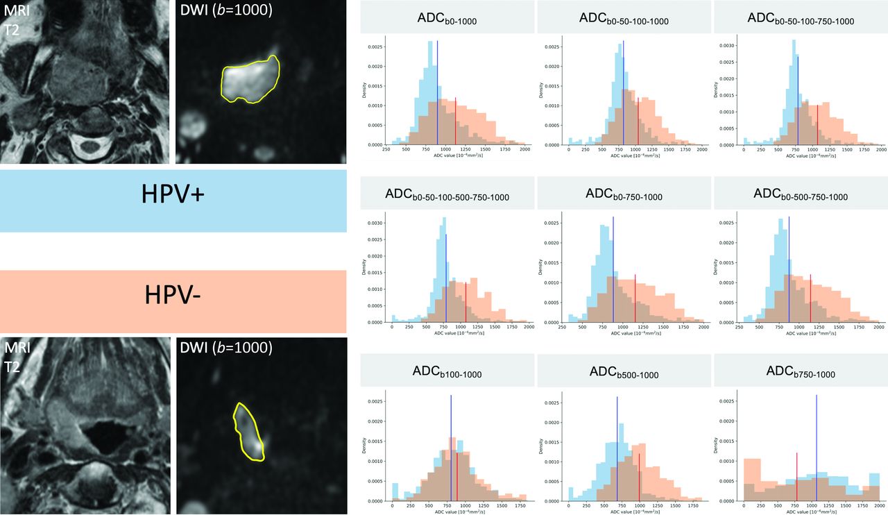
- FIG 1.
ADC histogram changes caused by b-value choice illustrated in 2 different patients with OPSCC. T2 and corresponding b=1000 images with tumor ROIs (in yellow) are shown on the left side of the figure. The histograms in blue were obtained from the pixel values of the HPV+ OPSCC ROI, and the histograms in orange, from the pixel values of the HPV– OPSCC ROI. ADC mean values are indicated in blue for the HPV+ OPSCC and in red for the HPV– OPSCC, respectively. On ADC maps with b=0, HPV+ OPSCC histograms have lower ADC mean values, a slender peak (leptokurtic shape), and a right skew, whereas HPV– OPSCC histograms have higher ADC mean values, lower kurtosis (flatter shape), and a more symmetric shape (Gaussian distribution). On ADC maps with b=0, the histograms of the 2 tumors can be easily distinguished one from another. This is hardly possible on perfusion-insensitive ADC maps (ADCb100-1000, ADCb500-1000, ADCb750-1000) due to overlapping metrics.
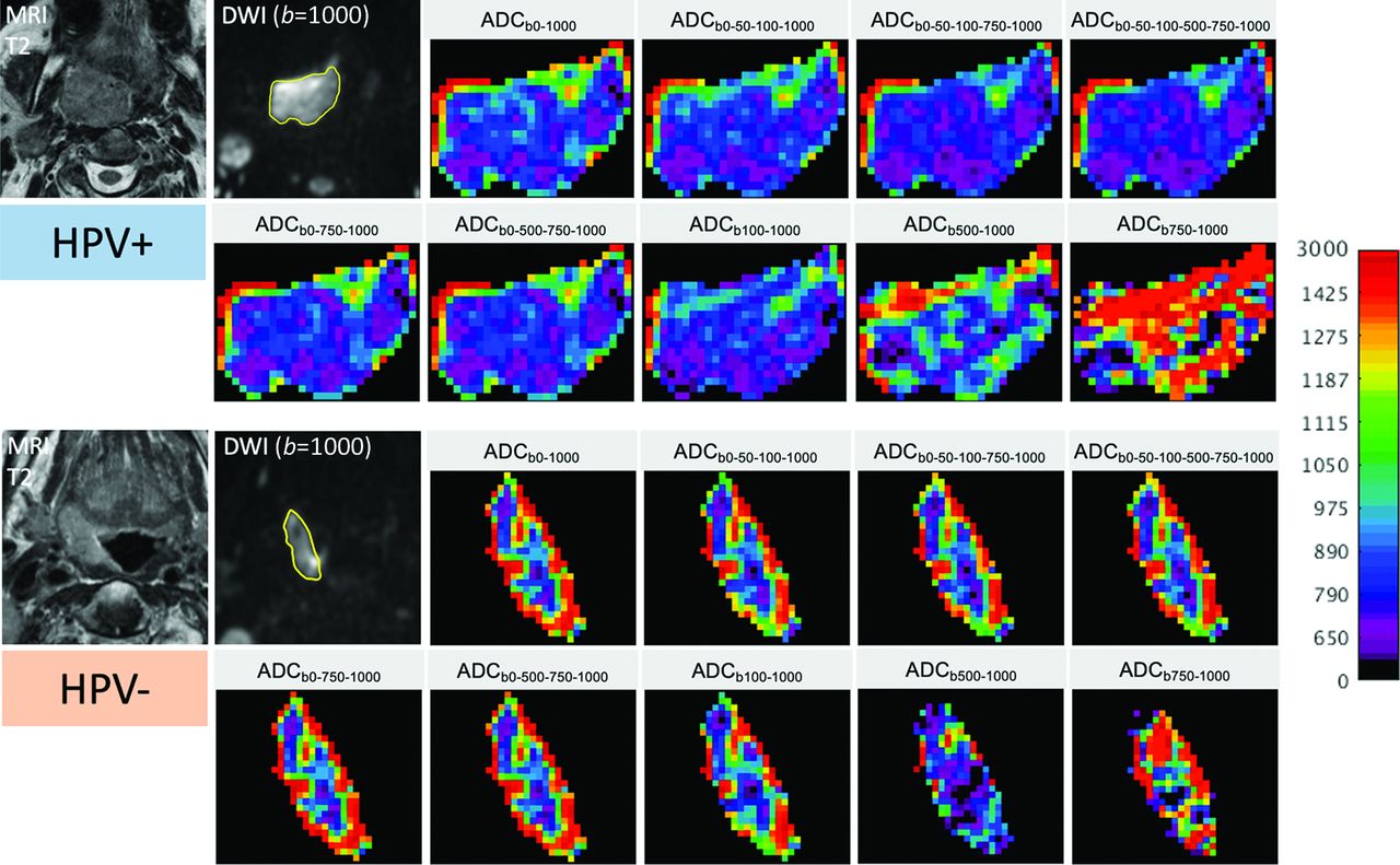
- FIG 2.
Color-coded maps in 2 different patients with OPSCC (same patients as in Fig 1) illustrating changes in ADC pixel values and distribution caused by the choice of b-values. T2 images and corresponding b=1000 images with tumor ROIs (in yellow) are shown on the left. The upper 2 rows on the right show the color-coded ADC maps of the HPV+ OPSCC, while the lower 2 rows show the respective maps of the HPV– OPSCC. For all images, the same color map with the same quantitative scale (0–3000 ×10−6mm2/s) was used to display the original gray levels. Note the clearly visible difference between HPV+ versus HPV– OPSCC on the ADC maps with b=0, with HPV+ OPSCC having lower ADCs and less ROI heterogeneity than HPV– OPSCC. This difference disappears on maps calculated with higher b-values only, and the distinction between the 2 tumor types is visually hardly possible on maps calculated with b ≥ 500. The 2 readers correctly identified the HPV+ and the HPV– OPSCC on the first 7 ADC maps; they failed, however, to correctly distinguish between the 2 tumor types on the last 2 ADC maps.


Tables
No. of b-Values Used b-Values Monoexponential Model 2 0 1000 ADCb0–1000 4 0 50 100 1000 ADCb0–50–100–1000 5 0 50 100 750 1000 ADCb0–50–100–750–1000 6 0 50 100 500 750 1000 ADCb0–50–100–500–750–1000 3 0 750 1000 ADCb0–750–1000 4 0 500 750 1000 ADCb0–500–750–1000 2 100 1000 ADCb100–1000 2 500 1000 ADCb500–1000 2 750 1000 ADC b750–1000 HPV– OPSCC (n = 23) HPV+ OPSCC (n = 11) P Value Average age (range) (yr) 62 (50–82) 62 (48–85) .986a Sex Women 7 (7/23, 30%) 6 (6/11, 55%) Men 16 (16/23, 70%) 5 (5/11, 45%) .329b Tumor location Oropharynx 23 (23/23, 100%) 11 (11/11, 100%) T classification according to AJCC 20184,5 .944c T1 3 (3/23, 13%) 0 (0/11, 0%) T2 3 (3/23, 13%) 2 (2/11, 18%) T3 5 (5/23, 22%) 2 (2/11, 18%) T4 12 (12/23, 52%) 7 (7/11, 64%) N classification according to AJCC 20184,5 .138c N0 5 (5/23, 22%) 0 (0/11, 0%) N1 3 (3/23, 13%) 5 (5/11, 45%) N2 15 (15/23, 65%) 6 (6/11, 55%) N3 0 (0/23, 0%) 0 (0/11, 0%) M classification according to AJCC 20184,5 .630c M0 22 (22/23, 96%) 11 (11/11, 100%) M1 1 (1/23, 4%) 0 (0/11, 0%) Tumor keratinization at histopathology .140b Present 14 (14/23, 61%) 7 (7/11, 64%) Absent 9 (9/23, 39%) 4 (4/11, 36%) Mean proliferation index, MIB-1 (range) (%) 55 (15–90) 69 (50–90) .126d - Table 3:
Comparison of ADC histograms in HPV+ versus HPV– OPSCC for each b-value combinationa
HPV– OPSCC HPV+ OPSCC P Valueb ADC Meanc ADCb0–1000 1117 (SD, 151) 977 (SD, 183) .038 ADCb0–50–100–1000 1029 (SD, 176) 924 (SD, 185) .164 ADCb0–50–100–750–1000 1051 (SD, 168) 925 (SD, 176) .077 ADCb0–50–100–500–750–1000 1061 (SD, 167) 935 (SD, 176) .084 ADCb0–750–1000 1141 (SD, 156) 983 (SD, 177) .017 ADCb0–500–750–1000 1127 (SD, 156) 970 (SD, 177) .017 ADCb100–1000 947 (SD, 245) 875 (SD, 206) .214 ADCb500–1000 838 (SD, 255) 758 (SD, 287) .176 ADCb750–1000 900 (SD, 360) 942 (SD, 340) .942 Skewness ADCb0–1000 0.156 (SD, 0.453) 0.486 (SD, 0.444) .031 ADCb0–50–100–1000 0.266 (SD, 0.442) 0.393 (SD, 0.48) .258 ADCb0–50–100–750–1000 0.331 (SD, 0.526) 0.363 (SD, 0.609) .445 ADCb0–50–100–500–750–1000 0.31 (SD, 0.542) 0.363 (SD, 0.649) .383 ADCb0–750–1000 0.177 (SD, 0.518) 0.506 (SD, 0.483) .046 ADCb0–500–750–1000 0.2 (SD, 0.496) 0.499 (SD, 0.458) .034 ADCb100-1000 0.136 (SD, 0.427) 0.069 (SD, 0.486) .468 ADCb500–1000 0.229 (SD, 0.431) 0.103 (SD, 0.47) .537 ADCb750–1000 0.672 (SD, 0.739) 0.435 (SD, 0.383) .214 Excess kurtosis ADCb0–1000 0.12 (SD, 0.71) 1.22 (SD, 0.45) < .001 ADCb0–50–100–1000 0.34 (SD, 0.81) 1.36 (SD, 0.69) < .001 ADCb0–50–100–750–1000 0.57 (SD, 1.25) 1.64 (SD, 1.15) .008 ADCb0–50–100–500–750–1000 0.55 (SD, 1.25) 1.74 (SD, 1.23) .007 ADCb0–750–1000 0.21 (SD, 0.81) 1.22 (SD, 0.58) .002 ADCb0–500–750–1000 0.2 (SD, 0.84) 1.19 (SD, 0.53) .002 ADCb100–1000 0.51 (SD, 1.2) 1.22 (SD, 1.09) .019 ADCb500–1000 0.19 (SD, 0.84) 0.7 (SD, 1.1) .188 ADCb750–1000 0.76 (SD, 2.18) 0.3 (SD, 1.09) .942 - Table 4:
Diagnostic performance of ADC maps capable of distinguishing HPV+ from HPV– OPSCC on the basis of ADC mean, skewness, and kurtosisa
Feature AUC P Value TP FP TN FN Sensitivity Specificity Accuracy Optimal Threshold ADCb0–1000 ADC mean 0.723 .019 8 7 16 3 0.727 0.695 0.706 1062.429b ADC skewness 0.731 .016 8 6 17 3 0.727 0.739 0.735 0.349 ADC kurtosisc 0.893 <.001 11 4 19 0 1.000 0.826 0.882 0.640 ADCb0–750–1000 ADC mean 0.755 .009 8 7 16 3 0.727 0.695 0.706 1088.702b ADC skewness 0.715 .023 8 5 18 3 0.727 0.783 0.765 0.500 ADC kurtosisc 0.826 .001 10 5 18 1 0.909 0.783 0.823 0.651 ADCb0–500–750–1000 ADC mean 0.755 .009 8 5 18 3 0.727 0.783 0.765 1034.222b ADC skewness 0.727 .009 8 4 19 3 0.727 0.826 0.794 0.525 ADC kurtosisc 0.826 .001 10 5 18 1 0.909 0.783 0.823 0.620 ADCb100–1000 ADC mean 0.636 .105 8 8 15 3 0.727 0.652 0.676 958.089b ADC skewness 0.581 .231 7 9 14 4 0.636 0.609 0.618 0.133 ADC kurtosisc 0.751 .010 9 7 16 2 0.818 0.696 0.735 0.484 Note:—TP indicates true-positive; FP, false-positive; TN, true-negative; FN, false-negative.
↵a For comparison, the diagnostic performance of the perfusion-insensitive map recommended in the literature14 is equally shown. P values to distinguish HPV+ from HPV– OPSCC were calculated with the Mann-Whitney-Wilcoxon test. Sensitivity, specificity, and accuracy were calculated using the optimal threshold (Youden index from receiver operating curve analysis).
↵b ADC mean thresholds in x10−6mm2/s.
↵c Excess kurtosis.


Jump to section
Related Articles
Cited By...
- No citing articles found.