Advertisement

Main menu

  • Home
  • Content
    • Current Issue
    • Accepted Manuscripts
    • Article Preview
    • Past Issue Archive
    • Video Articles
    • AJNR Case Collection
    • Case of the Week Archive
    • Case of the Month Archive
    • Classic Case Archive
  • Special Collections
    • AJNR Awards
    • Low-Field MRI
    • Alzheimer Disease
    • ASNR Foundation Special Collection
    • Photon-Counting CT
    • View All
  • Multimedia
    • AJNR Podcasts
    • AJNR SCANtastic
    • Trainee Corner
    • MRI Safety Corner
    • Imaging Protocols
  • For Authors
    • Submit a Manuscript
    • Submit a Video Article
    • Submit an eLetter to the Editor/Response
    • Manuscript Submission Guidelines
    • Statistical Tips
    • Fast Publishing of Accepted Manuscripts
    • Graphical Abstract Preparation
    • Imaging Protocol Submission
    • Author Policies
  • About Us
    • About AJNR
    • Editorial Board
    • Editorial Board Alumni
  • More
    • Become a Reviewer/Academy of Reviewers
    • Subscribers
    • Permissions
    • Alerts
    • Feedback
    • Advertisers
    • ASNR Home

User menu

  • Alerts
  • Log in

Search

  • Advanced search
American Journal of Neuroradiology

American Journal of Neuroradiology

ASHNR American Society of Functional Neuroradiology ASHNR American Society of Pediatric Neuroradiology ASSR
  • Alerts
  • Log in

Advanced Search

  • Home
  • Content
    • Current Issue
    • Accepted Manuscripts
    • Article Preview
    • Past Issue Archive
    • Video Articles
    • AJNR Case Collection
    • Case of the Week Archive
    • Case of the Month Archive
    • Classic Case Archive
  • Special Collections
    • AJNR Awards
    • Low-Field MRI
    • Alzheimer Disease
    • ASNR Foundation Special Collection
    • Photon-Counting CT
    • View All
  • Multimedia
    • AJNR Podcasts
    • AJNR SCANtastic
    • Trainee Corner
    • MRI Safety Corner
    • Imaging Protocols
  • For Authors
    • Submit a Manuscript
    • Submit a Video Article
    • Submit an eLetter to the Editor/Response
    • Manuscript Submission Guidelines
    • Statistical Tips
    • Fast Publishing of Accepted Manuscripts
    • Graphical Abstract Preparation
    • Imaging Protocol Submission
    • Author Policies
  • About Us
    • About AJNR
    • Editorial Board
    • Editorial Board Alumni
  • More
    • Become a Reviewer/Academy of Reviewers
    • Subscribers
    • Permissions
    • Alerts
    • Feedback
    • Advertisers
    • ASNR Home
  • Follow AJNR on Twitter
  • Visit AJNR on Facebook
  • Follow AJNR on Instagram
  • Join AJNR on LinkedIn
  • RSS Feeds

AJNR Awards, New Junior Editors, and more. Read the latest AJNR updates

Research ArticleAdult Brain

Volumetric Measurement of Relative CBV Using T1-Perfusion-Weighted MRI with High Temporal Resolution Compared with Traditional T2*-Perfusion-Weighted MRI in Postoperative Patients with High-Grade Gliomas

M. Seo, K.-J. Ahn, Y. Choi, N.-Y. Shin, J. Jang and B.-S. Kim
American Journal of Neuroradiology June 2022, 43 (6) 864-871; DOI: https://doi.org/10.3174/ajnr.A7527
M. Seo
aFrom the Department of Radiology, Seoul St. Mary's Hospital, College of Medicine, the Catholic University of Korea, Seoul, Republic of Korea
  • Find this author on Google Scholar
  • Find this author on PubMed
  • Search for this author on this site
  • ORCID record for M. Seo
K.-J. Ahn
aFrom the Department of Radiology, Seoul St. Mary's Hospital, College of Medicine, the Catholic University of Korea, Seoul, Republic of Korea
  • Find this author on Google Scholar
  • Find this author on PubMed
  • Search for this author on this site
  • ORCID record for K.-J. Ahn
Y. Choi
aFrom the Department of Radiology, Seoul St. Mary's Hospital, College of Medicine, the Catholic University of Korea, Seoul, Republic of Korea
  • Find this author on Google Scholar
  • Find this author on PubMed
  • Search for this author on this site
  • ORCID record for Y. Choi
N.-Y. Shin
aFrom the Department of Radiology, Seoul St. Mary's Hospital, College of Medicine, the Catholic University of Korea, Seoul, Republic of Korea
  • Find this author on Google Scholar
  • Find this author on PubMed
  • Search for this author on this site
  • ORCID record for N.-Y. Shin
J. Jang
aFrom the Department of Radiology, Seoul St. Mary's Hospital, College of Medicine, the Catholic University of Korea, Seoul, Republic of Korea
  • Find this author on Google Scholar
  • Find this author on PubMed
  • Search for this author on this site
  • ORCID record for J. Jang
B.-S. Kim
aFrom the Department of Radiology, Seoul St. Mary's Hospital, College of Medicine, the Catholic University of Korea, Seoul, Republic of Korea
  • Find this author on Google Scholar
  • Find this author on PubMed
  • Search for this author on this site
  • ORCID record for B.-S. Kim
  • Article
  • Figures & Data
  • Supplemental
  • Info & Metrics
  • Responses
  • References
  • PDF
Loading

Article Figures & Data

Figures

  • Tables
  • FIG 1.
    • Download figure
    • Open in new tab
    • Download powerpoint
    FIG 1.

    A flow chart outlining the selection of patients and examinations is shown.
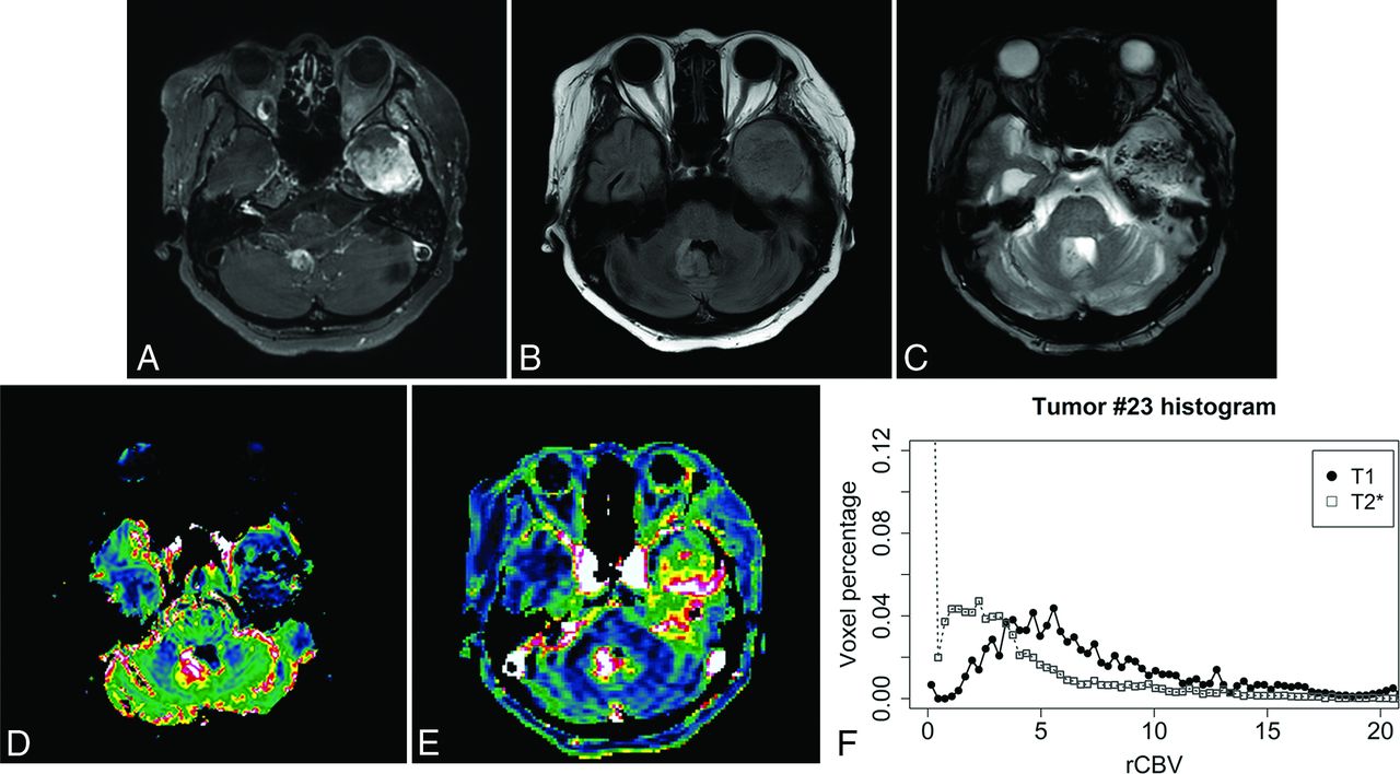
  • FIG 2.
    • Download figure
    • Open in new tab
    • Download powerpoint
    FIG 2.

    A 53-year-old woman with primary glioblastoma located in the left thalamus. On the follow-up image 10 months after the operation (7 months after the last radiation therapy), new enhancing lesions are noticed in the left temporal lobe base and fourth ventricle (A and B). Owing to its location and considerable amount of internal hemorrhage (C), signal loss shades the portions of the left temporal lobe lesion, resulting in grade 1 visualization on T2*-PWI (D, rCBV 90th percentile, 8.18). Grade 3 visualization is achieved on T1-PWI (E, rCBV 90th percentile, 23.92). Susceptibility-induced signal loss of the T2*-PWI is also well-noticed on the histogram, with the leftmost peak of near-zero voxels (F). The patient died in 1 month and was assigned to the progression group on ROC curve analysis.

  • FIG 3.
    • Download figure
    • Open in new tab
    • Download powerpoint
    FIG 3.

    Scatterplots of rCBV values derived from T1- and T2*-PWI. A, Ninetieth percentile of the whole lesion rCBV values, with fair consistency (ICC = 0.558) and a positive correlation (R = 0.614). B, Mean of the whole-lesion rCBV values, with fair consistency (ICC = 0.566) and a positive correlation (R = 0.663).

  • FIG 4.
    • Download figure
    • Open in new tab
    • Download powerpoint
    FIG 4.

    A Bland-Altman plot representing the 90th percentile of rCBV values derived from T1- and T2*-PWI. The upper and lower dashed lines represent the 1.96 and −1.96 limits of agreement, respectively (95% confidence interval not indicated).

  • FIG 5.
    • Download figure
    • Open in new tab
    • Download powerpoint
    FIG 5.

    Cumulative histogram of T1- and T2*-rCBV values in all included examinations. With T2*-rCBV measurement, the leftmost beginning point of the cumulative fraction is >0.2 (horizontal dashed line) in 17 lesions (37.8%), implying that in each entire VOI of those lesions, more than one-fifth of the voxels contained rCBV values of <0.3 (the first bin of histogram). With T1-PWI–based rCBV measurements, in contrast, only 2 lesions (4.4%) show an initial cumulative fraction above 0.2.
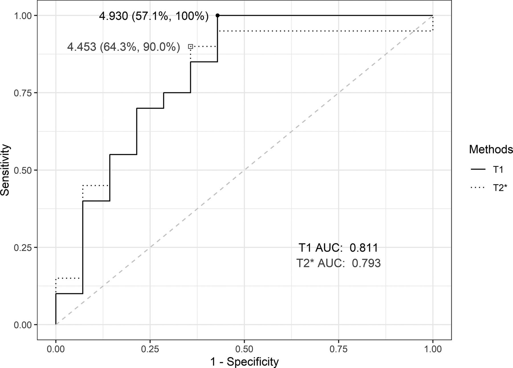
  • FIG 6.
    • Download figure
    • Open in new tab
    • Download powerpoint
    FIG 6.

    The ROC curves of T1- and T2*-rCBV using 90th percentile values. The AUC values of T1- and T2*-rCBV revealed no significant difference (P = .835).

Tables

  • Figures
    • View popup
    Table 1:

    Parameters of key MR imaging sequences

    T1-PWIT2*-PWIT2WIT2 FLAIR3D CE-T1WI
    Sequence3D T1-FFE3D T2-FFETSETSE3D TSE
    TR/TE (ms)4.2/2.31800/303000/809000/100550/30
    Flip angle8°40°90°90°90°
    FOV (mm2)220 × 220210 × 210230 × 230230 × 230240 × 240
    Matrix137 × 137128 × 128404 × 382308 × 290240 × 240
    Section thickness (mm)55551
    Slices30252626200
    Time resolution (sec)2.21.8
    Phases15050
    NoteBlack-blood, Dixon fat suppression
    • Note:—FFE indicates fast-field echo.

    • View popup
    Table 2:

    Summary of T1- and T2*-rCBV values distinguishing the progression group (n = 20) from the nonprogression group (n = 14)

    ValuesP Group (mean)NP Group (mean)P ValueaAUCCutoffbSensitivitySpecificityP Value versus T1-rCBVc
    T1-rCBV, 90th percentile13.0 (SD, 6.9)6.6 (SD, 6.1).0080.8114.930100.0%57.1%
    T2*-rCBV, 90th percentile7.9 (SD, 4.0)4.3 (SD, 3.1).0090.7934.45390.0%64.3%.835
    • Note:—P Group indicates progression group; NP Group, nonprogression group.

    • ↵a Independent t test.

    • ↵b Youden index.

    • ↵c DeLong test.

PreviousNext
Back to top

In this issue

American Journal of Neuroradiology: 43 (6)
American Journal of Neuroradiology
Vol. 43, Issue 6
1 Jun 2022
  • Table of Contents
  • Index by author
  • Complete Issue (PDF)
Advertisement
Print
Download PDF
Email Article

Thank you for your interest in spreading the word on American Journal of Neuroradiology.

NOTE: We only request your email address so that the person you are recommending the page to knows that you wanted them to see it, and that it is not junk mail. We do not capture any email address.

Enter multiple addresses on separate lines or separate them with commas.
Volumetric Measurement of Relative CBV Using T1-Perfusion-Weighted MRI with High Temporal Resolution Compared with Traditional T2*-Perfusion-Weighted MRI in Postoperative Patients with High-Grade Gliomas
(Your Name) has sent you a message from American Journal of Neuroradiology
(Your Name) thought you would like to see the American Journal of Neuroradiology web site.
CAPTCHA
This question is for testing whether or not you are a human visitor and to prevent automated spam submissions.
Cite this article
M. Seo, K.-J. Ahn, Y. Choi, N.-Y. Shin, J. Jang, B.-S. Kim
Volumetric Measurement of Relative CBV Using T1-Perfusion-Weighted MRI with High Temporal Resolution Compared with Traditional T2*-Perfusion-Weighted MRI in Postoperative Patients with High-Grade Gliomas
American Journal of Neuroradiology Jun 2022, 43 (6) 864-871; DOI: 10.3174/ajnr.A7527

Citation Manager Formats

  • BibTeX
  • Bookends
  • EasyBib
  • EndNote (tagged)
  • EndNote 8 (xml)
  • Medlars
  • Mendeley
  • Papers
  • RefWorks Tagged
  • Ref Manager
  • RIS
  • Zotero
0 Responses
Respond to this article
Share
Bookmark this article
Volumetric Measurement of Relative CBV Using T1-Perfusion-Weighted MRI with High Temporal Resolution Compared with Traditional T2*-Perfusion-Weighted MRI in Postoperative Patients with High-Grade Gliomas
M. Seo, K.-J. Ahn, Y. Choi, N.-Y. Shin, J. Jang, B.-S. Kim
American Journal of Neuroradiology Jun 2022, 43 (6) 864-871; DOI: 10.3174/ajnr.A7527
del.icio.us logo Twitter logo Facebook logo Mendeley logo
  • Tweet Widget
  • Facebook Like
  • Google Plus One
Purchase

Jump to section

  • Article
    • Abstract
    • ABBREVIATIONS:
    • MATERIALS AND METHODS
    • RESULTS
    • DISCUSSION
    • CONCLUSIONS
    • Footnotes
    • References
  • Figures & Data
  • Supplemental
  • Info & Metrics
  • Responses
  • References
  • PDF

Related Articles

  • PubMed
  • Google Scholar

Cited By...

  • No citing articles found.
  • Crossref
  • Google Scholar

This article has not yet been cited by articles in journals that are participating in Crossref Cited-by Linking.

More in this TOC Section

Adult Brain

  • Diagnostic Neuroradiology of Monoclonal Antibodies
  • Clinical Outcomes After Chiari I Decompression
  • Segmentation of Brain Metastases with BLAST
Show more Adult Brain

Functional

  • Kurtosis and Epileptogenic Tubers: A Pilot Study
  • Glutaric Aciduria Type 1: DK vs. Conventional MRI
  • Multiparametric MRI in PEDS Pontine Glioma
Show more Functional

Similar Articles

Advertisement

Indexed Content

  • Current Issue
  • Accepted Manuscripts
  • Article Preview
  • Past Issues
  • Editorials
  • Editor's Choice
  • Fellows' Journal Club
  • Letters to the Editor
  • Video Articles

Cases

  • Case Collection
  • Archive - Case of the Week
  • Archive - Case of the Month
  • Archive - Classic Case

More from AJNR

  • Trainee Corner
  • Imaging Protocols
  • MRI Safety Corner
  • Book Reviews

Multimedia

  • AJNR Podcasts
  • AJNR Scantastics

Resources

  • Turnaround Time
  • Submit a Manuscript
  • Submit a Video Article
  • Submit an eLetter to the Editor/Response
  • Manuscript Submission Guidelines
  • Statistical Tips
  • Fast Publishing of Accepted Manuscripts
  • Graphical Abstract Preparation
  • Imaging Protocol Submission
  • Evidence-Based Medicine Level Guide
  • Publishing Checklists
  • Author Policies
  • Become a Reviewer/Academy of Reviewers
  • News and Updates

About Us

  • About AJNR
  • Editorial Board
  • Editorial Board Alumni
  • Alerts
  • Permissions
  • Not an AJNR Subscriber? Join Now
  • Advertise with Us
  • Librarian Resources
  • Feedback
  • Terms and Conditions
  • AJNR Editorial Board Alumni

American Society of Neuroradiology

  • Not an ASNR Member? Join Now

© 2025 by the American Society of Neuroradiology All rights, including for text and data mining, AI training, and similar technologies, are reserved.
Print ISSN: 0195-6108 Online ISSN: 1936-959X

Powered by HighWire