Advertisement

Main menu

  • Home
  • Content
    • Current Issue
    • Accepted Manuscripts
    • Article Preview
    • Past Issue Archive
    • Video Articles
    • AJNR Case Collection
    • Case of the Week Archive
    • Case of the Month Archive
    • Classic Case Archive
  • Special Collections
    • AJNR Awards
    • Low-Field MRI
    • Alzheimer Disease
    • ASNR Foundation Special Collection
    • Photon-Counting CT
    • View All
  • Multimedia
    • AJNR Podcasts
    • AJNR SCANtastic
    • Trainee Corner
    • MRI Safety Corner
    • Imaging Protocols
  • For Authors
    • Submit a Manuscript
    • Submit a Video Article
    • Submit an eLetter to the Editor/Response
    • Manuscript Submission Guidelines
    • Statistical Tips
    • Fast Publishing of Accepted Manuscripts
    • Graphical Abstract Preparation
    • Imaging Protocol Submission
    • Author Policies
  • About Us
    • About AJNR
    • Editorial Board
    • Editorial Board Alumni
  • More
    • Become a Reviewer/Academy of Reviewers
    • Subscribers
    • Permissions
    • Alerts
    • Feedback
    • Advertisers
    • ASNR Home

User menu

  • Alerts
  • Log in

Search

  • Advanced search
American Journal of Neuroradiology

American Journal of Neuroradiology

ASHNR American Society of Functional Neuroradiology ASHNR American Society of Pediatric Neuroradiology ASSR
  • Alerts
  • Log in

Advanced Search

  • Home
  • Content
    • Current Issue
    • Accepted Manuscripts
    • Article Preview
    • Past Issue Archive
    • Video Articles
    • AJNR Case Collection
    • Case of the Week Archive
    • Case of the Month Archive
    • Classic Case Archive
  • Special Collections
    • AJNR Awards
    • Low-Field MRI
    • Alzheimer Disease
    • ASNR Foundation Special Collection
    • Photon-Counting CT
    • View All
  • Multimedia
    • AJNR Podcasts
    • AJNR SCANtastic
    • Trainee Corner
    • MRI Safety Corner
    • Imaging Protocols
  • For Authors
    • Submit a Manuscript
    • Submit a Video Article
    • Submit an eLetter to the Editor/Response
    • Manuscript Submission Guidelines
    • Statistical Tips
    • Fast Publishing of Accepted Manuscripts
    • Graphical Abstract Preparation
    • Imaging Protocol Submission
    • Author Policies
  • About Us
    • About AJNR
    • Editorial Board
    • Editorial Board Alumni
  • More
    • Become a Reviewer/Academy of Reviewers
    • Subscribers
    • Permissions
    • Alerts
    • Feedback
    • Advertisers
    • ASNR Home
  • Follow AJNR on Twitter
  • Visit AJNR on Facebook
  • Follow AJNR on Instagram
  • Join AJNR on LinkedIn
  • RSS Feeds

AJNR Awards, New Junior Editors, and more. Read the latest AJNR updates

Research ArticleAdult Brain

Prevalence of Intracranial Aneurysms in Hereditary Hemorrhagic Telangiectasia: Report from a Single Reference Center

M.S. Perez Akly, C. Vazquez, C.H. Besada, M.J. Rodriguez, M.F. Conde, A.R. Cajal, V.A. Peuchot, D. Dardik, M.M. Baccanelli and M.M. Serra
American Journal of Neuroradiology June 2022, 43 (6) 844-849; DOI: https://doi.org/10.3174/ajnr.A7505
M.S. Perez Akly
aFrom the Department of Radiology (M.S.P.A., C.H.B., M.J.R., C.M.F.), Hospital Italiano de Buenos Aires, Buenos Aires, Argentina
bA.R.G. Argentine Rendu Study Group (M.S.P.A., C.V., C.H.B., A.R.C., VA.P., D.D., M.M.B., M.M.S.), Buenos Aires, Argentina
eHereditary Hemorrhagic Telangiectasia Unit (M.S.P.A., C.H.B., A.R.C., M.M.B., M.M.S.) Hospital Italiano, Buenos Aires, Argentina
  • Find this author on Google Scholar
  • Find this author on PubMed
  • Search for this author on this site
  • ORCID record for M.S. Perez Akly
C. Vazquez
bA.R.G. Argentine Rendu Study Group (M.S.P.A., C.V., C.H.B., A.R.C., VA.P., D.D., M.M.B., M.M.S.), Buenos Aires, Argentina
cDepartment of Internal Medicine (C.V., M.M.S.), Hospital Italiano, Buenos Aires, Argentina
  • Find this author on Google Scholar
  • Find this author on PubMed
  • Search for this author on this site
  • ORCID record for C. Vazquez
C.H. Besada
aFrom the Department of Radiology (M.S.P.A., C.H.B., M.J.R., C.M.F.), Hospital Italiano de Buenos Aires, Buenos Aires, Argentina
bA.R.G. Argentine Rendu Study Group (M.S.P.A., C.V., C.H.B., A.R.C., VA.P., D.D., M.M.B., M.M.S.), Buenos Aires, Argentina
eHereditary Hemorrhagic Telangiectasia Unit (M.S.P.A., C.H.B., A.R.C., M.M.B., M.M.S.) Hospital Italiano, Buenos Aires, Argentina
  • Find this author on Google Scholar
  • Find this author on PubMed
  • Search for this author on this site
  • ORCID record for C.H. Besada
M.J. Rodriguez
aFrom the Department of Radiology (M.S.P.A., C.H.B., M.J.R., C.M.F.), Hospital Italiano de Buenos Aires, Buenos Aires, Argentina
  • Find this author on Google Scholar
  • Find this author on PubMed
  • Search for this author on this site
  • ORCID record for M.J. Rodriguez
M.F. Conde
aFrom the Department of Radiology (M.S.P.A., C.H.B., M.J.R., C.M.F.), Hospital Italiano de Buenos Aires, Buenos Aires, Argentina
  • Find this author on Google Scholar
  • Find this author on PubMed
  • Search for this author on this site
  • ORCID record for M.F. Conde
A.R. Cajal
bA.R.G. Argentine Rendu Study Group (M.S.P.A., C.V., C.H.B., A.R.C., VA.P., D.D., M.M.B., M.M.S.), Buenos Aires, Argentina
eHereditary Hemorrhagic Telangiectasia Unit (M.S.P.A., C.H.B., A.R.C., M.M.B., M.M.S.) Hospital Italiano, Buenos Aires, Argentina
fTranslational Medicine and Biomedical Engineering Institute (A.R.C.), Buenos Aires, Argentina
gUniversity Institute (A.R.C., M.M.B., M.M.S.), Hospital Italiano, Buenos Aires, Argentina
  • Find this author on Google Scholar
  • Find this author on PubMed
  • Search for this author on this site
  • ORCID record for A.R. Cajal
V.A. Peuchot
bA.R.G. Argentine Rendu Study Group (M.S.P.A., C.V., C.H.B., A.R.C., VA.P., D.D., M.M.B., M.M.S.), Buenos Aires, Argentina
hInternal Medicine Research Area (V.A.P.), Hospital Italiano, Buenos Aires, Argentina
  • Find this author on Google Scholar
  • Find this author on PubMed
  • Search for this author on this site
  • ORCID record for V.A. Peuchot
D. Dardik
bA.R.G. Argentine Rendu Study Group (M.S.P.A., C.V., C.H.B., A.R.C., VA.P., D.D., M.M.B., M.M.S.), Buenos Aires, Argentina
iFrom the Department of Radiology (D.D.), Clínica Instituto de Diagnóstico Sociedad Anónima (INDISA), Santiago, Chile
  • Find this author on Google Scholar
  • Find this author on PubMed
  • Search for this author on this site
  • ORCID record for D. Dardik
M.M. Baccanelli
bA.R.G. Argentine Rendu Study Group (M.S.P.A., C.V., C.H.B., A.R.C., VA.P., D.D., M.M.B., M.M.S.), Buenos Aires, Argentina
dDepartment of Neurosurgery (M.M.B.), Hospital Italiano, Buenos Aires, Argentina
eHereditary Hemorrhagic Telangiectasia Unit (M.S.P.A., C.H.B., A.R.C., M.M.B., M.M.S.) Hospital Italiano, Buenos Aires, Argentina
gUniversity Institute (A.R.C., M.M.B., M.M.S.), Hospital Italiano, Buenos Aires, Argentina
  • Find this author on Google Scholar
  • Find this author on PubMed
  • Search for this author on this site
  • ORCID record for M.M. Baccanelli
M.M. Serra
bA.R.G. Argentine Rendu Study Group (M.S.P.A., C.V., C.H.B., A.R.C., VA.P., D.D., M.M.B., M.M.S.), Buenos Aires, Argentina
cDepartment of Internal Medicine (C.V., M.M.S.), Hospital Italiano, Buenos Aires, Argentina
eHereditary Hemorrhagic Telangiectasia Unit (M.S.P.A., C.H.B., A.R.C., M.M.B., M.M.S.) Hospital Italiano, Buenos Aires, Argentina
gUniversity Institute (A.R.C., M.M.B., M.M.S.), Hospital Italiano, Buenos Aires, Argentina
  • Find this author on Google Scholar
  • Find this author on PubMed
  • Search for this author on this site
  • ORCID record for M.M. Serra
  • Article
  • Figures & Data
  • Supplemental
  • Info & Metrics
  • Responses
  • References
  • PDF
Loading

Abstract

BACKGROUND AND PURPOSE: Neurologic manifestations in hereditary hemorrhagic telangiectasia include an increased incidence of brain abscesses and ischemic strokes due to paradoxic embolization in addition to a wide spectrum of symptoms and complications due to typical brain vascular malformations. Intracranial aneurysms are not part of this brain vascular malformation spectrum. The aim of this study was to determine their prevalence in patients with hereditary hemorrhagic telangiectasia.

MATERIALS AND METHODS: This was a single-center, retrospective study. Adult patients from the institutional Hereditary Hemorrhagic Telangiectasia registry with a definitive diagnosis of hereditary hemorrhagic telangiectasia and an available report or angiographic imaging study were included and reviewed to determine the intracranial aneurysm prevalence. In addition, the morphologic characteristics of intracranial aneurysms and possible associated risk factors were collected.

RESULTS: Two hundred twenty-eight patients were analyzed. Thirty-seven aneurysms in 33 patients (14.5%; 95% CI, 9.9%–19%) were found. The median diameter of intracranial aneurysms was 3.2 mm (interquartile range, 2.6–4.4 mm). No association between intracranial aneurysm and sex, age, or genetic background was noted. There were no subarachnoid hemorrhagic events due to intracranial aneurysm rupture.

CONCLUSIONS: Due to the high prevalence of intracranial aneurysms in adult patients with hereditary hemorrhagic telangiectasia, further studies regarding bleeding risks and monitoring should be addressed.

ABBREVIATIONS:

ACVRL1
activin receptor-like kinase 1 or ALK-1
BVM
brain vascular malformation
ENG
endoglin
HHT
hereditary hemorrhagic telangiectasia
IA
intracranial aneurysm
IQR
25–75 interquartile range
MADH4
mothers against decapentaplegic drosophila homolog 4
MMP
matrix metalloproteinase
TGFβ/BMP
Transforming Growth Factorβ/bone morphogenetic protein

Hereditary hemorrhagic telangiectasia (HHT) is a multisystemic vascular disorder characterized by the presence of mucocutaneous telangiectasias and vascular malformations in organs including the central nervous system, liver, lungs, and the digestive tract.1 It is a rare autosomal dominant disease that affects 1 in 5000–8000 people worldwide.2,3 The diagnosis is based on the Curaçao criteria and/or genetic testing.3 The genes affected in HHT encode receptor proteins involved in the TGFβ/BMP signaling pathway, which plays an important role in angiogenesis and vascular remodeling.4 Mutations in the ENG or ACVRL1 genes are found in approximately 85% of individuals with HHT and up to 96% when the Curaçao criteria are strictly applied.5 Approximately 15% of patients diagnosed with HHT do not present with any of these gene mutations, suggesting that other genes might be involved. The MADH4 gene is mutated in patients with a combined syndrome of juvenile polyposis and HHT, representing 1%–3% of HHT cases.6

Neurologic manifestations of HHT include a higher incidence of brain abscesses and ischemic strokes due to paradoxic embolization through pulmonary arteriovenous fistulas, intracranial hemorrhages caused by BVMs, and mild or moderate psychomotor disturbances in some patients with manganese deposits in the basal ganglia.7⇓⇓-10 The spectrum of BVMs in HHT includes classic AVMs, capillary malformations, cavernous malformations, venous angiomas/developmental venous anomalies, vein of Galen malformations, telangiectasias, enlarged capillary-sized vessels, high-flow pial fistulas, and mixed malformations. Most classic presentations in HHT include multiple or single micro-AVMs or arteriovenous fistulas.7,10 The prevalence of BVMs is around 23%,11 with an estimated risk of hemorrhage that ranges from 0.27% to 0.46% annually.12 Screening with MR imaging is mandatory in children and adults, but a definitive diagnosis needs conventional angiography. Once a BVM is diagnosed, the management should be discussed with an expert vascular neurosurgery team.7,10,13

Intracranial aneurysms are acquired vascular lesions with an estimated prevalence ranging from 2% to 5% in the general population.14 The exact etiopathogenesis of intracranial aneurysms (IAs) still remains uncertain. It may result from congenital vascular wall defects, atheromatous disease, trauma or infectious emboli, or other systemic or local inflammatory processes.15 IAs are believed to be associated with the presence of AVMs,16 and IA development may be related to hemodynamic changes caused by the presence of shunting in AVMs.17

Although cases of IA have been reported in patients with HHT, the association between HHT and the existence of IA is still unclear.10 The main aim of this study was to describe the prevalence of IAs and their morphologic characteristics and explore possible risk factors associated with them.

MATERIALS AND METHODS

A descriptive/analytic cross-sectional study was designed and conducted at an HHT referral center in Buenos Aires, Argentina. Patients were eligible if they were older than 17 years of age, had definite HHT based on the presence of ≥3 Curaçao criteria (epistaxis: spontaneous and recurrent nasal bleeding; telangiectasias: multiple, at characteristic sites; visceral lesions: pulmonary, liver, gastrointestinal, cerebral, and spinal vascular malformations; family history: a first-degree relative with HHT according to these criteria) or a confirmed genetic test and had available angiographic studies (MRA, CTA, and/or cerebral angiography). The availability of these studies was defined as either images being uploaded to the hospital's medical record system or an available reliable imaging report with detailed data, in case the angiographic study was performed externally. The main outcome was to determine the prevalence of IAs within the study population. In addition, clinical and genetic factors would be analyzed to determine their possible association with an IA diagnosis. Data were obtained until December 2021 from the institutional HHT registry (ClinicalTrials.gov identifier: NCT01761981), which was started in 2012. This registry includes patients with confirmed HHT and/or who are in the process of being diagnosed. This study was approved by the local ethics review board, and each included patient consented to the use of data for research purposes. All the data analyzed in this study were properly anonymized.

Imaging Protocol and Analysis

CTA scans were obtained with a 64-detector-row CT system (Aquilion; Toshiba) or a 320-detector-row system (Aquilion One; Toshiba), including a nonenhanced phase and a contrast-enhanced phase, with 0.5-mm-thick sections every 0.3 mm, 0.641 pitch, 300 mAs, 120 kV, and 0.5-second scan time. For the contrast-enhanced phase, we used a 1-mL/kg dose of nonionic contrast medium (Iobitridol, Xenetix 350; Guerbet) delivered intravenously through an automatic injection pump at 4 mL/s. The DICOM data from the CT scanner were sent from a local computer to a Vitrea 2 workstation (Vital Images), where images are evaluated and 2D multiplanar reformations, 3D volume-rendering, maximum intensity projections, and minimum intensity projections were used. MRA images were obtained on 1.5T Achieva (Philips Healthcare), Magnetom Essenza (Siemens), or Magnetom Avanto (Siemens) magnets, or a 3T Ingenia (Philips Healthcare) system. The protocol included T2, FLAIR, gradient recalled-echo, DWI, and T1-weighted images in an axial plane, axial TOF volume, and contrast-enhanced T1-weighted spoiled gradient echo sagittal volume. Cerebral DSAs were performed using Integris V5000 (Phillips Healthcare), BV 300 (Phillips Healthcare), or Artis Zeego (Siemens) systems. The angiographic studies that were part of the initial clinical assessment of the included patients were retrospectively reviewed by 2 senior neuroradiologists. In case of disagreement, consensus was reached by a third neuroradiologist. The presence and number of aneurysms per patient and their imaging characteristics including geometric measurements (width, height, and neck diameter) and anatomic location were registered.

Other Registered Variables

We registered key clinical variables: age, sex, Curaçao criteria, the presence of other vascular malformations in each enrolled patient, and the results of mutations on target genes (ENG, ACVRL1, and MADH4) when available. The presence of other risk factors associated with IAs in the general population such as hypertension and tobacco use were also taken into account.

Statistical Analysis

Continuous variables are described as the mean and SD or median and interquartile range (IQR) according to distribution. Categoric variables are described as absolute frequency and percentage. The IA prevalence was determined as the proportion of patients having at least 1 IA within the total population of patients with confirmed HHT who had at least 1 suitable angiographic study available for revision. For analyzing the association between baseline clinical and genetic characteristics with the outcome, categoric data were compared using the Fisher exact test, and continuous data were compared using the Mann-Whitney test. All statistical analyses were performed using STATA, Version 14 (StataCorp). One- and 2-sided P values < .05 were considered statistically significant.

RESULTS

Of the 707 patients currently enrolled in the registry, 566 had confirmed HHT. Of these, 228 (40.3%) subjects had an angiographic study and were finally included in the study. Reliable imaging reports were the only source of data in 38 (16.7%) of the included patients. Female patients represented 61.8% of the included population (Online Supplemental Data). The median age was 49.0 years (IQR, 38.4–64.1 years). Demographic characteristics are detailed in Table 1.

View this table:
  • View inline
  • View popup
Table 1:

Population characteristics

Thirty-seven aneurysms were identified in 33 patients (14.5%; 95% CI, 9.9%–19%), with 2 patients having 2 and another single patient having 3 IAs. The most frequent locations of IAs were the following: 9 in the middle cerebral artery, 9 in the carotid intracavernous segment, and 8 in the ophthalmic segment of the ICA. The median diameter of IAs was 3.2 mm (IQR, 2.6–4.4 mm). Further IA characteristics are mentioned in Table 2. Angiographic images of IAs from 2 different patients are shown in Figs 1 and 2.

FIG 1.
  • Download figure
  • Open in new tab
  • Download powerpoint
FIG 1.

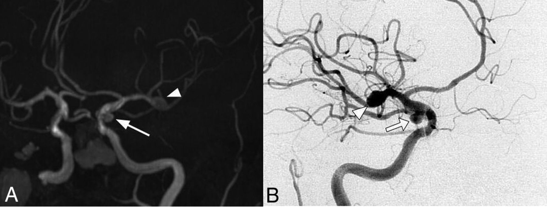
Cerebral MRA (A) and DSA (B) images of a 39-year-old female patient that meet 4 of 4 Curaçao criteria show an ophthalmic segment of a left carotid artery aneurysm (arrowhead) and AVMs in the parietal and occipital contralateral lobes (arrows).

FIG 2.
  • Download figure
  • Open in new tab
  • Download powerpoint
FIG 2.

Cerebral MRA (A) and DSA (B) images of a 78-year-old female patient that meet 4 of 4 Curaçao criteria show a right MCA bifurcation aneurysm (arrowhead) and another one located in the right posterior communicating artery (arrow).

View this table:
  • View inline
  • View popup
Table 2:

Intracranial aneurysm characteristics

AVMs were found in 39 patients; within this subgroup, 7 patients also had IAs, with no statistical association between both vascular malformations. Only 2 IAs were located in the same vascular territory as the AVM. Furthermore, among those patients who had available images for studying aneurysms outside the CNS, no association between IAs and aneurysms (mostly splanchnic) in other body segments was found. There were no differences in either age, sex, Curaçao criteria, hypertension, or tobacco use between patients with and without IAs. Genetic results were available in 81 patients (35.5%), with 39 patients having ENG mutations, 25 having ACVRL1 mutations, and 10 with MADH4 mutations. In 7 patients, genetic tests were noninformative. No statistically significant association was found between patients with and without IAs regarding the distribution of genetic results (Table 3).

View this table:
  • View inline
  • View popup
Table 3:

Bivariate analysis between baseline variables and intracranial aneurysms

DISCUSSION

A high prevalence of IAs among patients with HHT was found in this study. The IA prevalence was also increased in patients with a family history of IA18 and in certain inherited diseases, including autosomal dominant polycystic kidney disease and some connective tissue disorders.19 A retrospective study by Kim et al20 showed that the prevalence of IAs was as follows: 14% among patients with Marfan syndrome, 12% among patients with Ehlers-Danlos syndrome, 11% among patients with neurofibromatosis type 1, and 28% among patients with Loeys-Dietz syndrome. The Loeys-Dietz syndrome is associated with genetic mutations, also involving the TGF-β/BMP pathway, especially in the SMAD2/3 proteins, a pathway that is also altered in HHT.20⇓-22 Moreover, the combined syndrome of juvenile polyposis and HHT syndrome, caused by MADH4 mutations, is also associated with aortic root aneurysms and joint hypermobility.23,24

Numerous reports describe the presence of IAs in patients with HHT,25⇓⇓⇓⇓⇓⇓⇓-33 and many authors include HHT in a group of hereditary diseases that present with a higher prevalence of IAs, such as Marfan, Ehlers-Danlos, and polycystic kidney syndromes and neurofibromatosis,19,34,35 but these studies do not specifically analyze the prevalence of IAs in patients with HHT in a quantitative manner. Several authors have studied the neurologic manifestations of HHT.11,36⇓-38 These researchers describe multiple kinds of BVMs and, second, mention the frequency of IAs, such as Brinjikji et al,38,39 whose findings indicated that the prevalence of IAs is around 2%, similar to that of the general population. There is a specific study that looked for the prevalence of aneurysms in all locations in HHT, which reported a higher prevalence of IAs of around 4%.40 The number of aneurysms in these populations might have been underestimated due to the lack of angiographic studies to evaluate many of these patients.

The prevalence of IAs in the present HHT cohort was 14.5%, whereas the reported prevalence in the general population is close to 3%–5%.18 Most of the aneurysms were small, with a predilection for the anterior circulation, similar to those observed in the general population.15 These findings could correlate with the vascular malformations, which these patients have in multiple organs, even aneurysms in the splanchnic circulation. Considering that BVMs are more frequent in patients with HHT having the ENG mutation,41 a possible link between IAs and genotypes arises, but no statistically significant association was found.

The coexistence of IA and brain AVMs in patients without HHT is estimated to be between 5% and 20%.42 Although the pathogenesis of IAs in these patients is not fully understood, it is believed that it may be related to hemodynamic factors triggered by the presence of a shunt in the AVM.17 AVMs and IAs were both found in 7 patients, but only 2 were in the same vascular territory as the AVM, which may be interpreted as flow-related. Even subtracting these 2 patients, the prevalence would still be high, so the increased prevalence of IAs in HHT could be explained by vascular alterations caused by mutations in genes of the TGFβ/BMP pathway mentioned above. There is evidence pointing to inflammation as a mechanism of aneurysm formation in patients without HHT. The process begins with a hemodynamic or inflammatory insult, with a local increase in matrix metalloproteinases (MMPs) released by macrophages and mast cells, with intimal migration and shape changes of the smooth-muscle cells.43 In HHT, a state of chronic inflammation of the vascular wall, induced by an imbalance between pro- and anti-inflammatory factors of mononuclear cells, has been widely reported. This would explain one of the possible biologic bases to suspect a higher incidence of IAs in HHT.44 In addition, endoglin is involved in cell migration, proliferation, and extracellular matrix remodeling, mechanisms that involve MMPs and macrophages.44,45 Furthermore, endoglin and TGFβ/BMP are involved in inflammation and the tissue-repair processes as well as in angiogenesis. The vascular wall of AVMs in HHT is abnormal and lacks organized mural cell layers such as pericytes and smooth-muscle cells.46 Finally, drugs that inhibit MMPs such as doxycycline are proposed for the treatment of epistaxis in HHT and unruptured IAs.43,47

The present study has research limitations to be acknowledged. First, it is a retrospective study and, thus, though only patients with angiographic studies were included, not all of them were evaluated with the same angiographic imaging technique, and alternative modalities have different sensitivities and specificities to detect IAs. In addition, a small proportion of these patients had a reliable imaging report but no available image for us to re-evaluate. Along the same lines, there were missing data regarding other risk factors for aneurysm development in some patients. Patient genotypes were not available for the entire cohort, and the relationship between IAs and mutations could not be fully studied; therefore, the lack of an association found should be viewed with caution until further studies are developed. Only adult patients were enrolled, as for other BVMs, the prevalence could be similar in the pediatric population, but this issue should be addressed in future studies. Finally, because this study does not have a control group and there is a lack of regional data on IA prevalence, these results have been compared with those reported in other populations.

Patients with HHT are usually evaluated for BVMs with contrast-enhanced MR imaging; however, the results found in this study may suggest adding an angiography sequence to the current screening methodology in search of IAs. The largest meta-analysis on IA rupture risk included data from 8382 patients. This study identified 6 independent risk factors for aneurysm rupture: older than 70 years of age, hypertension, previous SAH from another aneurysm, the size and site of the aneurysm, and the patient's geographic region. The subsequently developed score for prediction of aneurysm rupture risk is based on these 6 key risk factors and provides absolute estimates for a 5-year rupture risk, ranging from 0.3% to ≥15%.48 There were no SAH events in this cohort due to IA rupture, and all patients are under close follow-up and/or have already been treated according to the usual standards of care. Although there is no prospective information about the rate of rupture of IA in patients with HHT, medical follow-up on these patients should be considered to prevent SAH with its high morbimortality rate.

CONCLUSIONS

The present study suggests an increased prevalence of IA in HHT compared with the general population. Considering the morbimortality associated with aneurysm rupture, these findings highlight the importance of conducting larger studies in the future; including pediatric population, evaluating the etiopathogenesis of IAs associated with HHT and discussing the clinical relevance and usefulness of different detection methods, follow-up and currently available treatments.

Footnotes

  • Disclosure forms provided by the authors are available with the full text and PDF of this article at www.ajnr.org.

References

  1. 1.↵
    1. Shovlin CL,
    2. Letarte M
    . Hereditary haemorrhagic telangiectasia and pulmonary arteriovenous malformations: issues in clinical management and review of pathogenic mechanisms. Thorax 1999;54:714–29 doi:10.1136/thx.54.8.714 pmid:10413726
    FREE Full Text
  2. 2.↵
    1. Govani FS,
    2. Shovlin CL
    . Hereditary haemorrhagic telangiectasia: a clinical and scientific review. Eur J Hum Genet 2009;17:860–71 doi:10.1038/ejhg.2009.35 pmid:19337313
    CrossRefPubMed
  3. 3.↵
    1. Shovlin CL
    . Hereditary haemorrhagic telangiectasia: pathophysiology, diagnosis and treatment. Blood Rev 2010;24:203–19 doi:10.1016/j.blre.2010.07.001 pmid:20870325
    CrossRefPubMed
  4. 4.↵
    1. Bernabeu C,
    2. Bayrak-Toydemir P,
    3. McDonald J, et al
    . Potential second-hits in hereditary hemorrhagic telangiectasia. J Clin Med 2020;9:3571 doi:10.3390/jcm9113571 pmid:33167572
    CrossRefPubMed
  5. 5.↵
    1. McDonald J,
    2. Wooderchak-Donahue W,
    3. VanSant Webb C, et al
    . Hereditary hemorrhagic telangiectasia: genetics and molecular diagnostics in a new era. Front Genet 2015;6:1 doi:10.3389/fgene.2015.00001 pmid:25674101
    CrossRefPubMed
  6. 6.↵
    1. Abdalla SA,
    2. Letarte M
    . Hereditary haemorrhagic telangiectasia: current views on genetics and mechanisms of disease. J Med Genet 2006;43:97–110 doi:10.1136/jmg.2005.030833 pmid:15879500
    Abstract/FREE Full Text
  7. 7.↵
    1. Faughnan ME,
    2. Mager JJ,
    3. Hetts SW, et al
    . Second International Guidelines for the Diagnosis and Management of Hereditary Hemorrhagic Telangiectasia. Ann Intern Med 2020;173:989–1001 doi:10.7326/M20-1443 pmid:32894695
    CrossRefPubMed
  8. 8.↵
    1. Serra MM,
    2. Besada CH,
    3. Cabana Cal A, et al
    . Central nervous system manganese induced lesions and clinical consequences in patients with hereditary hemorrhagic telangiectasia. Orphanet J Rare Dis 2017;12:92 doi:10.1186/s13023-017-0632-2 pmid:28521822
    CrossRefPubMed
  9. 9.↵
    1. Klostranec JM,
    2. Chen L,
    3. Mathur S, et al
    . A theory for polymicrogyria and brain arteriovenous malformations in HHT. Neurology 2019;92:34–42 doi:10.1212/WNL.0000000000006686 pmid:30584075
    Abstract/FREE Full Text
  10. 10.↵
    1. Faughnan ME,
    2. Palda VA,
    3. Garcia-Tsao G, et al
    ; HHT Foundation International Guidelines Working Group. International guidelines for the diagnosis and management of hereditary haemorrhagic telangiectasia. J Med Genet 2011;48:73–87 doi:10.1136/jmg.2009.069013 pmid:19553198
    Abstract/FREE Full Text
  11. 11.↵
    1. Fulbright RK,
    2. Chaloupka JC,
    3. Putman CM, et al
    . MR of hereditary hemorrhagic telangiectasia: prevalence and spectrum of cerebrovascular malformations. AJNR Am J Neuroradiol 1998;19:477–84 pmid:9541302
    PubMed
  12. 12.↵
    1. Willemse RB,
    2. Mager JJ,
    3. Westermann CJ, et al
    . Bleeding risk of cerebrovascular malformations in hereditary hemorrhagic telangiectasia. J Neurosurg 2000;92:779–84 doi:10.3171/jns.2000.92.5.0779 pmid:10794291
    CrossRefPubMed
  13. 13.↵
    1. Meybodi AT,
    2. Kim H,
    3. Nelson J, et al
    ; Malformation Consortium HHT Investigator Group. Surgical treatment vs nonsurgical treatment for brain arteriovenous malformations in patients with hereditary hemorrhagic telangiectasia: a retrospective multicenter consortium study. Neurosurgery 2018;82:253 doi:10.1093/neuros/nyx16 pmid:28973426
    CrossRefPubMed
  14. 14.↵
    1. Amenta PS,
    2. Yadla S,
    3. Campbell PG, et al
    . Analysis of nonmodifiable risk factors for intracranial aneurysm rupture in a large, retrospective cohort. Neurosurgery 2012;70:693–701 doi:10.1227/NEU.0b013e3182354d68 pmid:21904261
    CrossRefPubMed
  15. 15.↵
    1. Grobelny TJ
    . Brain aneurysms: epidemiology, treatment options, and milestones of endovascular treatment evolution. Dis Mon 2011;57:647–55 doi:10.1016/j.disamonth.2011.08.022 pmid:22036120
    CrossRefPubMed
  16. 16.↵
    1. Brown RD Jr.,
    2. Wiebers DO,
    3. Forbes GS
    . Unruptured intracranial aneurysms and arteriovenous malformations: frequency of intracranial hemorrhage and relationship of lesions. J Neurosurg 1990;73:859–63 doi:10.3171/jns.1990.73.6.0859 pmid:2230969
    CrossRefPubMed
  17. 17.↵
    1. Rammos SK,
    2. Gardenghi B,
    3. Bortolotti C, et al
    . Aneurysms associated with brain arteriovenous malformations. AJNR Am J Neuroradiol 2016;37:1966–71 doi:10.3174/ajnr.A4869 pmid:27339951
    Abstract/FREE Full Text
  18. 18.↵
    1. Vlak MH,
    2. Algra A,
    3. Brandenburg R, et al
    . Prevalence of unruptured intracranial aneurysms, with emphasis on sex, age, comorbidity, country, and time period: a systematic review and meta-analysis. Lancet Neurol 2011;10:626–36 doi:10.1016/S1474-4422(11)70109-0 pmid:21641282
    CrossRefPubMed
  19. 19.↵
    1. Mohan D,
    2. Munteanu V,
    3. Coman T, et al
    . Genetic factors involves in intracranial aneurysms–actualities. J Med Life 2015;8:336–41 pmid:26351537
    PubMed
  20. 20.↵
    1. Kim ST,
    2. Brinjikji W,
    3. Lanzino G, et al
    . Neurovascular manifestations of connective-tissue diseases: a review. Interv Neuroradiol 2016;22:624–37 doi:10.1177/1591019916659262 pmid:27511817
    CrossRefPubMed
  21. 21.↵
    1. Maleszewski JJ,
    2. Miller DV,
    3. Lu J, et al
    . Histopathologic findings in ascending aortas from individuals with Loeys-Dietz syndrome (LDS). Am J Surg Pathol 2009;33:194–201 doi:10.1097/PAS.0b013e31817f3661 pmid:18852674
    CrossRefPubMed
  22. 22.↵
    1. Kim ST,
    2. Brinjikji W,
    3. Kallmes DF
    . Prevalence of intracranial aneurysms in patients with connective tissue diseases: a retrospective study. AJNR Am J Neuroradiol 2016;37:1422–26 doi:10.3174/ajnr.A4718 pmid:26992822
    Abstract/FREE Full Text
  23. 23.↵
    1. Vorselaars VM,
    2. Diederik A,
    3. Prabhudesai V, et al
    . SMAD4 gene mutation increases the risk of aortic dilation in patients with hereditary haemorrhagic telangiectasia. Int J Cardiol 2017;245:114–18 doi:10.1016/j.ijcard.2017.06.059 pmid:28874282
    CrossRefPubMed
  24. 24.↵
    1. Cavalli G
    . SMAD4 gene mutation and risk of aortic dilation: lessons from hereditary hemorrhagic telangiectasia. Int J Cardiol 2017;245:145–46 doi:10.1016/j.ijcard.2017.07.046 pmid:28874286
    CrossRefPubMed
  25. 25.↵
    1. Ishii D,
    2. Takechi A,
    3. Seyama G, et al
    . Ruptured large basilar artery aneurysm associated with an arteriovenous malformation in hereditary hemorrhagic telangiectasia. Neurol Med Chir (Tokyo) 2012;52:502–05 doi:10.2176/nmc.52.502 pmid:22850500
    CrossRefPubMed
  26. 26.↵
    1. Chick JF,
    2. Sheehan SE,
    3. Chauhan NR
    . The many faces of hereditary hemorrhagic telangiectasia: median arcuate ligament syndrome, arteriovenous malformations, and cerebral aneurysms. Intern Emerg Med 2012;7:385–86 doi:10.1007/s11739-012-0810-0 pmid:22782335
    CrossRefPubMed
  27. 27.↵
    1. Helmchen C,
    2. Nahser HC,
    3. Yousry T, et al
    . Therapy of cerebral aneurysms and arteriovenous vascular malformations in hereditary hemorrhagic telangiectasia (Rendu-Osler-Weber disease) (in German). Nervenarzt 1995;66:124–28 pmid:7715751
    PubMed
  28. 28.↵
    1. Roy C,
    2. Noseda G,
    3. Arzimanoglou A, et al
    . Rendu Osler disease revealed by ruptured cerebral arterial aneurysm in an infant. Arch Fr Pediatr 1990;47:741–42 pmid:2082850
    PubMed
  29. 29.↵
    1. Sobel D,
    2. Norman D
    . CNS manifestations of hereditary hemorrhagic telangiectasia. AJNR Am J Neuroradiol 1984;5:569–73 pmid:6435422
    Abstract/FREE Full Text
  30. 30.↵
    1. Fisher M,
    2. Zito JL
    . Focal cerebral ischemia distal to a cerebral aneurysm in hereditary hemorrhagic telangiectasia. Stroke 1983;14:419–21 doi:10.1161/01.str.14.3.419 pmid:6658911
    Abstract/FREE Full Text
  31. 31.↵
    1. Kamiyama K,
    2. Okada H,
    3. Niizuma H, et al
    . A case report: Osler-Weber-Rendu disease with cerebral aneurysm, cerebral arteriovenous malformation and pulmonary arteriovenous fistula (author's transl). No Shinkei Geka 1981;9:67–72 pmid:7231629
    PubMed
  32. 32.↵
    1. Román G,
    2. Fisher M,
    3. Perl DP, et al
    . Neurological manifestations of hereditary hemorrhagic telangiectasia (Rendu-Osler-Weber disease): report of 2 cases and review of the literature. Ann Neurol 1978;4:130–44 doi:10.1002/ana.410040207 pmid:707984
    CrossRefPubMed
  33. 33.↵
    1. Grollmus J,
    2. Hoff J
    . Multiple aneurysms associated with Osler-Weber-Rendu disease. Surg Neurol 1973;1:91–93 pmid:4772800
    PubMed
  34. 34.↵
    1. Brown RD Jr.,
    2. Huston J,
    3. Hornung R, et al
    . Screening for brain aneurysm in the Familial Intracranial Aneurysm study: frequency and predictors of lesion detection. J Neurosurg 2008;108:1132–38 doi:10.3171/JNS/2008/108/6/1132 pmid:18518716
    CrossRefPubMed
  35. 35.↵
    1. Caranci F,
    2. Briganti F,
    3. Cirillo L, et al
    . Epidemiology and genetics of intracranial aneurysms. Eur J Radiol 2013;82:1598–605 doi:10.1016/j.ejrad.2012.12.026 pmid:23399038
    CrossRefPubMed
  36. 36.↵
    1. Maher CO,
    2. Piepgras DG,
    3. Brown RD Jr., et al
    . Cerebrovascular manifestations in 321 cases of hereditary hemorrhagic telangiectasia. Stroke 2001;32:877–82 doi:10.1161/01.str.32.4.877 pmid:11283386
    Abstract/FREE Full Text
  37. 37.↵
    1. Krings T,
    2. Ozanne A,
    3. Chng SM, et al
    . Neurovascular phenotypes in hereditary haemorrhagic telangiectasia patients according to age: review of 50 consecutive patients aged 1 day-60 years. Neuroradiology 2005;47:711–20 doi:10.1007/s00234-005-1390-8 pmid:16136265
    CrossRefPubMed
  38. 38.↵
    1. Brinjikji W,
    2. Iyer VN,
    3. Yamaki V, et al
    . Neurovascular manifestations of hereditary hemorrhagic telangiectasia: a consecutive series of 376 patients during 15 years. AJNR Am J Neuroradiol 2016;37:1479–86 doi:10.3174/ajnr.A4762 pmid:27012295
    Abstract/FREE Full Text
  39. 39.↵
    1. Woodall MN,
    2. McGettigan M,
    3. Figueroa R, et al
    . Cerebral vascular malformations in hereditary hemorrhagic telangiectasia. J Neurosurg 2014;120:87–92 doi:10.3171/2013.10.JNS122402 pmid:24236651
    CrossRefPubMed
  40. 40.↵
    1. Ring NY,
    2. Latif MA,
    3. Hafezi-Nejad N, et al
    . Prevalence of and factors associated with arterial aneurysms in patients with hereditary hemorrhagic telangiectasia: 17-year retrospective series of 418 patients. J Vasc Interv Radiol 2021;32:1661–69 doi:10.1016/j.jvir.2021.08.018 pmid:34478850
    CrossRefPubMed
  41. 41.↵
    1. Brinjikji W,
    2. Iyer VN,
    3. Wood CP, et al
    . Prevalence and characteristics of brain arteriovenous malformations in hereditary hemorrhagic telangiectasia: a systematic review and meta-analysis. J Neurosurg 2017;127:302–10 doi:10.3171/2016.7.JNS16847 pmid:27767404
    CrossRefPubMed
  42. 42.↵
    1. Cagnazzo F,
    2. Brinjikji W,
    3. Lanzino G
    . Arterial aneurysms associated with arteriovenous malformations of the brain: classification, incidence, risk of hemorrhage, and treatment-a systematic review. Acta Neurochir (Wien) 2016;158:2095–104 doi:10.1007/s00701-016-2957-3 pmid:27644700
    CrossRefPubMed
  43. 43.↵
    1. Chalouhi N,
    2. Hoh BL,
    3. Hasan D
    . Review of cerebral aneurysm formation, growth, and rupture. Stroke 2013;44:3613–22 doi:10.1161/STROKEAHA.113.002390 pmid:24130141
    FREE Full Text
  44. 44.↵
    1. Dingenouts CK,
    2. Goumans M-J,
    3. Bakker W
    . Mononuclear cells and vascular repair in HHT. Front Genet 2015;6:114 doi:10.3389/fgene.2015.00114 pmid:25852751
    CrossRefPubMed
  45. 45.↵
    1. Aristorena M,
    2. Gallardo-Vara E,
    3. Vicen M, et al
    . MMP-12, secreted by pro-inflammatory macrophages, targets endoglin in human macrophages and endothelial cells. Int J Mol Sci 2019;20:3107 doi:10.3390/ijms20123107 pmid:31242676
    CrossRefPubMed
  46. 46.↵
    1. Lebrin F,
    2. Srun S,
    3. Raymond K, et al
    . Thalidomide stimulates vessel maturation and reduces epistaxis in individuals with hereditary hemorrhagic telangiectasia. Nat Med 2010;16:420–48 doi:10.1038/nm.2131 pmid:20364125
    CrossRefPubMed
  47. 47.↵
    1. Wu V,
    2. Lee JM,
    3. Vozoris NT, et al
    . Weekly epistaxis duration as an indicator of epistaxis severity in hereditary hemorrhagic telangiectasia: preliminary results from a randomized controlled trial. Laryngoscope Investig Otolaryngol 2021;6:370–75 doi:10.1002/lio2.561 pmid:34195356
    CrossRefPubMed
  48. 48.↵
    1. Greving JP,
    2. Wermer MJH,
    3. Brown RD Jr., et al
    . Development of the PHASES score for prediction of risk of rupture of intracranial aneurysms: a pooled analysis of six prospective cohort studies. Lancet Neurol 2014;13:59–66 doi:10.1016/S1474-4422(13)70263-1 pmid:24290159
    CrossRefPubMed
  • Received January 30, 2022.
  • Accepted after revision March 17, 2022.
  • © 2022 by American Journal of Neuroradiology
PreviousNext
Back to top

In this issue

American Journal of Neuroradiology: 43 (6)
American Journal of Neuroradiology
Vol. 43, Issue 6
1 Jun 2022
  • Table of Contents
  • Index by author
  • Complete Issue (PDF)
Advertisement
Print
Download PDF
Email Article

Thank you for your interest in spreading the word on American Journal of Neuroradiology.

NOTE: We only request your email address so that the person you are recommending the page to knows that you wanted them to see it, and that it is not junk mail. We do not capture any email address.

Enter multiple addresses on separate lines or separate them with commas.
Prevalence of Intracranial Aneurysms in Hereditary Hemorrhagic Telangiectasia: Report from a Single Reference Center
(Your Name) has sent you a message from American Journal of Neuroradiology
(Your Name) thought you would like to see the American Journal of Neuroradiology web site.
CAPTCHA
This question is for testing whether or not you are a human visitor and to prevent automated spam submissions.
Cite this article
M.S. Perez Akly, C. Vazquez, C.H. Besada, M.J. Rodriguez, M.F. Conde, A.R. Cajal, V.A. Peuchot, D. Dardik, M.M. Baccanelli, M.M. Serra
Prevalence of Intracranial Aneurysms in Hereditary Hemorrhagic Telangiectasia: Report from a Single Reference Center
American Journal of Neuroradiology Jun 2022, 43 (6) 844-849; DOI: 10.3174/ajnr.A7505

Citation Manager Formats

  • BibTeX
  • Bookends
  • EasyBib
  • EndNote (tagged)
  • EndNote 8 (xml)
  • Medlars
  • Mendeley
  • Papers
  • RefWorks Tagged
  • Ref Manager
  • RIS
  • Zotero
0 Responses
Respond to this article
Share
Bookmark this article
Intracranial Aneurysms in HTT
M.S. Perez Akly, C. Vazquez, C.H. Besada, M.J. Rodriguez, M.F. Conde, A.R. Cajal, V.A. Peuchot, D. Dardik, M.M. Baccanelli, M.M. Serra
American Journal of Neuroradiology Jun 2022, 43 (6) 844-849; DOI: 10.3174/ajnr.A7505
del.icio.us logo Twitter logo Facebook logo Mendeley logo
  • Tweet Widget
  • Facebook Like
  • Google Plus One
Purchase

Jump to section

  • Article
    • Abstract
    • ABBREVIATIONS:
    • MATERIALS AND METHODS
    • RESULTS
    • DISCUSSION
    • CONCLUSIONS
    • Footnotes
    • References
  • Figures & Data
  • Supplemental
  • Info & Metrics
  • Responses
  • References
  • PDF

Related Articles

  • PubMed
  • Google Scholar

Cited By...

  • Prevalence and Characteristics of Intracranial Aneurysms in Hereditary Hemorrhagic Telangiectasia
  • Crossref (6)
  • Google Scholar

This article has been cited by the following articles in journals that are participating in Crossref Cited-by Linking.

  • De novo brain vascular malformation in an adult with hereditary hemorrhagic telangiectasia and juvenile polyposis overlap syndrome
    Elisa Guan, Ana Braslavsky, Carolina Vazquez, Cristina H. Besada, Manuel S. Pérez Akly, Oscar Peralta, Ricardo García Mónaco, Nicolás M. Ciarrocchi, Matteo Baccanelli, Marcelo M. Serra
    Journal of Stroke and Cerebrovascular Diseases 2024 33 6
  • Prevalence and Characteristics of Intracranial Aneurysms in Hereditary Hemorrhagic Telangiectasia
    How-Chung Cheng, Marie E. Faughnan, Karel G. terBrugge, Hon-Man Liu, Timo Krings
    American Journal of Neuroradiology 2023 44 12
  • Advances in stem cell therapy for stroke: mechanisms, challenges, and future directions
    Abdullah Md. Sheikh, Sayeed Hossain, Shatera Tabassum
    Regenerative Medicine Reports 2024 1 1
  • Update August 2022
    Francine Blei
    Lymphatic Research and Biology 2022 20 4
  • A Rare Intersection: Mosaic Turner Syndrome and Hereditary Hemorrhagic Telangiectasia
    Diego Mateo Cornejo Gonzalez, Imran Naeem Aziz, Shreeya Atul Sawant, Reem Yousef Darwish, Joseph Guan
    Annals of Internal Medicine: Clinical Cases 2025 4 1
  • Exploring the Landscape of Intracranial Aneurysms in South America: A Comprehensive Narrative Review Intracranial Aneurysms in South America
    Mohammed Dheyaa Marsool Marsool, Hareesha Rishab Bharadwaj, Syed Hasham Ali, Nicholas Aderinto, Muhammad Hamza Shah, Nathanael Shing, Priyal Dalal, Helen Huang, Jack Wellington, Talhah Chaudri, Wireko Andrew Awuah, Niels Pacheco-Barrios, Luis Macha-Quillama, Daniel Fernandez-Guzman, Khabab Abbasher Hussien Mohamed Ahmed
    World Neurosurgery 2024 185

More in this TOC Section

  • Diagnostic Neuroradiology of Monoclonal Antibodies
  • Cerebral ADC Changes in Fabry Disease
  • ML for Glioma Molecular Subtype Prediction
Show more ADULT BRAIN

Similar Articles

Advertisement

Indexed Content

  • Current Issue
  • Accepted Manuscripts
  • Article Preview
  • Past Issues
  • Editorials
  • Editor's Choice
  • Fellows' Journal Club
  • Letters to the Editor
  • Video Articles

Cases

  • Case Collection
  • Archive - Case of the Week
  • Archive - Case of the Month
  • Archive - Classic Case

More from AJNR

  • Trainee Corner
  • Imaging Protocols
  • MRI Safety Corner

Multimedia

  • AJNR Podcasts
  • AJNR Scantastics

Resources

  • Turnaround Time
  • Submit a Manuscript
  • Submit a Video Article
  • Submit an eLetter to the Editor/Response
  • Manuscript Submission Guidelines
  • Statistical Tips
  • Fast Publishing of Accepted Manuscripts
  • Graphical Abstract Preparation
  • Imaging Protocol Submission
  • Evidence-Based Medicine Level Guide
  • Publishing Checklists
  • Author Policies
  • Become a Reviewer/Academy of Reviewers
  • News and Updates

About Us

  • About AJNR
  • Editorial Board
  • Editorial Board Alumni
  • Alerts
  • Permissions
  • Not an AJNR Subscriber? Join Now
  • Advertise with Us
  • Librarian Resources
  • Feedback
  • Terms and Conditions
  • AJNR Editorial Board Alumni

American Society of Neuroradiology

  • Not an ASNR Member? Join Now

© 2025 by the American Society of Neuroradiology All rights, including for text and data mining, AI training, and similar technologies, are reserved.
Print ISSN: 0195-6108 Online ISSN: 1936-959X

Powered by HighWire