Advertisement

Main menu

  • Home
  • Content
    • Current Issue
    • Accepted Manuscripts
    • Article Preview
    • Past Issue Archive
    • Video Articles
    • AJNR Case Collection
    • Case of the Week Archive
    • Case of the Month Archive
    • Classic Case Archive
  • Special Collections
    • AJNR Awards
    • Low-Field MRI
    • Alzheimer Disease
    • ASNR Foundation Special Collection
    • Photon-Counting CT
    • View All
  • Multimedia
    • AJNR Podcasts
    • AJNR SCANtastic
    • Trainee Corner
    • MRI Safety Corner
    • Imaging Protocols
  • For Authors
    • Submit a Manuscript
    • Submit a Video Article
    • Submit an eLetter to the Editor/Response
    • Manuscript Submission Guidelines
    • Statistical Tips
    • Fast Publishing of Accepted Manuscripts
    • Graphical Abstract Preparation
    • Imaging Protocol Submission
    • Author Policies
  • About Us
    • About AJNR
    • Editorial Board
    • Editorial Board Alumni
  • More
    • Become a Reviewer/Academy of Reviewers
    • Subscribers
    • Permissions
    • Alerts
    • Feedback
    • Advertisers
    • ASNR Home

User menu

  • Alerts
  • Log in

Search

  • Advanced search
American Journal of Neuroradiology

American Journal of Neuroradiology

ASHNR American Society of Functional Neuroradiology ASHNR American Society of Pediatric Neuroradiology ASSR
  • Alerts
  • Log in

Advanced Search

  • Home
  • Content
    • Current Issue
    • Accepted Manuscripts
    • Article Preview
    • Past Issue Archive
    • Video Articles
    • AJNR Case Collection
    • Case of the Week Archive
    • Case of the Month Archive
    • Classic Case Archive
  • Special Collections
    • AJNR Awards
    • Low-Field MRI
    • Alzheimer Disease
    • ASNR Foundation Special Collection
    • Photon-Counting CT
    • View All
  • Multimedia
    • AJNR Podcasts
    • AJNR SCANtastic
    • Trainee Corner
    • MRI Safety Corner
    • Imaging Protocols
  • For Authors
    • Submit a Manuscript
    • Submit a Video Article
    • Submit an eLetter to the Editor/Response
    • Manuscript Submission Guidelines
    • Statistical Tips
    • Fast Publishing of Accepted Manuscripts
    • Graphical Abstract Preparation
    • Imaging Protocol Submission
    • Author Policies
  • About Us
    • About AJNR
    • Editorial Board
    • Editorial Board Alumni
  • More
    • Become a Reviewer/Academy of Reviewers
    • Subscribers
    • Permissions
    • Alerts
    • Feedback
    • Advertisers
    • ASNR Home
  • Follow AJNR on Twitter
  • Visit AJNR on Facebook
  • Follow AJNR on Instagram
  • Join AJNR on LinkedIn
  • RSS Feeds

AJNR Awards, New Junior Editors, and more. Read the latest AJNR updates

Research ArticleSpine

Same-Day Bilateral Decubitus CT Myelography for Detecting CSF-Venous Fistulas in Spontaneous Intracranial Hypotension

L. Carlton Jones and P.J. Goadsby
American Journal of Neuroradiology April 2022, 43 (4) 645-648; DOI: https://doi.org/10.3174/ajnr.A7476
L. Carlton Jones
aFrom the Department of Radiology (L.C.J.), Guy’s and St. Thomas’s Hospitals National Health Service Foundation Trust, St. Thomas’ Hospital, London, UK
bDepartment of Neuroradiology (L.C.J.), King’s College Hospital National Health Service Foundation Trust, London, UK
  • Find this author on Google Scholar
  • Find this author on PubMed
  • Search for this author on this site
  • ORCID record for L. Carlton Jones
P.J. Goadsby
cNational Institute for Health Research Wellcome Trust King’s Clinical Research Facility (P.J.G.), King’s College Hospital, London, UK
  • Find this author on Google Scholar
  • Find this author on PubMed
  • Search for this author on this site
  • ORCID record for P.J. Goadsby
  • Article
  • Figures & Data
  • Info & Metrics
  • Responses
  • References
  • PDF
Loading

Article Figures & Data

Figures

  • FIG 1.
    • Download figure
    • Open in new tab
    • Download powerpoint
    FIG 1.

    Left T5 CVF with contralateral venous drainage and subsequent treatment. A, Axial right lateral decubitus CTM image shows dependent layering of contrast on the right side of the subarachnoid space but no leak. B, Axial left lateral decubitus CTM image shows more uniform distribution of contrast within the subarachnoid space following turning of the patient but also abnormal left radicular veins opacified by dense contrast (dashed arrow) and a hyperdense right paravertebral vein (arrow). C, Axial left lateral decubitus CTM MIP image shows transvertebral intraosseous drainage of the CVF to the right side (arrow). D, Axial posttreatment CT shows contrast-opacified fibrin sealant filling the foramen (arrow) and extending into the epidural space of vertebral canal.
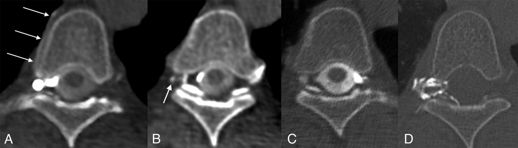
  • FIG 2.
    • Download figure
    • Open in new tab
    • Download powerpoint
    FIG 2.

    Right T5 CVF and subsequent treatment. A, Axial right lateral decubitus CTM image shows a right foraminal meningeal diverticulum and a hyperdense right paraspinal vein (arrows). B, Axial right lateral decubitus CTM image obtained immediately caudal to (A) shows opacification of a small radicular vein (arrow). C, Axial left lateral decubitus CTM image shows no abnormality. D, Axial posttreatment CT shows contrast-opacified fibrin sealant filling the foramen and extending into the epidural space of vertebral canal.
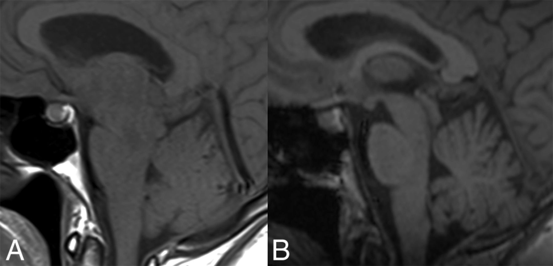
  • FIG 3.
    • Download figure
    • Open in new tab
    • Download powerpoint
    FIG 3.

    Pre- and posttreatment MRIs of the patient in Fig 2. A, Sagittal T1-weighted MR imaging shows brain sag with narrowing of the mamillopontine distance, narrowing of the prepontine cistern, and inferior sloping of the floor of the third ventricle. There is also pituitary enlargement and distension of the straight sinus. B, Resolution of these changes following CT-guided injection of fibrin sealant.

PreviousNext
Back to top

In this issue

American Journal of Neuroradiology: 43 (4)
American Journal of Neuroradiology
Vol. 43, Issue 4
1 Apr 2022
  • Table of Contents
  • Index by author
  • Complete Issue (PDF)
Advertisement
Print
Download PDF
Email Article

Thank you for your interest in spreading the word on American Journal of Neuroradiology.

NOTE: We only request your email address so that the person you are recommending the page to knows that you wanted them to see it, and that it is not junk mail. We do not capture any email address.

Enter multiple addresses on separate lines or separate them with commas.
Same-Day Bilateral Decubitus CT Myelography for Detecting CSF-Venous Fistulas in Spontaneous Intracranial Hypotension
(Your Name) has sent you a message from American Journal of Neuroradiology
(Your Name) thought you would like to see the American Journal of Neuroradiology web site.
CAPTCHA
This question is for testing whether or not you are a human visitor and to prevent automated spam submissions.
Cite this article
L. Carlton Jones, P.J. Goadsby
Same-Day Bilateral Decubitus CT Myelography for Detecting CSF-Venous Fistulas in Spontaneous Intracranial Hypotension
American Journal of Neuroradiology Apr 2022, 43 (4) 645-648; DOI: 10.3174/ajnr.A7476

Citation Manager Formats

  • BibTeX
  • Bookends
  • EasyBib
  • EndNote (tagged)
  • EndNote 8 (xml)
  • Medlars
  • Mendeley
  • Papers
  • RefWorks Tagged
  • Ref Manager
  • RIS
  • Zotero
0 Responses
Respond to this article
Share
Bookmark this article
Same-Day Bilateral Decubitus CT Myelography for Detecting CSF-Venous Fistulas in Spontaneous Intracranial Hypotension
L. Carlton Jones, P.J. Goadsby
American Journal of Neuroradiology Apr 2022, 43 (4) 645-648; DOI: 10.3174/ajnr.A7476
del.icio.us logo Twitter logo Facebook logo Mendeley logo
  • Tweet Widget
  • Facebook Like
  • Google Plus One
Purchase

Jump to section

  • Article
    • Abstract
    • ABBREVIATIONS:
    • MATERIALS AND METHODS
    • RESULTS
    • DISCUSSION
    • CONCLUSIONS
    • Footnotes
    • References
  • Figures & Data
  • Info & Metrics
  • Responses
  • References
  • PDF

Related Articles

  • No related articles found.
  • PubMed
  • Google Scholar

Cited By...

  • Photon-Counting CT Myelography for the Detection of Spinal CSF Leaks
  • Safety and Technical Performance of Bilateral Decubitus CT Myelography Using Standard versus Increased Intrathecal Iodinated Contrast Volume
  • Spinal CSF Leaks: The Neuroradiologist Transforming Care
  • Myelographic Techniques for the Localization of CSF-Venous Fistulas: Updates in 2024
  • Direct comparison of digital subtraction myelography versus CT myelography in lateral decubitus position: evaluation of diagnostic yield for cerebrospinal fluid-venous fistulas
  • Diagnostic Yield of Decubitus CT Myelography for Detection of CSF-Venous Fistulas
  • Lateral Decubitus Dynamic CT Myelography with Real-Time Bolus Tracking (dCTM-BT) for Evaluation of CSF-Venous Fistulas: Diagnostic Yield Stratified by Brain Imaging Findings
  • Diagnostic Performance of Decubitus Photon-Counting Detector CT Myelography for the Detection of CSF-Venous Fistulas
  • Utility of Photon-Counting Detector CT Myelography for the Detection of CSF-Venous Fistulas
  • Utility of Photon-Counting Detector CT Myelography for the Detection of CSF-Venous Fistulas
  • Surgical Ligation of Spinal CSF-Venous Fistulas after Transvenous Embolization in Patients with Spontaneous Intracranial Hypotension
  • Crossref (24)
  • Google Scholar

This article has been cited by the following articles in journals that are participating in Crossref Cited-by Linking.

  • Utility of Photon-Counting Detector CT Myelography for the Detection of CSF-Venous Fistulas
    A.A. Madhavan, L. Yu, W. Brinjikji, J.K. Cutsforth-Gregory, F.R. Schwartz, I.T. Mark, J.C. Benson, T.J. Amrhein
    American Journal of Neuroradiology 2023 44 6
  • Spontaneous intracranial hypotension: updates from diagnosis to treatment
    Paul J. Farnsworth, Ajay A. Madhavan, Jared T. Verdoorn, Darya P. Shlapak, Derek R. Johnson, Jeremy K. Cutsforth-Gregory, Waleed Brinjikji, Vance T. Lehman
    Neuroradiology 2023 65 2
  • Diagnostic Performance of Decubitus Photon-Counting Detector CT Myelography for the Detection of CSF-Venous Fistulas
    Ajay A. Madhavan, Jeremy K. Cutsforth-Gregory, Waleed Brinjikji, Girish Bathla, John C. Benson, Felix E. Diehn, Laurence J. Eckel, Ian T. Mark, Pearse P. Morris, Melissa A. Payne, Jared T. Verdoorn, Nikkole M. Weber, Lifeng Yu, Francis Baffour, Joel G. Fletcher, Cynthia H. McCollough
    American Journal of Neuroradiology 2023 44 12
  • Direct comparison of digital subtraction myelography versus CT myelography in lateral decubitus position: evaluation of diagnostic yield for cerebrospinal fluid-venous fistulas
    Niklas Lützen, Theo Demerath, Urs Würtemberger, Nebiyat Filate Belachew, Enrique Barvulsky Aleman, Katharina Wolf, Amir El Rahal, Florian Volz, Christian Fung, Jürgen Beck, Horst Urbach
    Journal of NeuroInterventional Surgery 2024 16 10
  • Lateral Decubitus Dynamic CT Myelography with Real-Time Bolus Tracking (dCTM-BT) for Evaluation of CSF-Venous Fistulas: Diagnostic Yield Stratified by Brain Imaging Findings
    Thien J. Huynh, Donna Parizadeh, Ahmed K. Ahmed, Christopher T. Gandia, Hal C. Davison, John V. Murray, Ian T. Mark, Ajay A. Madhavan, Darya Shlapak, Todd D. Rozen, Waleed Brinjikji, Prasanna Vibhute, Vivek Gupta, Kacie Brewer, Olga Fermo
    American Journal of Neuroradiology 2024 45 1
  • Surgical Ligation of Spinal CSF-Venous Fistulas after Transvenous Embolization in Patients with Spontaneous Intracranial Hypotension
    W.I. Schievink, R.B. Tache, M.M. Maya
    American Journal of Neuroradiology 2022 43 7
  • Diagnostic Yield of Decubitus CT Myelography for Detection of CSF-Venous Fistulas
    Jacob T. Gibby, Timothy J. Amrhein, Derek S. Young, Jessica L. Houk, Peter G. Kranz
    American Journal of Neuroradiology 2024 45 10
  • Myelographic Techniques for the Localization of CSF-Venous Fistulas: Updates in 2024
    Ajay A. Madhavan, Waleed Brinjikji, Jeremy K. Cutsforth-Gregory, Timothy J. Amrhein, Peter G. Kranz, John C. Benson, Felix E. Diehn, Ben A. Johnson-Tesch, Greta B. Liebo, Vance T. Lehman, Ian T. Mark, Pearse P. Morris, Michael P. Oien, Darya P. Shlapak, Jared T. Verdoorn
    American Journal of Neuroradiology 2024 45 10
  • CSF-venous leak responsible for spontaneous intracranial hypotension treated by endovascular venous route: First cases in Italy
    Mauro Bergui, Francesco Mistretta, Giovanni Bosco, Giacomo Cester, Franco Chioffi, Andrea Gambino, Stefano Molinaro, Riccardo Russo, Gianni Sorarù, Francesco Causin
    Interventional Neuroradiology 2024 30 3
  • Cerebrospinal Fluid Leaks: Challenges in Localizing Spontaneous Spinal Leak Sites and Minimally Invasive Treatment
    Jeffrey Sung, Matthew Lin, Alexandra Giantini-Larsen, Andrew Kim, David Edasery, Michelle Roytman, Sara Strauss, Andrew D. Schweitzer, John K. Park, Gayle Salama
    World Neurosurgery 2024 187

More in this TOC Section

  • Bern Score Validity for SIH
  • MP2RAGE 7T in MS Lesions of the Cervical Spine
  • Resisted Inspiration for CSF-Venous Fistula
Show more Spine

Similar Articles

Advertisement

Indexed Content

  • Current Issue
  • Accepted Manuscripts
  • Article Preview
  • Past Issues
  • Editorials
  • Editor's Choice
  • Fellows' Journal Club
  • Letters to the Editor
  • Video Articles

Cases

  • Case Collection
  • Archive - Case of the Week
  • Archive - Case of the Month
  • Archive - Classic Case

More from AJNR

  • Trainee Corner
  • Imaging Protocols
  • MRI Safety Corner
  • Book Reviews

Multimedia

  • AJNR Podcasts
  • AJNR Scantastics

Resources

  • Turnaround Time
  • Submit a Manuscript
  • Submit a Video Article
  • Submit an eLetter to the Editor/Response
  • Manuscript Submission Guidelines
  • Statistical Tips
  • Fast Publishing of Accepted Manuscripts
  • Graphical Abstract Preparation
  • Imaging Protocol Submission
  • Evidence-Based Medicine Level Guide
  • Publishing Checklists
  • Author Policies
  • Become a Reviewer/Academy of Reviewers
  • News and Updates

About Us

  • About AJNR
  • Editorial Board
  • Editorial Board Alumni
  • Alerts
  • Permissions
  • Not an AJNR Subscriber? Join Now
  • Advertise with Us
  • Librarian Resources
  • Feedback
  • Terms and Conditions
  • AJNR Editorial Board Alumni

American Society of Neuroradiology

  • Not an ASNR Member? Join Now

© 2025 by the American Society of Neuroradiology All rights, including for text and data mining, AI training, and similar technologies, are reserved.
Print ISSN: 0195-6108 Online ISSN: 1936-959X

Powered by HighWire