
Article Figures & Data
Figures
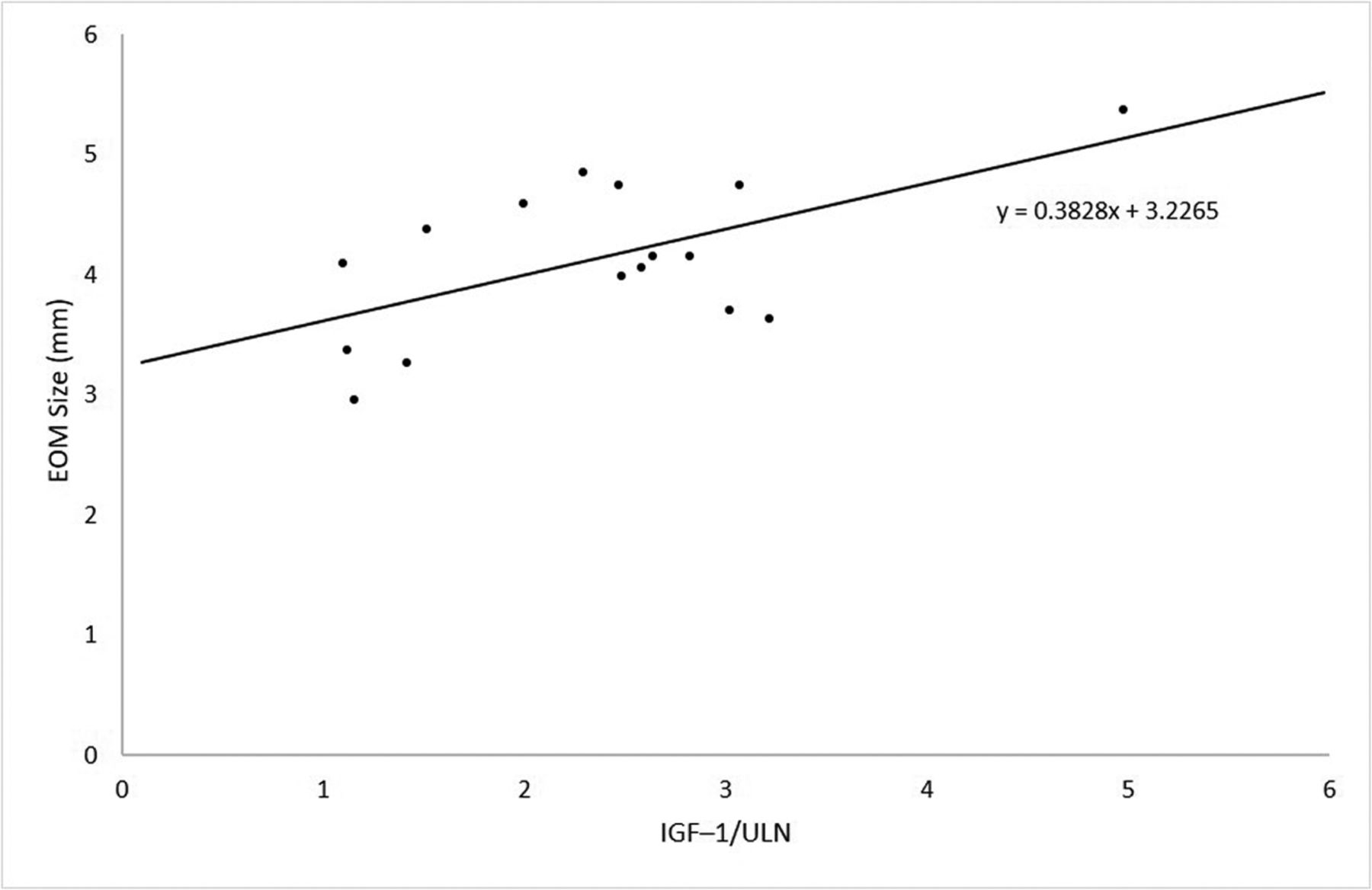
- FIG 1.
In patients with GH-secretory adenoma, EOM size is positively correlated with IGF-1/ULN at diagnosis. The Pearson correlation of the relationship is 0.599 (P = .014).
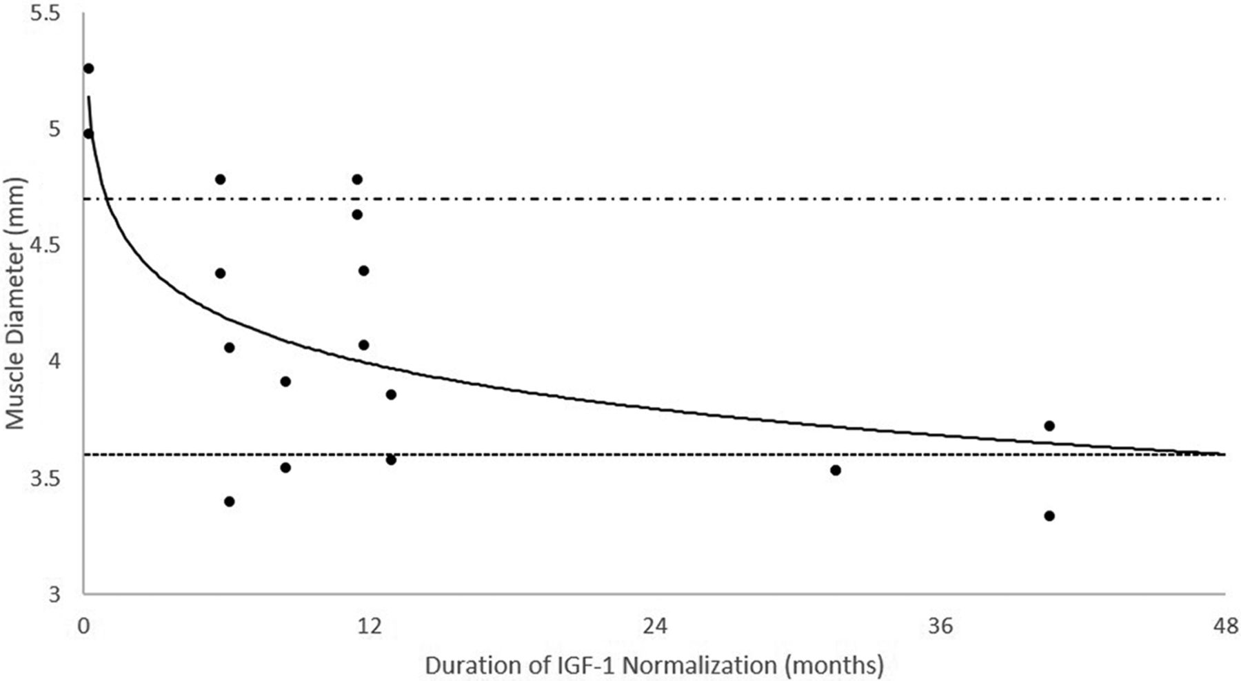
- FIG 2.
EOM size in patients who achieved normalization of the IGF-1 before their most recent MR imaging. The average EOM size appears to decrease as the duration (months) of IGF-1 normalization increases. The alternating dashed/dotted line represents the average EOM size (4.7 mm) in patients who did not achieve IGF-1 normalization before their most recent MR imaging. The dashed line represents that average EOM size (3.6 mm) in patients without acromegaly at the time of their most recent MR imaging.


Tables
- Table 1:
EOM sizes at the time of diagnosis and at time of most recent MR imaging evaluation of patients with GH-secreting pituitary adenomas compared with nonsecretory pituitary adenomas
Characteristic GH-secretory (95% CI) Nonsecretory (95% CI) Difference (95% CI) P Value Initial size (mm) Superior rectus 4.7 (4.4–5.0) 4.0 (3.6–4.3) 0.7 (0.3–1.2) .002 Medial rectus 4.8 (4.4–5.2) 3.7 (3.4–3.9) 1.2 (0.7–1.6) <.001 Lateral rectus 4.5 (4.1–4.8) 3.6 (3.3–3.9) 0.9 (0.5–1.4) <.001 Inferior rectus 4.8 (4.4–5.3) 4.0 (3.8–4.3) 0.8 (0.3–1.3) .003 Average 4.7 (4.4–4.9) 3.8 (3.6–3.9) 0.9 (0.6–1.2) <.001 Final size (mm) Superior rectus 4.3 (4.0–4.6) 3.9 (3.5–4.2) 0.4 (−0.1–0.9) .088 Medial rectus 4.6 (4.3–5.0) 3.5 (3.2–3.7) 1.1 (0.8–1.6) <.001 Lateral rectus 3.9 (3.6–4.2) 3.5 (3.2–3.7) 0.4 (0.1–0.8) .026 Inferior rectus 4.2 (3.8–4.6) 3.7 (3.5–4.0) 0.5 (0.0–0.9) .048 Average 4.3 (4.0–4.5) 3.6 (3.5–3.8) 0.7 (0.4–0.9) <.001 - Table 2:
Change in size of EOMs with time of patients with GH-secreting pituitary adenomas
Muscle Initial Size (95% CI) Final Size (95% CI) Change (95% CI) P Value Superior rectus (mm) 4.7 (4.4–5.0) 4.3 (4.0–4.6) −0.4 (−0.9–0.0) .075 Medial rectus (mm) 4.8 (4.4–5.2) 4.6 (4.3–5.0) −0.2 (−0.7–0.4) .546 Lateral rectus (mm) 4.5 (4.1–4.8) 3.9 (3.6–4.2) −0.6 (−1.0–−0.1) .013 Inferior rectus (mm) 4.8 (4.4–5.3) 4.2 (3.8–4.6) −0.7 (−1.2–−0.1) .024 Average (mm) 4.7 (4.4–4.9) 4.3 (4.0–4.5) −0.4 (−0.7–0.0) .037 - Table 3:
Patients with GH-secretory pituitary adenomas who did or did not achieve a normalized IGF-1 level before their most recent MR imaging
Characteristic Normalized IGF-1 (95% CI) (n = 9) Elevated IGF-1 (95% CI) (n = 7) Difference (95% CI) P Value Extraocular muscle size at most recent MR imaging (mm) 3.9 (3.6–4.2) 4.7 (4.3–5.0) 0.7 (0.3–1.2) <.001 Average IGF-1/ULN at diagnosis 1.9 (1.5–2.3) 2.8 (2.3–3.4) 0.9 (0.2–1.5) .010 Size of pituitary adenoma (mm) 19.1 (15.9–22.3) 23.9 (17.4–30.3) 4.7 (−2.1–11.6) .170 Average duration of elevated IGF-1 (mo) 20.4 (12.4–28.5) 39.3 (18.8–59.9) 18.9 (−2.3–40.1) .078 Average time between initial and final MR imaging (mo) 52.8 (26.1–79.4) 39.9 (19.7–60.1) −12.9 (−45.0–19.1) .417


