Advertisement

Main menu

  • Home
  • Content
    • Current Issue
    • Accepted Manuscripts
    • Article Preview
    • Past Issue Archive
    • Video Articles
    • AJNR Case Collection
    • Case of the Week Archive
    • Case of the Month Archive
    • Classic Case Archive
  • Special Collections
    • AJNR Awards
    • Low-Field MRI
    • Alzheimer Disease
    • ASNR Foundation Special Collection
    • Photon-Counting CT
    • View All
  • Multimedia
    • AJNR Podcasts
    • AJNR SCANtastic
    • Trainee Corner
    • MRI Safety Corner
    • Imaging Protocols
  • For Authors
    • Submit a Manuscript
    • Submit a Video Article
    • Submit an eLetter to the Editor/Response
    • Manuscript Submission Guidelines
    • Statistical Tips
    • Fast Publishing of Accepted Manuscripts
    • Graphical Abstract Preparation
    • Imaging Protocol Submission
    • Author Policies
  • About Us
    • About AJNR
    • Editorial Board
    • Editorial Board Alumni
  • More
    • Become a Reviewer/Academy of Reviewers
    • Subscribers
    • Permissions
    • Alerts
    • Feedback
    • Advertisers
    • ASNR Home

User menu

  • Alerts
  • Log in

Search

  • Advanced search
American Journal of Neuroradiology

American Journal of Neuroradiology

ASHNR American Society of Functional Neuroradiology ASHNR American Society of Pediatric Neuroradiology ASSR
  • Alerts
  • Log in

Advanced Search

  • Home
  • Content
    • Current Issue
    • Accepted Manuscripts
    • Article Preview
    • Past Issue Archive
    • Video Articles
    • AJNR Case Collection
    • Case of the Week Archive
    • Case of the Month Archive
    • Classic Case Archive
  • Special Collections
    • AJNR Awards
    • Low-Field MRI
    • Alzheimer Disease
    • ASNR Foundation Special Collection
    • Photon-Counting CT
    • View All
  • Multimedia
    • AJNR Podcasts
    • AJNR SCANtastic
    • Trainee Corner
    • MRI Safety Corner
    • Imaging Protocols
  • For Authors
    • Submit a Manuscript
    • Submit a Video Article
    • Submit an eLetter to the Editor/Response
    • Manuscript Submission Guidelines
    • Statistical Tips
    • Fast Publishing of Accepted Manuscripts
    • Graphical Abstract Preparation
    • Imaging Protocol Submission
    • Author Policies
  • About Us
    • About AJNR
    • Editorial Board
    • Editorial Board Alumni
  • More
    • Become a Reviewer/Academy of Reviewers
    • Subscribers
    • Permissions
    • Alerts
    • Feedback
    • Advertisers
    • ASNR Home
  • Follow AJNR on Twitter
  • Visit AJNR on Facebook
  • Follow AJNR on Instagram
  • Join AJNR on LinkedIn
  • RSS Feeds

AJNR Awards, New Junior Editors, and more. Read the latest AJNR updates

Research ArticleExtracranial Vascular
Open Access

Proximal Region of Carotid Atherosclerotic Plaque Shows More Intraplaque Hemorrhage: The Plaque at Risk Study

G.A.J.C. Crombag, M. Aizaz, F.H.B.M. Schreuder, F. Benali, D.H.K. van Dam-Nolen, M.I. Liem, C. Lucci, A.F. van der Steen, M.J.A.P. Daemen, W.H. Mess, A. van der Lugt, P.J. Nederkoorn, J. Hendrikse, P.A.M. Hofman, R.J. van Oostenbrugge, J.E. Wildberger and M.E. Kooi
American Journal of Neuroradiology February 2022, 43 (2) 265-271; DOI: https://doi.org/10.3174/ajnr.A7384
G.A.J.C. Crombag
aFrom the Departments of Radiology and Nuclear Medicine (G.A.J.C.C., M.A., F.B., P.A.M.H., J.E.W., M.E.K.)
dCARIM School for Cardiovascular Diseases (G.A.J.C.C., M.A., R.J.v.O., J.E.W., M.E.K.), Maastricht University, Maastricht, the Netherlands
  • Find this author on Google Scholar
  • Find this author on PubMed
  • Search for this author on this site
  • ORCID record for G.A.J.C. Crombag
M. Aizaz
aFrom the Departments of Radiology and Nuclear Medicine (G.A.J.C.C., M.A., F.B., P.A.M.H., J.E.W., M.E.K.)
dCARIM School for Cardiovascular Diseases (G.A.J.C.C., M.A., R.J.v.O., J.E.W., M.E.K.), Maastricht University, Maastricht, the Netherlands
  • Find this author on Google Scholar
  • Find this author on PubMed
  • Search for this author on this site
  • ORCID record for M. Aizaz
F.H.B.M. Schreuder
eDepartment of Neurology & Donders Institute for Brain Cognition & Behaviour (F.H.B.M.S.), Radboud University Medical Center, Nijmegen, the Netherlands
  • Find this author on Google Scholar
  • Find this author on PubMed
  • Search for this author on this site
  • ORCID record for F.H.B.M. Schreuder
F. Benali
aFrom the Departments of Radiology and Nuclear Medicine (G.A.J.C.C., M.A., F.B., P.A.M.H., J.E.W., M.E.K.)
  • Find this author on Google Scholar
  • Find this author on PubMed
  • Search for this author on this site
  • ORCID record for F. Benali
D.H.K. van Dam-Nolen
fDepartments of Radiology and Nuclear Medicine (D.H.K.v.D.-N., A.v.d.L.)
  • Find this author on Google Scholar
  • Find this author on PubMed
  • Search for this author on this site
  • ORCID record for D.H.K. van Dam-Nolen
M.I. Liem
hDepartments of Neurology (M.I.L., P.J.N.)
  • Find this author on Google Scholar
  • Find this author on PubMed
  • Search for this author on this site
  • ORCID record for M.I. Liem
C. Lucci
jDepartment of Radiology (C.L., J.H.), University Medical Center Utrecht, Utrecht, the Netherlands
  • Find this author on Google Scholar
  • Find this author on PubMed
  • Search for this author on this site
  • ORCID record for C. Lucci
A.F. van der Steen
gBiomedical Engineering (A.F.v.d.S.), Erasmus University Medical Center, University Medical Center Rotterdam, Rotterdam, the Netherlands
  • Find this author on Google Scholar
  • Find this author on PubMed
  • Search for this author on this site
  • ORCID record for A.F. van der Steen
M.J.A.P. Daemen
iPathology (M.J.A.P.D.), Amsterdam University Medical Centres, University of Amsterdam, Amsterdam, the Netherlands
  • Find this author on Google Scholar
  • Find this author on PubMed
  • Search for this author on this site
  • ORCID record for M.J.A.P. Daemen
W.H. Mess
bClinical Neurophysiology (W.H.M.)
  • Find this author on Google Scholar
  • Find this author on PubMed
  • Search for this author on this site
  • ORCID record for W.H. Mess
A. van der Lugt
fDepartments of Radiology and Nuclear Medicine (D.H.K.v.D.-N., A.v.d.L.)
  • Find this author on Google Scholar
  • Find this author on PubMed
  • Search for this author on this site
  • ORCID record for A. van der Lugt
P.J. Nederkoorn
hDepartments of Neurology (M.I.L., P.J.N.)
  • Find this author on Google Scholar
  • Find this author on PubMed
  • Search for this author on this site
  • ORCID record for P.J. Nederkoorn
J. Hendrikse
jDepartment of Radiology (C.L., J.H.), University Medical Center Utrecht, Utrecht, the Netherlands
  • Find this author on Google Scholar
  • Find this author on PubMed
  • Search for this author on this site
  • ORCID record for J. Hendrikse
P.A.M. Hofman
aFrom the Departments of Radiology and Nuclear Medicine (G.A.J.C.C., M.A., F.B., P.A.M.H., J.E.W., M.E.K.)
  • Find this author on Google Scholar
  • Find this author on PubMed
  • Search for this author on this site
  • ORCID record for P.A.M. Hofman
R.J. van Oostenbrugge
dCARIM School for Cardiovascular Diseases (G.A.J.C.C., M.A., R.J.v.O., J.E.W., M.E.K.), Maastricht University, Maastricht, the Netherlands
cNeurology (R.J.v.O.), Maastricht University Medical Center, Maastricht, the Netherlands
  • Find this author on Google Scholar
  • Find this author on PubMed
  • Search for this author on this site
  • ORCID record for R.J. van Oostenbrugge
J.E. Wildberger
aFrom the Departments of Radiology and Nuclear Medicine (G.A.J.C.C., M.A., F.B., P.A.M.H., J.E.W., M.E.K.)
dCARIM School for Cardiovascular Diseases (G.A.J.C.C., M.A., R.J.v.O., J.E.W., M.E.K.), Maastricht University, Maastricht, the Netherlands
  • Find this author on Google Scholar
  • Find this author on PubMed
  • Search for this author on this site
  • ORCID record for J.E. Wildberger
M.E. Kooi
aFrom the Departments of Radiology and Nuclear Medicine (G.A.J.C.C., M.A., F.B., P.A.M.H., J.E.W., M.E.K.)
dCARIM School for Cardiovascular Diseases (G.A.J.C.C., M.A., R.J.v.O., J.E.W., M.E.K.), Maastricht University, Maastricht, the Netherlands
  • Find this author on Google Scholar
  • Find this author on PubMed
  • Search for this author on this site
  • ORCID record for M.E. Kooi
  • Article
  • Figures & Data
  • Supplemental
  • Info & Metrics
  • Responses
  • References
  • PDF
Loading

Abstract

BACKGROUND AND PURPOSE: Intraplaque hemorrhage contributes to lipid core enlargement and plaque progression, leading to plaque destabilization and stroke. The mechanisms that contribute to the development of intraplaque hemorrhage are not completely understood. A higher incidence of intraplaque hemorrhage and thin/ruptured fibrous cap (upstream of the maximum stenosis in patients with severe [≥70%] carotid stenosis) has been reported. We aimed to noninvasively study the distribution of intraplaque hemorrhage and a thin/ruptured fibrous cap in patients with mild-to-moderate carotid stenosis.

MATERIALS AND METHODS: Eighty-eight symptomatic patients with stroke (<70% carotid stenosis included in the Plaque at Risk study) demonstrated intraplaque hemorrhage on MR imaging in the carotid artery plaque ipsilateral to the side of TIA/stroke. The intraplaque hemorrhage area percentage was calculated. A thin/ruptured fibrous cap was scored by comparing pre- and postcontrast black-blood TSE images. Differences in mean intraplaque hemorrhage percentages between the proximal and distal regions were compared using a paired-samples t test. The McNemar test was used to reveal differences in proportions of a thin/ruptured fibrous cap.

RESULTS: We found significantly larger areas of intraplaque hemorrhage in the proximal part of the plaque at 2, 4, and 6 mm from the maximal luminal narrowing, respectively: 14.4% versus 9.6% (P = .04), 14.7% versus 5.4% (P < .001), and 11.1% versus 2.2% (P = .001). Additionally, we found an increased proximal prevalence of a thin/ruptured fibrous cap on MR imaging at 2, 4, 6, and 8 mm from the MR imaging section with the maximal luminal narrowing, respectively: 33.7% versus 18.1%, P = .007; 36.1% versus 7.2%, P < .001; 33.7% versus 2.4%, P = .001; and 30.1% versus 3.6%, P = .022.

CONCLUSIONS: We demonstrated that intraplaque hemorrhage and a thin/ruptured fibrous cap are more prevalent on the proximal side of the plaque compared with the distal side in patients with mild-to-moderate carotid stenosis.

ABBREVIATIONS:

FC
fibrous cap
IPH
intraplaque hemorrhage
QIR TSE
quadruple inversion recovery turbo spin-echo
TRFC
thin/ruptured fibrous cap

Rupture of a vulnerable atherosclerotic plaque is an important underlying cause of myocardial infarction and stroke.1 Noninvasive visualization of plaque vulnerability has demonstrated that intraplaque hemorrhage (IPH) contributes to enlargement of the lipid core and plaque progression, leading to plaque destabilization.2,3 Indeed, we and others have demonstrated that IPH on MR imaging is a strong predictor of future cerebrovascular events.4⇓-6 This finding has led to the recognition of IPH as a key marker and pathologic factor contributing to plaque vulnerability.

However, the underlying mechanisms of IPH development are not completely understood. Most often, leakage of erythrocytes from intraplaque microvessels is proposed as a cause for IPH.7,8 In contrast, we recently observed fewer microvessels in plaques with IPH,9 suggesting that damage to the plaque luminal surface such as fissures and rupture of the fibrous cap (FC) could contribute to IPH.8,10 It was demonstrated in histologic specimens that IPH occurs more frequently in the proximal part of the plaque,11,12 where the biomechanical wall stress is usually higher,13⇓⇓-16 due to arterial pressure wave reflection. In a histopathologic study, we demonstrated that IPH occurred in regions with FC fissures and juxtaluminal calcifications. Juxtaluminal calcifications may lead to increased biomechanical wall stress.17 Fissures were found in 58% of plaques with grossly intact luminal surfaces. Most of the fissures (88%) occurred in the proximal region of the plaque. The fissures were connected to IPH (92%) and calcifications (43%), while they were connected to microvessels in only 25% of cases.17

Previous histologic studies were dependent on patient populations that underwent carotid endarterectomy, usually patients with severe carotid stenosis. MR imaging allows investigation of carotid plaques, even in patients with a lower grade of stenosis in which the arterial pressure wave reflection will be less severe.18⇓-20

This study aimed to investigate, noninvasively, whether there is a difference in volume of IPH and the status of the FC in the proximal-versus-distal regions in a relatively large group of patients with stroke with mild-to-moderate carotid stenosis and IPH.

MATERIALS AND METHODS

Study Design

Baseline MR imaging data were analyzed from patients included in the Plaque at Risk (PARISK) study that demonstrated IPH. PARISK is a large, prospective, multicenter cohort study investigating whether plaque imaging enables us to better identify patients with carotid stenosis who have an increased stroke risk.21 Patients with a recent (<3 months) TIA or ischemic stroke in the anterior circulation and carotid artery plaque of >2 mm but <70% stenosis ipsilateral to the side of TIA/stroke were prospectively included. The degree of stenosis was determined with Doppler sonography or with CTA. An upper cutoff value of 70% was used on the basis of the NASCET criteria.22 Institutional medical ethical committee approval was obtained, and all patients provided written informed consent.

MRI

The MR imaging protocol has been described in detail previously.21

In brief, multisequence carotid MR imaging was performed on a 3T whole-body scanner (Achieva or Ingenia, Philips Healthcare, or Discovery MR 750, GE Healthcare). A dedicated 8-channel phased-array coil (Shanghai Chenguang Medical Technologies) or a 4-channel carotid coil (PACC-ST30, Machnet B.V. Roden, the Netherlands) was used. Dedicated vessel wall image-analysis software (VesselMass; Leiden University Medical Center, the Netherlands) was used to analyze the MR images of the ipsilateral carotid plaque. After an extensive training period and demonstrating good interobserver agreement with a validation set that was previously delineated in consensus by experts (with >7 and >10 years of experience, respectively), the trained observers manually delineated the outer vessel wall, luminal area, and plaque components as previously described.23 Image quality was rated on a 5-point scale.24 Patients were excluded if the mean image quality score was <2.

The vessel wall and luminal area were delineated on the precontrast T1-weighted double inversion recovery FSE images (center 2) or the quadruple inversion recovery turbo spin-echo (QIR TSE) (center 1, 3, and 4) images. IPH delineation was performed on T1-weighted inversion recovery turbo field echo images (center 1, 3, and 4) or 3D T1-weighted fat-suppressed spoiled gradient-echo images (center 2).

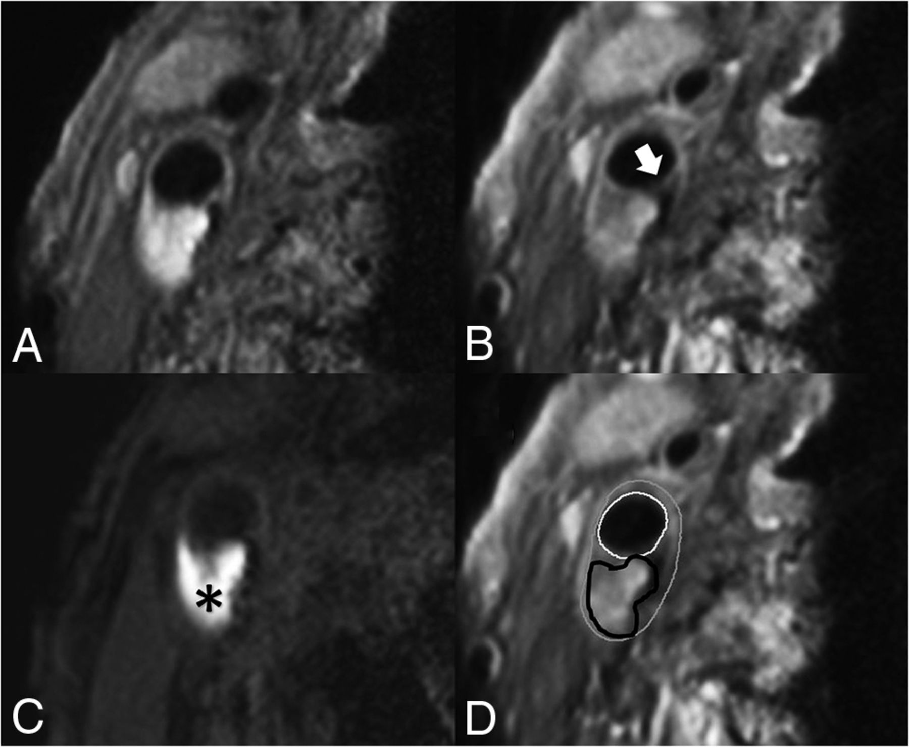
IPH was delineated by visual observation and was defined as hyperintense signal within the bulk of the plaque compared with the adjacent sternocleidomastoid muscle and was manually delineated per section by the trained observers. FC status was dichotomized as thick versus thin or ruptured.25 When a continuous signal enhancement on the postcontrast images between the lipid rich necrotic core and the lumen was identified, the FC status was classified as “intact and thick.” When no or an interrupted area of enhancement was identified, the FC status was classified as “thin and/or ruptured.” In slices without a lipid rich necrotic core, there is no interrupted area of signal enhancement; therefore, the status of the FC is scored by definition as “intact and thick” (Fig 1).

FIG 1.
  • Download figure
  • Open in new tab
  • Download powerpoint
FIG 1.

Transversal MR images of carotid plaque in the right carotid artery. We acquired the following MR images: precontrast T1-weighted QIR TSE (A), postcontrast T1-weighted QIR TSE (B), T1-weighted inversion recovery turbo field echo (C). D, The same image as in B, including the contours delineating intraplaque hemorrhage and the inner and outer vessel wall: white = lumen, gray = outer vessel wall, black = intraplaque hemorrhage. C, A hyperintense signal in the bulk of the plaque can be clearly observed, indicating the presence of intraplaque hemorrhage (black asterisk). B, An interruption of the signal enhancement adjoining the lumen can be observed, indicating a TRFC (white arrow).

Previously, we have shown that our method of scoring IPH demonstrated good agreement between MR imaging and histology.26 Interobserver reproducibility for the detection of IPH was very good (κ = 0.86). We also demonstrated an overall very good interobserver reproducibility of MR image analysis (for carotid lumen volume, wall volume, and total vessel volume: intraclass correlation coefficient/coefficient of variation = 0.96/7.7%, 0.95/8.8%, and 0.92/12.8%, respectively).27 Interobserver reproducibility of FC status assessment was good (κ = 0.60–0.71).28 Also, we have previously shown a very good overall interscan/intraobserver reproducibility in a study in which patients were scanned twice within a mean of 4 (SD, 2.9)  days (for carotid lumen volume and wall volume: intraclass coefficient/coefficient of variation  = 0.99/7.2% and 0.99/7.1%, respectively).27 The interscan/intraobserver reproducibility for the detection of IPH and FC status were very good (κ =1.00).27 On the basis of these delineations, the dedicated vessel wall imaging software package automatically calculates the luminal and vessel wall area and the areas of each plaque component per section. Using the vessel wall and IPH areas, we calculated the percentage of IPH of the total vessel wall area per section. Starting at the section with the narrowest lumen, the IPH area and the presence of a thin/ruptured fibrous cap (TRFC) were determined for the neighboring proximal and distal slices at an interval of 2 mm (ie, the MR imaging section thickness) (Fig 2).

FIG 2.
  • Download figure
  • Open in new tab
  • Download powerpoint
FIG 2.

A patient’s dataset acquired with an inversion recovery turbo field echo sequence. Delineation of the inner and outer vessel walls and IPH in every 2-mm-thick MR imaging section proximal and distal to the section with the narrowest lumen (0 mm). Gray outline = outer vessel wall, white outline = inner vessel wall, black outline= IPH, white arrow (−6 mm to +2 mm) = MR imaging slices in which the FC status is scored as thin or ruptured. The status of the FC was determined by using the postcontrast T1-weighted QIR TSE sequence. There is a larger volume of IPH at the proximal side of the section with the narrowest lumen (0 mm), and most of the slices with a TRFC cap are also located at the proximal side.

Plaque Composition in Proximal-versus-Distal Regions

The proximal region of the plaque is defined as the region proximal to the imaging section with the narrowest lumen (lumen with the absolute lowest area based on luminal contour). The vessel wall area was calculated for each MR imaging section. We generated a histogram of the distribution of the mean IPH area percentage. In addition, we also calculated differences in absolute areas of IPH between the proximal and distal parts of the plaque. The distribution of MR imaging slices with a TRFC at each MR imaging section position proximal and distal to the MR imaging section with the narrowest lumen was also assessed. Section positions with data from <25 patients are excluded in these histograms because of the small statistical power.

Statistical Analyses

Statistical analyses were performed using SPSS 24.0 (IBM). A P value < .05 was considered statistically significant. To analyze the differences between proximal and distal areas of IPH, we used a paired t test to compare the slices proximal (–n) and distal (+n) to the section with the narrowest lumen (ie, Section 0). The paired t test was also used to test the overall difference in absolute IPH area between the proximal and distal regions. A McNemar test was used to analyze differences in proportions of the FC status between the proximal and distal slices, per section and overall per patient.

RESULTS

Of 244 included patients, 6 patients withdrew from the study. Of the remaining 238 patients, another 14 patients were excluded due to bad quality scores of the MR imaging (n = 4), incomplete MR imaging protocol (n = 2), or absence of MR imaging data due to claustrophobia (n = 6) or obesity (n = 2). Of the remaining 224 patients, 88 patients (39%) demonstrated IPH on the ipsilateral side. Baseline patient characteristics are shown in the Online Supplemental Data. For the analysis of the FC status, another 5 patients were excluded because no postcontrast MR imaging was available (Online Supplemental Data).

The mean IPH area percentage was significantly larger in the proximal part of the plaque (Fig 3A). The largest mean IPH area percentage is observed 4 mm proximal to the narrowest lumen. The proximal-versus-distal area percentages of IPH are at 2, 4, and 6 mm from the narrowest lumen, respectively: 14.4% versus 9.6% (P = .04), 14.7% versus 5.4% (P < .001), and 11.1% versus 2.2% (P = .001). The MR images at 8 and 10 mm from the narrowest lumen also show a higher mean area percentage of IPH proximal compared with distal; however, this difference was not significant (7.5% versus 2.2%, P = .056; 5% versus 1.1%, P = .077) (Table 1). Also, the absolute IPH area in the proximal region was significantly higher compared with the distal region at 2, 4, 6, 8, and 10 mm from the narrowest lumen, respectively (0.11 versus 0.07 cm2, P = .019; 0.12 versus 0.04 cm2, P < .001; 0.10 versus 0.01 cm2, P < .001; 0.06 versus 0.01 cm2, P = .005; and 0.03 versus 0.0003 cm2, P = .004).

FIG 3.
  • Download figure
  • Open in new tab
  • Download powerpoint
FIG 3.

A, Histogram showing significantly larger mean IPH areas in the proximal region of the plaque compared with the distal region. Mean IPH area percentages are shown for each section in relation to the smallest lumen. The white bar indicates the section with the narrowest lumen (distance = 0 mm), the bars on the left with the negative numbers are slices proximal (proximally), and the bars on the right with the positive numbers indicate the slices distal to the smallest lumen. Each section has a thickness of 2 mm. An asterisk indicates a statistically significant difference (P < .05) between the proximal and distal slices using a paired t test. B, Prevalence of a TRFC plotted for each section position with respect to the section with the smallest lumen (0). The bars on the left with the negative numbers are slices proximal, and the bars on the right with the positive numbers indicate the slices distal to the smallest lumen. An asterisk indicates a statistically significant difference (P < .05) between the proximal and distal slices using a McNemar test.

View this table:
  • View inline
  • View popup
Table 1:

Differences in mean IPH area (% of total vessel wall) between proximal and distal regions

In these patients with IPH, 76% demonstrated a TRFC. The proximal region of the plaque in patients with IPH showed a higher prevalence of a TRFC compared with the distal region at 2, 4, 6, and 8 mm from the MR imaging section with the narrowest lumen, respectively: 33.7% versus 18.1%, P = .007; 36.1% versus 7.2%, P < .001; 33.7% versus 2.4%, P = .001; and 30.1% versus 3.6%, P = .022. At 10 mm from the narrowest lumen, the prevalence of a TRFC was also higher in the proximal region; however, it was not significant (19.3% versus 0%; P = 1.0) (Table 2). Overall, 66 (75%) patients demonstrated a TRFC on the proximal side, while this number was 19 (22%) on the distal side (P < .001) (Fig 3B).

View this table:
  • View inline
  • View popup
Table 2:

Differences in prevalence of a TRFC between proximal and distal regions

To investigate whether the position of the stenosis relative to the carotid bifurcation affects the distribution of intraplaque hemorrhage, we divided the patients into 2 groups, ie, 50% of the patients with the most distal position of the stenosis and 50% of the patients with the most proximal position. The median difference in the position of the narrowest lumen with respect to the bifurcation was 10 mm between these 2 groups. Both groups showed more IPH at the proximal side of the carotid plaque (data not shown).

DISCUSSION

The present study demonstrates larger mean IPH areas on the proximal region compared with the distal region of carotid plaques assessed with MR imaging in patients with TIA and stroke with mild-to-moderate carotid artery stenosis. Additionally, we found that TRFCs are more frequently located in the proximal region.

The higher prevalence of a TRFC on the proximal side of the plaques was also reported in a previous study with symptomatic and asymptomatic individuals.29 This could be caused by differences in the wall stress and wall shear stress between the upstream and downstream parts of the plaque. Similarly, increased prevalence of ulcerations in the proximal region of the carotid atherosclerotic plaque has been reported.30,31 The increased prevalence of a TRFC on the proximal side could be related to the increased amount of intraplaque hemorrhage in this region because IPH might also develop from the luminal side via fissures or rupture of the FC.17 A previous PARISK substudy has shown an association between the disrupted plaque surface and intraplaque hemorrhage on MR imaging.10

Previously, Fagerberg et al11 also described a higher incidence of severe carotid atherosclerotic lesions (containing IPH, macrophages, TRFC) proximal compared with distal in histopathologic specimens. Their population consisted of 40 patients scheduled for carotid endarterectomy with severe stenosis, while patients included in our analysis had a mild-to-moderate stenosis. Yilmaz et al29 and Dirksen et al32 also demonstrated significantly more macrophages and rupture sites at the proximal side in patients with carotid stenosis who underwent carotid endarterectomy. Yilmaz et al found an equal prevalence of intraplaque hemorrhage between the proximal and distal regions (22% versus 23%) in their study of symptomatic and asymptomatic patients with carotid endarterectomy with >70% carotid stenosis.29 However, they did not investigate differences in the area of IPH. Our results also demonstrate that in a group of patients with a mild-to-moderate degree of stenosis in which the arterial pressure wave reflection is less severe, the proximal region still shows significantly more IPH. Thus, also in this population, biomechanical or hemodynamic factors may relate to IPH.

The larger areas of IPH on the proximal part of the plaque may be related to differences in wall stress, which is often increased in the proximal part of the plaque13⇓⇓-16 due to arterial pressure wave reflection at the stenosis. The local blood pressure is typically 3 orders of magnitude larger than wall shear stress33 and, therefore, is the dominant factor for plaque deformation and plaque rupture.34 A large population study, eg, the Rotterdam Study, demonstrated that pulse pressure, the driving force for plaque deformation, is the strongest determinant of IPH, independent of cardiovascular disease risk factors and other blood pressure components.35 Plaque deformation could lead to local tissue damage such as fissures in the FC or rupture of microvessels, causing development or progression of IPH. Most interesting, population studies also showed that pulse pressure is an independent predictor of coronary heart disease,35 cardiovascular mortality in men,36 multiple adverse cardiovascular outcomes,37 and stroke.38 The latter meta-analysis demonstrated that a relatively small increase in pulse pressure (10 mm Hg) was associated with the occurrence of stroke.38 Therefore, studies have recently been advocated to assess therapies targeted at a reduction of pulse pressure.38

Previous studies have assessed plaque biomechanics using computational models with inherent assumptions. Most of these studies focused on plaque rupture rather than IPH development.39,40 Huang et al41 showed that by using an image-based computational model in 5 patients, the IPH is associated with higher structural wall stress. Teng et al42 demonstrated that by using a numeric model based on 4 surgery specimens, local tissue deformations were larger around microvasculature surrounded by red blood cells, indicative of IPH.

Wall stress is not to be confused with wall shear stress, caused by frictional (eg, hemodynamic) forces due to blood flow. Wall shear stress is usually also higher in the upstream region with laminar flow, while the turbulent flow downstream of the maximal stenosis is associated with low shear stress levels.11,12,14,33,43⇓⇓⇓-47 High wall shear stress promotes apoptosis of vascular smooth-muscle cells.48 It also leads to an increased expression of endothelial adhesion molecules, resulting in an accumulation of macrophages,32 which will result in an increased amount of metalloproteinases, which can break down the matrix of the overlying FC and, therefore, contribute to destabilization of the plaque.12,30,32 This will tip the scale toward matrix breakdown instead of cap-reinforcing matrix synthesis by smooth-muscle cells.14 Thus, increased wall shear stress in the upstream region may lead to degradation of the FC, which can also contribute to the development of IPH.

A limitation of the present study is the lack of hemodynamic and biomechanical data (ie, the distribution of wall stress and wall shear stress). Computational fluid dynamics and 4D flow MR imaging can be used to determine wall stress and wall shear stress.46,47 Future studies are warranted that include computational fluid dynamics or 4D flow MR imaging to directly relate the local distribution of IPH and TRFC to wall stress and wall shear stress. If a causal relation is proved, it may lead to evaluation of new treatment options, such as pulse-pressure reduction.

Another limitation in the PARISK study is that different scanners and coils were used at different centers. Consequently, there were differences in the image quality of datasets coming from different centers.

CONCLUSIONS

We demonstrated that IPH is more prevalent on the proximal side of the plaque compared with the distal side in patients with mild-to-moderate stenosis. This prevalence may indicate that biomechanical and hemodynamic factors play an important role in the development of IPH. The results of our study suggest that radiologists could pay attention to whether they observe abnormal soft tissue in the proximal region of the plaque when reviewing carotid MRA and CTA examinations.

Acknowledgments

Participating centers: Academic Medical Center, Amsterdam (P.J. Nederkoorn); Erasmus Medical Center, Rotterdam (A. van der Lugt, P.J. Koudstaal); Almere (M. Limburg); Kennemer Gasthuis, Haarlem (M. Weisfelt); Laurentius Ziekenhuis, Roermond (A.G. Korten); Maasstad Ziekenhuis, Rotterdam (R. Saxena); Maastricht University Medical Center (M.E. Kooi, R.J. van Oostenbrugge, W.H. Mess); Sint Antonius Ziekenhuis, Nieuwegein (S.C. Tromp); Sint Franciscus Gasthuis, Rotterdam (S.L.M. Bakker); Slotervaartziekenhuis, Amsterdam (N.D. Kruyt); Tergooi Ziekenhuizen, Hilversum/Blaricum (J.R. de Kruijk); University Medical Center Utrecht (J. Hendrikse, G.J. de Borst); Viecuri Medisch Centrum, Venlo (B.J. Meems); Vlietland Ziekenhuis, Schiedam (J.C.B. Verhey); IJsselland Ziekenhuis, Capelle a/day IJsel (A.D. Wijnhoud); Zuyderland, Sittard (N.P. van Orshoven)/Heerlen (A.H.C.M.L. Schreuder).

Footnotes

  • This work was supported by the Center for Translational Molecular Medicine (www.ctmm.nl), project PARISK (grant 01C-202), and the Netherlands Heart Foundation. This project has received funding from the European Union Horizon 2020 research and innovation program under the Marie Skłodowska-Curie grant agreement No 722609. M.E. Kooi is supported by an Aspasia Grant 2018/SGw/00460457 from Nederlandse Organisatie voor Wetenschappelijk Onderzoek. J. Hendrikse received funding from the European Research Council under the European Union’s Horizon 2020 program (H2020)/European Research Council grant agreement No. 637024. F.H.B.M. Schreuder is supported by the Netherlands Heart Foundation (2019T060).

  • Disclosure forms provided by the authors are available with the full text and PDF of this article at www.ajnr.org.

Indicates open access to non-subscribers at www.ajnr.org

References

  1. 1.↵
    1. Bentzon JF,
    2. Otsuka F,
    3. Virmani R, et al
    . Mechanisms of plaque formation and rupture. Circ Res 2014;114:1852–66 doi:10.1161/CIRCRESAHA.114.302721 pmid:24902970
    Abstract/FREE Full Text
  2. 2.↵
    1. Daemen MJ,
    2. Kooi ME
    . Intraplaque hemorrhage as a stimulator of episodic growth of advanced, but nonsymptomatic atherosclerotic lesions bridging the gap. JACC Cardiovasc Imaging 2009;2:1390–92 doi:10.1016/j.jcmg.2009.09.009 pmid:20083073
    FREE Full Text
  3. 3.↵
    1. Underhill HR,
    2. Yuan C,
    3. Yarnykh VL, et al
    . Arterial remodeling in [corrected] subclinical carotid artery disease. JACC Cardiovasc Imaging 2009;2:1381–89 doi:10.1016/j.jcmg.2009.08.007 pmid:20083072
    Abstract/FREE Full Text
  4. 4.↵
    1. Gupta A,
    2. Baradaran H,
    3. Schweitzer AD, et al
    . Carotid plaque MRI and stroke risk: a systematic review and meta-analysis. Stroke 2013;44:3071–77 doi:10.1161/STROKEAHA.113.002551 pmid:23988640
    Abstract/FREE Full Text
  5. 5.↵
    1. Kwee RM,
    2. van Oostenbrugge RJ,
    3. Mess WH, et al
    . MRI of carotid atherosclerosis to identify TIA and stroke patients who are at risk of a recurrence. J Magn Reson Imaging 2013;37:1189–94 doi:10.1002/jmri.23918 pmid:23166040
    CrossRefPubMed
  6. 6.↵
    1. Schindler A,
    2. Schinner R,
    3. Altaf N, et al
    . Prediction of stroke risk by detection of hemorrhage in carotid plaques: meta-analysis of individual patient data. JACC Cardiovasc Imaging 2019;13(2 Pt 2)395–406 doi:10.1016/j.jcmg.2019.03.028pmid:31202755
    Abstract/FREE Full Text
  7. 7.↵
    1. Virmani R,
    2. Kolodgie FD,
    3. Burke AP, et al
    . Atherosclerotic plaque progression and vulnerability to rupture: angiogenesis as a source of intraplaque hemorrhage. Arterioscler Thromb Vasc Biol 2005;25:2054–61 doi:10.1161/01.ATV.0000178991.71605.18 pmid:16037567
    Abstract/FREE Full Text
  8. 8.↵
    1. Falk E,
    2. Nakano M,
    3. Bentzon JF, et al
    . Update on acute coronary syndromes: the pathologist’s view. Eur Heart J 2013;34:719–728 doi:10.1093/eurheartj/ehs411 pmid:23242196
    CrossRefPubMed
  9. 9.↵
    1. Crombag G,
    2. Schreuder F,
    3. van Hoof RH, et al
    . Microvasculature and intraplaque hemorrhage in atherosclerotic carotid lesions: a cardiovascular magnetic resonance imaging study. J Cardiovasc Magn Reson 2019;21:15 doi:10.1186/s12968-019-0524-9 pmid:30832656
    CrossRefPubMed
  10. 10.↵
    1. van Dijk AC,
    2. Truijman MT,
    3. Hussain B, et al
    . Intraplaque hemorrhage and the plaque surface in carotid atherosclerosis: the Plaque at Risk study (PARISK). AJNR Am J Neuroradiol 2015;36:2127– 33 doi:10.3174/ajnr.A4414 pmid:26251429
    Abstract/FREE Full Text
  11. 11.↵
    1. Fagerberg B,
    2. Ryndel M,
    3. Kjelldahl J, et al
    . Differences in lesion severity and cellular composition between in vivo assessed upstream and downstream sides of human symptomatic carotid atherosclerotic plaques. J Vasc Res 2010;47:221–30 doi:10.1159/000255965 pmid:19893319
    CrossRefPubMed
  12. 12.↵
    1. Cicha I,
    2. Worner A,
    3. Urschel K, et al
    . Carotid plaque vulnerability: a positive feedback between hemodynamic and biochemical mechanisms. Stroke 2011;42:3502–10 doi:10.1161/STROKEAHA.111.627265 pmid:21998063
    Abstract/FREE Full Text
  13. 13.↵
    1. Gao H,
    2. Long Q,
    3. Kumar Das S, et al
    . Study of carotid arterial plaque stress for symptomatic and asymptomatic patients. J Biomech 2011;44:2551–57 doi:10.1016/j.jbiomech.2011.07.012 pmid:21824619
    CrossRefPubMed
  14. 14.↵
    1. Slager CJ,
    2. Wentzel JJ,
    3. Gijsen FJ, et al
    . The role of shear stress in the destabilization of vulnerable plaques and related therapeutic implications. Nat Clin Pract Cardiovasc Med 2005;2:456–64 doi:10.1038/ncpcardio0298 pmid:16265586
    CrossRefPubMed
  15. 15.↵
    1. Wenk JF
    . Numerical modeling of stress in stenotic arteries with microcalcifications: a parameter sensitivity study. J Biomech Eng 2011;133:014503 doi:10.1115/1.4003128 pmid:21186905
    CrossRefPubMed
  16. 16.↵
    1. Thrysoe SA,
    2. Oikawa M,
    3. Yuan C, et al
    . Longitudinal distribution of mechanical stresses in carotid plaques of symptomatic patients. Stroke 2010;41:1041–43 doi:10.1161/STROKEAHA.109.571588 pmid:20224059
    Abstract/FREE Full Text
  17. 17.↵
    1. Daemen MJ,
    2. Ferguson MS,
    3. Gijsen FJ, et al
    . Carotid plaque fissure: an underestimated source of intraplaque hemorrhage. Atherosclerosis 2016;254:102–08 doi:10.1016/j.atherosclerosis.2016.09.069 pmid:27718372
    CrossRefPubMed
  18. 18.↵
    1. Saba L,
    2. Yuan C,
    3. Hatsukami TS, et al
    ; Vessel Wall Imaging Study Group of the American Society of Neuroradiology. Carotid Artery Wall Imaging: Perspective and Guidelines from the ASNR Vessel Wall Imaging Study Group and Expert Consensus Recommendations of the American Society of Neuroradiology. AJNR Am J Neuroradiol 2018;39:E9–31 doi:10.3174/ajnr.A5488 pmid:29326139
    Abstract/FREE Full Text
  19. 19.↵
    1. Truijman MT,
    2. Kwee RM,
    3. van Hoof RH, et al
    . Combined 18F-FDG PET-CT and DCE-MRI to assess inflammation and microvascularization in atherosclerotic plaques. Stroke 2013;44:3568–70 doi:10.1161/STROKEAHA.113.003140 pmid:24114456
    Abstract/FREE Full Text
  20. 20.↵
    1. Ota H,
    2. Yarnykh VL,
    3. Ferguson MS, et al
    . Carotid intraplaque hemorrhage imaging at 3.0-T MR imaging: comparison of the diagnostic performance of three T1-weighted sequences. Radiology 2010;254:551–63 doi:10.1148/radiol.09090535 pmid:20093526
    CrossRefPubMed
  21. 21.↵
    1. Truijman MT,
    2. Kooi ME,
    3. van Dijk AC, et al
    . Plaque at Risk (PARISK): prospective multicenter study to improve diagnosis of high-risk carotid plaques. Int J Stroke 2014;9:747–54 doi:10.1111/ijs.12167 pmid:24138596
    CrossRefPubMed
  22. 22.↵
    1. Kim S,
    2. Kwak HS,
    3. Hwang SB, et al
    . Dynamic change of carotid intraplaque hemorrhage volume in subjects with mild carotid stenosis. Eur J Radiol 2018;105:15–19 doi:10.1016/j.ejrad.2018.05.018 pmid:30017272
    CrossRefPubMed
  23. 23.↵
    1. Crombag G,
    2. Spronk HM,
    3. Nelemans P, et al
    . No association between thrombin generation and intra-plaque haemorrhage in symptomatic carotid atherosclerotic plaques: the Plaque at RISK (PARISK) study. Thromb Haemost 2018;118:1461–69 doi:10.1055/s-0038-1666858 pmid:29972860
    CrossRefPubMed
  24. 24.↵
    1. Yuan C,
    2. Mitsumori LM,
    3. Ferguson MS, et al
    . In vivo accuracy of multispectral magnetic resonance imaging for identifying lipid-rich necrotic cores and intraplaque hemorrhage in advanced human carotid plaques. Circulation 2001;104:2051–56 doi:10.1161/hc4201.097839 pmid:11673345
    Abstract/FREE Full Text
  25. 25.↵
    1. Takaya N,
    2. Yuan C,
    3. Chu B, et al
    . Association between carotid plaque characteristics and subsequent ischemic cerebrovascular events: a prospective assessment with MRI: initial results. Stroke 2006;37:818–23 doi:10.1161/01.STR.0000204638.91099.91 pmid:16469957
    Abstract/FREE Full Text
  26. 26.↵
    1. Cappendijk VC,
    2. Cleutjens KB,
    3. Heeneman S, et al
    . In vivo detection of hemorrhage in human atherosclerotic plaques with magnetic resonance imaging. J Magn Reson Imaging 2004;20:105–10 doi:10.1002/jmri.20060 pmid:15221815
    CrossRefPubMed
  27. 27.↵
    1. Kwee RM,
    2. van Oostenbrugge RJ,
    3. Mess WH, et al
    . Carotid plaques in transient ischemic attack and stroke patients: one-year follow-up study by magnetic resonance imaging. Invest Radiol 2010;45:803–09 doi:10.1097/RLI.0b013e3181ed15ff pmid:20829705
    CrossRefPubMed
  28. 28.↵
    1. Kwee RM,
    2. van Engelshoven JM,
    3. Mess WH, et al
    . Reproducibility of fibrous cap status assessment of carotid artery plaques by contrast-enhanced MRI. Stroke 2009;40:3017–21 doi:10.1161/STROKEAHA.109.555052 pmid:19556528
    Abstract/FREE Full Text
  29. 29.↵
    1. Yilmaz A,
    2. Lipfert B,
    3. Cicha I, et al
    . Accumulation of immune cells and high expression of chemokines/chemokine receptors in the upstream shoulder of atherosclerotic carotid plaques. Exp Mol Pathol 2007;82:245–55 doi:10.1016/j.yexmp.2006.10.008 pmid:17222820
    CrossRefPubMed
  30. 30.↵
    1. Lovett JK,
    2. Rothwell PM
    . Site of carotid plaque ulceration in relation to direction of blood flow: an angiographic and pathological study. Cerebrovasc Dis 2003;16:369–75 doi:10.1159/000072559 pmid:13130178
    CrossRefPubMed
  31. 31.↵
    1. de Weert TT,
    2. Cretier S,
    3. Groen HC, et al
    . Atherosclerotic plaque surface morphology in the carotid bifurcation assessed with multidetector computed tomography angiography. Stroke 2009;40:1334–40 doi:10.1161/STROKEAHA.108.538439 pmid:19265048
    Abstract/FREE Full Text
  32. 32.↵
    1. Dirksen MT,
    2. van der Wal AC,
    3. van den Berg FM, et al
    . Distribution of inflammatory cells in atherosclerotic plaques relates to the direction of flow. Circulation 1998;98:2000–03 doi:10.1161/01.cir.98.19.2000 pmid:9808596
    Abstract/FREE Full Text
  33. 33.↵
    1. Gijsen FJ,
    2. Wentzel JJ,
    3. Thury A, et al
    . Strain distribution over plaques in human coronary arteries relates to shear stress. Am J Physiol Heart Circ Physiol 2008;295:H1608–14 doi:10.1152/ajpheart.01081.2007 pmid:18621851
    CrossRefPubMed
  34. 34.↵
    1. Hoeks AP,
    2. Reesink KD,
    3. Hermeling E, et al
    . Local blood pressure rather than shear stress should be blamed for plaque rupture. J Am Coll Cardiol 2008;52:1107–08; author reply 1108–09 doi:10.1016/j.jacc.2008.04.071, doi:10.1016/j.jacc.2008.06.033 pmid:18848149
    FREE Full Text
  35. 35.↵
    1. Selwaness M,
    2. van den Bouwhuijsen QJ,
    3. Verwoert GC, et al
    . Blood pressure parameters and carotid intraplaque hemorrhage as measured by magnetic resonance imaging: the Rotterdam Study. Hypertension 2013;61:76–81 doi:10.1161/HYPERTENSIONAHA.112.198267 pmid:23213192
    CrossRefPubMed
  36. 36.↵
    1. Benetos A,
    2. Rudnichi A,
    3. Safar M, et al
    . Pulse pressure and cardiovascular mortality in normotensive and hypertensive subjects. Hypertension 1998;32:560–64 doi:10.1161/01.HYP.32.3.560 pmid:9740626
    Abstract/FREE Full Text
  37. 37.↵
    1. Selvaraj S,
    2. Steg PG,
    3. Elbez Y, et al
    . Pulse pressure and risk for cardiovascular events in patients with atherothrombosis: from the REACH Registry. J Am Coll Cardiol 2016;67:392–403 doi:10.1016/j.jacc.2015.10.084 pmid:26821627
    FREE Full Text
  38. 38.↵
    1. Liu FD,
    2. Shen XL,
    3. Zhao R, et al
    . Pulse pressure as an independent predictor of stroke: a systematic review and a meta-analysis. Clin Res Cardiol 2016;105:677–86 doi:10.1007/s00392-016-0972-2 pmid:26902972
    CrossRefPubMed
  39. 39.↵
    1. Holzapfel GA,
    2. Mulvihill JJ,
    3. Cunnane EM, et al
    . Computational approaches for analyzing the mechanics of atherosclerotic plaques: a review. J Biomech 2014;47:859–69 doi:10.1016/j.jbiomech.2014.01.011 pmid:24491496
    CrossRefPubMed
  40. 40.↵
    1. Cardoso L,
    2. Weinbaum S
    . Changing views of the biomechanics of vulnerable plaque rupture: a review. Ann Biomed Eng 2014;42:415–31 doi:10.1007/s10439-013-0855-x pmid:23842694
    CrossRefPubMed
  41. 41.↵
    1. Huang X,
    2. Teng Z,
    3. Canton G, et al
    . Intraplaque hemorrhage is associated with higher structural stresses in human atherosclerotic plaques: an in vivo MRI-based 3D fluid-structure interaction study. Biomed Eng Online 2010;9:86 doi:10.1186/1475-925X-9-86 pmid:21194481
    CrossRefPubMed
  42. 42.↵
    1. Teng Z,
    2. He J,
    3. Degnan AJ, et al
    . Critical mechanical conditions around neovessels in carotid atherosclerotic plaque may promote intraplaque hemorrhage. Atherosclerosis 2012;223:321–26 doi:10.1016/j.atherosclerosis.2012.06.015 pmid:22762729
    CrossRefPubMed
  43. 43.↵
    1. Siegel JM,
    2. Markou CP,
    3. Ku DN, et al
    . A scaling law for wall shear rate through an arterial stenosis. J Biomech Eng 1994;116:446–51 doi:10.1115/1.2895795 pmid:7869720
    CrossRefPubMed
  44. 44.↵
    1. Stroud JS,
    2. Berger SA,
    3. Saloner D
    . Numerical analysis of flow through a severely stenotic carotid artery bifurcation. J Biomech Eng 2002;124:9–20 doi:10.1115/1.1427042 pmid:11871610
    CrossRefPubMed
  45. 45.↵
    1. Fukumoto Y,
    2. Hiro T,
    3. Fujii T, et al
    . Localized elevation of shear stress is related to coronary plaque rupture: a 3-dimensional intravascular ultrasound study with in-vivo color mapping of shear stress distribution. J Am Coll Cardiol 2008;51:645–50 doi:10.1016/j.jacc.2007.10.030 pmid:18261684
    FREE Full Text
  46. 46.↵
    1. Tuenter A,
    2. Selwaness M,
    3. Arias Lorza A, et al
    . High shear stress relates to intraplaque haemorrhage in asymptomatic carotid plaques. Atherosclerosis 2016;251:348–54 doi:10.1016/j.atherosclerosis.2016.05.018 pmid:27263078
    CrossRefPubMed
  47. 47.↵
    1. Zhang G,
    2. Zhang S,
    3. Qin Y, et al
    . Differences in wall shear stress between high-risk and low-risk plaques in patients with moderate carotid artery stenosis: a 4D flow MRI study. Front Neurosci 2021;15:678358 doi:10.3389/fnins.2021.678358 pmid:34456667
    CrossRefPubMed
  48. 48.↵
    1. Wang Y,
    2. Qiu J,
    3. Luo S, et al
    . High shear stress induces atherosclerotic vulnerable plaque formation through angiogenesis. Regen Biomater 2016;3:257–67 doi:10.1093/rb/rbw021 pmid:27482467
    CrossRefPubMed
  • Received July 20, 2021.
  • Accepted after revision October 14, 2021.
  • © 2022 by American Journal of Neuroradiology
PreviousNext
Back to top

In this issue

American Journal of Neuroradiology: 43 (2)
American Journal of Neuroradiology
Vol. 43, Issue 2
1 Feb 2022
  • Table of Contents
  • Index by author
  • Complete Issue (PDF)
Advertisement
Print
Download PDF
Email Article

Thank you for your interest in spreading the word on American Journal of Neuroradiology.

NOTE: We only request your email address so that the person you are recommending the page to knows that you wanted them to see it, and that it is not junk mail. We do not capture any email address.

Enter multiple addresses on separate lines or separate them with commas.
Proximal Region of Carotid Atherosclerotic Plaque Shows More Intraplaque Hemorrhage: The Plaque at Risk Study
(Your Name) has sent you a message from American Journal of Neuroradiology
(Your Name) thought you would like to see the American Journal of Neuroradiology web site.
CAPTCHA
This question is for testing whether or not you are a human visitor and to prevent automated spam submissions.
Cite this article
G.A.J.C. Crombag, M. Aizaz, F.H.B.M. Schreuder, F. Benali, D.H.K. van Dam-Nolen, M.I. Liem, C. Lucci, A.F. van der Steen, M.J.A.P. Daemen, W.H. Mess, A. van der Lugt, P.J. Nederkoorn, J. Hendrikse, P.A.M. Hofman, R.J. van Oostenbrugge, J.E. Wildberger, M.E. Kooi
Proximal Region of Carotid Atherosclerotic Plaque Shows More Intraplaque Hemorrhage: The Plaque at Risk Study
American Journal of Neuroradiology Feb 2022, 43 (2) 265-271; DOI: 10.3174/ajnr.A7384

Citation Manager Formats

  • BibTeX
  • Bookends
  • EasyBib
  • EndNote (tagged)
  • EndNote 8 (xml)
  • Medlars
  • Mendeley
  • Papers
  • RefWorks Tagged
  • Ref Manager
  • RIS
  • Zotero
0 Responses
Respond to this article
Share
Bookmark this article
Proximal Region of Carotid Atherosclerotic Plaque Shows More Intraplaque Hemorrhage: The Plaque at Risk Study
G.A.J.C. Crombag, M. Aizaz, F.H.B.M. Schreuder, F. Benali, D.H.K. van Dam-Nolen, M.I. Liem, C. Lucci, A.F. van der Steen, M.J.A.P. Daemen, W.H. Mess, A. van der Lugt, P.J. Nederkoorn, J. Hendrikse, P.A.M. Hofman, R.J. van Oostenbrugge, J.E. Wildberger, M.E. Kooi
American Journal of Neuroradiology Feb 2022, 43 (2) 265-271; DOI: 10.3174/ajnr.A7384
del.icio.us logo Twitter logo Facebook logo Mendeley logo
  • Tweet Widget
  • Facebook Like
  • Google Plus One
Purchase

Jump to section

  • Article
    • Abstract
    • ABBREVIATIONS:
    • MATERIALS AND METHODS
    • RESULTS
    • DISCUSSION
    • CONCLUSIONS
    • Acknowledgments
    • Footnotes
    • References
  • Figures & Data
  • Supplemental
  • Info & Metrics
  • Responses
  • References
  • PDF

Related Articles

  • PubMed
  • Google Scholar

Cited By...

  • No citing articles found.
  • Crossref (9)
  • Google Scholar

This article has been cited by the following articles in journals that are participating in Crossref Cited-by Linking.

  • Hegemony of inflammation in atherosclerosis and coronary artery disease
    Ali Attiq, Sheryar Afzal, Waqas Ahmad, Mahmoud Kandeel
    European Journal of Pharmacology 2024 966
  • Imaging Carotid Plaque Burden in Living Mice via Hybrid Semiconducting Polymer Nanoparticles-Based Near-Infrared-II Fluorescence and Magnetic Resonance Imaging
    Li Xu, Zhe Li, Yuan Ma, Lingling Lei, Renye Yue, Hui Cao, Shuangyan Huan, Wei Sun, Guosheng Song
    Research 2023 6
  • A Spontaneous Extracranial Internal Carotid Artery Dissection with Autosomal Dominant Polycystic Kidney Disease: A Case Report and Literature Review
    Tsuyoshi Izumo, Yuka Ogawa, Ayaka Matsuo, Kazuaki Okamura, Ryotaro Takahira, Eisaku Sadakata, Michiharu Yoshida, Susumu Yamaguchi, Yohei Tateishi, Shiro Baba, Yoichi Morofuji, Takeshi Hiu, Takeo Anda, Takayuki Matsuo
    Medicina 2022 58 5
  • Carotid geometry is independently associated with complicated carotid artery plaques
    Christoph Strecker, Anna Kopczak, Tobias Saam, Dominik Sepp, Anja Hennemuth, Ernst Mayerhofer, Sven Poli, Ulf Ziemann, Holger Poppert, Andreas Schindler, Andreas Harloff
    Frontiers in Cardiovascular Medicine 2023 10
  • Lipid Content Distribution and its Clinical Implication in Patients with Acute Myocardial Infarction-Plaque Erosion: Results from the Prospective OCTAMI Study
    Jiannan Li, Runzhen Chen, Jinying Zhou, Ying Wang, Xiaoxiao Zhao, Chen Liu, Peng Zhou, Yi Chen, Li Song, Shaodi Yan, Hongbing Yan, Hanjun Zhao
    Journal of Atherosclerosis and Thrombosis 2024 31 1
  • The role and mechanism of various trace elements in atherosclerosis
    Tao Zeng, Guan-Lan Lei, Mei-Ling Yu, Ting-Yu Zhang, Zong-Bao Wang, Shu-Zhi Wang
    International Immunopharmacology 2024 142
  • Development and Validation of a Fusion Model Based on Carotid Plaques and White Matter Lesion Burden Imaging Characteristics to Evaluate Ischemic Stroke Severity in Symptomatic Patients
    Zhimeng Cui, Siting Xu, Jiali Miu, Ye Tang, Lei Pan, Xin Cao, Jun Zhang
    Journal of Magnetic Resonance Imaging 2025 61 2
  • Elastographic Assessment of Atherosclerotic Plaques and Determination of Vascular Risk in Patients with Rheumatoid Arthritis
    Velichka Popova, Stanislava Popova-Belova, Mariela Geneva-Popova, Rositsa Karalilova, Zguro Batalov, Konstantin Batalov, Mladen Doykov, Vesela Mitkova-Hristova
    Diagnostics 2024 14 21
  • Proximale Plaquebereiche häufiger von Blutungen betroffen als distale
    Neuroradiologie Scan 2022 12 04

More in this TOC Section

  • Proximal Vertebral Artery Variants and Embryology
  • High-Risk Plaque Features in Carotid MRI
  • Nonstenotic Carotid Plaques and Stroke Review
Show more EXTRACRANIAL VASCULAR

Similar Articles

Advertisement

Indexed Content

  • Current Issue
  • Accepted Manuscripts
  • Article Preview
  • Past Issues
  • Editorials
  • Editor's Choice
  • Fellows' Journal Club
  • Letters to the Editor
  • Video Articles

Cases

  • Case Collection
  • Archive - Case of the Week
  • Archive - Case of the Month
  • Archive - Classic Case

More from AJNR

  • Trainee Corner
  • Imaging Protocols
  • MRI Safety Corner
  • Book Reviews

Multimedia

  • AJNR Podcasts
  • AJNR Scantastics

Resources

  • Turnaround Time
  • Submit a Manuscript
  • Submit a Video Article
  • Submit an eLetter to the Editor/Response
  • Manuscript Submission Guidelines
  • Statistical Tips
  • Fast Publishing of Accepted Manuscripts
  • Graphical Abstract Preparation
  • Imaging Protocol Submission
  • Evidence-Based Medicine Level Guide
  • Publishing Checklists
  • Author Policies
  • Become a Reviewer/Academy of Reviewers
  • News and Updates

About Us

  • About AJNR
  • Editorial Board
  • Editorial Board Alumni
  • Alerts
  • Permissions
  • Not an AJNR Subscriber? Join Now
  • Advertise with Us
  • Librarian Resources
  • Feedback
  • Terms and Conditions
  • AJNR Editorial Board Alumni

American Society of Neuroradiology

  • Not an ASNR Member? Join Now

© 2025 by the American Society of Neuroradiology All rights, including for text and data mining, AI training, and similar technologies, are reserved.
Print ISSN: 0195-6108 Online ISSN: 1936-959X

Powered by HighWire