
Article Figures & Data
Figures
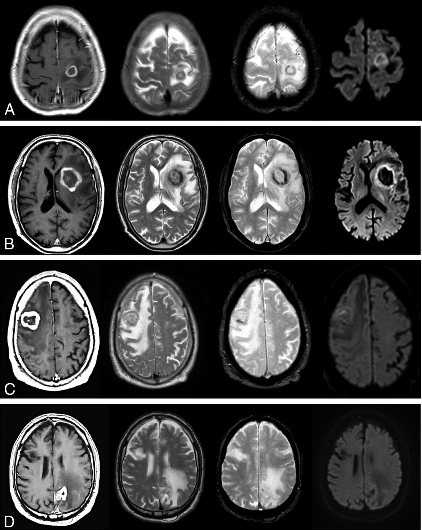
- FIG 1.
Visual summary of MR imaging features in 4 patients with pathology-confirmed DLBC EBV-positive CNS lymphoma. One patient in each row: CE-T1WI, TSE-T2WI, GE-T2*WI, and DWI at b = 1000. Regular thin ring enhancement of a subcortical lesion in A, irregular thick ring enhancement of a basal-ganglia lesion in B. The solid walls of lesions show homogeneous TSE-T2WI low signal and restricted diffusion. Incidental right frontal chronic infarct in D. Irregular thick ring enhancement in cortico-subcortical lesions: frontal in C, parietal in D. Heterogeneous signal on TSE-T2WI: hypointense in C, iso- to hyperintense in D. Intermediate heterogeneous signal on DWI. Different amounts of hemorrhage in all cases are depicted by the GE-T2*WI. Note the TSE-T2WI heterogeneous iso- to hypointense signal of the nonenhancing central content of tumors in A–C, especially in B and C.
- FIG 2.
Average nTIC of DLBC EBV-positive CNS lymphoma, PCNSL (EBV-negative), metastasis, and glioblastoma. Few differences may be seen between DLBC EBV-positive CNS lymphoma and PCNSL. The most relevant visual differences between DLBC EBV-positive CNS lymphoma and metastasis or glioblastoma are seen around the maximal-signal-intensity drop and the signal-recovery segments of the curves.
- FIG 3.
Boxplots depicting the results of the nTIC algorithms to differentiate PCNSL versus glioblastoma (upper left) and PCNSL versus metastasis (upper right) for each tumor subtype. Lower row: Boxplots depicting PSR and rCBV values for each tumor subtype.


Tables
- Table 1:
Clinical overview of the included patients with pathology-confirmed DLBC EBV-positive CNS lymphoma
Age (yr) Sex Underlying Conditions Radiologic Diagnosisa Diagnostic-Therapeutic Initial Management P 1 66 Male Kidney transplant Multiple metastasesb Biopsy: reason, multiple lesions P 2 76 Male Chronic myeloproliferative disorder; essential thrombocythemia Glioblastoma Biopsy: reason, patient basal clinical status P 3 74 Male Liver transplant Single metastasisc Biopsy: reason, second-look radiologic opinion P 4 62 Female Systemic sclerosis and discoid cutaneous lupus Glioblastoma Maximal safe surgical resection P 5 70 Female Immunosenescence Multiple metastases or multifocal glioblastoma Biopsy: reason, multiple lesions P 6 63 Male Autoimmune hepatitis Metastasis or glioblastoma Maximal safe surgical resection P 7 78 Female Kidney transplant Single metastasis Maximal safe surgical resection - Table 2:
Radiologic overview of the included patients with pathology-confirmed large B-cell EBV-positive primary CNS lymphoma
No. Location Necrosis CE-T1WI Ring T2WI Solid Parts T2*WI Hemorrhage DWI Solid Parts P 1 Multiple Bilateral basal ganglia and cortico-subcortical Yes Irregular thick and nodular Heterogeneous hyperintense Moderate Heterogeneous restricted P 2 Single Parietal corticosubcortical Yes Irregular thick Heterogeneous intermediate Subtle Heterogeneous intermediate P 3 Single Frontal cortico-subcortical Yes Irregular thick Heterogeneous hypointense Moderate Heterogeneous restricted P 4 Single Parietal cortico-subcortical Yes Irregular thick Heterogeneous hyperintense Prominent Heterogeneous restricted P 5 Multiple Cortico-subcortical unilateral Yes Irregular thin Heterogeneous hypointense Prominent Heterogeneous intermediate P 6 Single Basal ganglia Yes Irregular thick Homogenous hypointense Prominent Heterogeneous restricted P 7 Single Frontal subcortical Yes Regular thin Homogenous hypointense Moderate Homogeneous restricted


