Advertisement

Main menu

  • Home
  • Content
    • Current Issue
    • Accepted Manuscripts
    • Article Preview
    • Past Issue Archive
    • Video Articles
    • AJNR Case Collection
    • Case of the Week Archive
    • Case of the Month Archive
    • Classic Case Archive
  • Special Collections
    • AJNR Awards
    • Low-Field MRI
    • Alzheimer Disease
    • ASNR Foundation Special Collection
    • Photon-Counting CT
    • View All
  • Multimedia
    • AJNR Podcasts
    • AJNR SCANtastic
    • Trainee Corner
    • MRI Safety Corner
    • Imaging Protocols
  • For Authors
    • Submit a Manuscript
    • Submit a Video Article
    • Submit an eLetter to the Editor/Response
    • Manuscript Submission Guidelines
    • Statistical Tips
    • Fast Publishing of Accepted Manuscripts
    • Graphical Abstract Preparation
    • Imaging Protocol Submission
    • Author Policies
  • About Us
    • About AJNR
    • Editorial Board
    • Editorial Board Alumni
  • More
    • Become a Reviewer/Academy of Reviewers
    • Subscribers
    • Permissions
    • Alerts
    • Feedback
    • Advertisers
    • ASNR Home

User menu

  • Alerts
  • Log in

Search

  • Advanced search
American Journal of Neuroradiology

American Journal of Neuroradiology

ASHNR American Society of Functional Neuroradiology ASHNR American Society of Pediatric Neuroradiology ASSR
  • Alerts
  • Log in

Advanced Search

  • Home
  • Content
    • Current Issue
    • Accepted Manuscripts
    • Article Preview
    • Past Issue Archive
    • Video Articles
    • AJNR Case Collection
    • Case of the Week Archive
    • Case of the Month Archive
    • Classic Case Archive
  • Special Collections
    • AJNR Awards
    • Low-Field MRI
    • Alzheimer Disease
    • ASNR Foundation Special Collection
    • Photon-Counting CT
    • View All
  • Multimedia
    • AJNR Podcasts
    • AJNR SCANtastic
    • Trainee Corner
    • MRI Safety Corner
    • Imaging Protocols
  • For Authors
    • Submit a Manuscript
    • Submit a Video Article
    • Submit an eLetter to the Editor/Response
    • Manuscript Submission Guidelines
    • Statistical Tips
    • Fast Publishing of Accepted Manuscripts
    • Graphical Abstract Preparation
    • Imaging Protocol Submission
    • Author Policies
  • About Us
    • About AJNR
    • Editorial Board
    • Editorial Board Alumni
  • More
    • Become a Reviewer/Academy of Reviewers
    • Subscribers
    • Permissions
    • Alerts
    • Feedback
    • Advertisers
    • ASNR Home
  • Follow AJNR on Twitter
  • Visit AJNR on Facebook
  • Follow AJNR on Instagram
  • Join AJNR on LinkedIn
  • RSS Feeds

AJNR Awards, New Junior Editors, and more. Read the latest AJNR updates

Research ArticleExtracranial Vascular

Nonstenotic Carotid Plaques in Ischemic Stroke: Analysis of the STRATIS Registry

N. Singh, J. Ospel, A. Mayank, M. Marko, O.O. Zaidat, N.H. Mueller-Kronast, D.S. Liebeskind, M. Goyal and on behalf of the STRATIS Investigators
American Journal of Neuroradiology September 2021, 42 (9) 1645-1652; DOI: https://doi.org/10.3174/ajnr.A7218
N. Singh
aFrom the Departments of Clinical Neurosciences (N.S., A.M., M.G.)
  • Find this author on Google Scholar
  • Find this author on PubMed
  • Search for this author on this site
  • ORCID record for N. Singh
J. Ospel
cDepartment of Radiology (J.O.), University Hospital of Basel, Basel, Switzerland
  • Find this author on Google Scholar
  • Find this author on PubMed
  • Search for this author on this site
  • ORCID record for J. Ospel
A. Mayank
aFrom the Departments of Clinical Neurosciences (N.S., A.M., M.G.)
  • Find this author on Google Scholar
  • Find this author on PubMed
  • Search for this author on this site
M. Marko
dDepartment of Neurology (M.M.), Medical University of Vienna, Vienna, Austria
  • Find this author on Google Scholar
  • Find this author on PubMed
  • Search for this author on this site
  • ORCID record for M. Marko
O.O. Zaidat
eNeuroscience Institute (O.O.Z.), Mercy Health St. Vincent Medical Center, Toledo, Ohio
  • Find this author on Google Scholar
  • Find this author on PubMed
  • Search for this author on this site
  • ORCID record for O.O. Zaidat
N.H. Mueller-Kronast
fDepartment of Neurology (N.H.M.-K.), Delray Medical Center, Delray Beach, Florida
  • Find this author on Google Scholar
  • Find this author on PubMed
  • Search for this author on this site
  • ORCID record for N.H. Mueller-Kronast
D.S. Liebeskind
gUCLA Comprehensive Stroke Center (D.S.L.), Los Angeles, California
  • Find this author on Google Scholar
  • Find this author on PubMed
  • Search for this author on this site
  • ORCID record for D.S. Liebeskind
M. Goyal
aFrom the Departments of Clinical Neurosciences (N.S., A.M., M.G.)
bDiagnostic Imaging (M.G.), Foothills Medical Center, University of Calgary, Calgary, Alberta, Canada
  • Find this author on Google Scholar
  • Find this author on PubMed
  • Search for this author on this site
  • ORCID record for M. Goyal
  • Article
  • Figures & Data
  • Info & Metrics
  • Responses
  • References
  • PDF
Loading

Article Figures & Data

Figures

  • Tables
  • FIG 1.
    • Download figure
    • Open in new tab
    • Download powerpoint
    FIG 1.

    Sagittal oblique reformatted and axial CT angiographic images of exemplary cases of nonstenotic carotid plaques with varying morphology in patients with ipsilateral ischemic stroke. All plaques were classified as predominantly calcified. A, Predominantly calcified plaque. B, C, and D, Predominantly calcified plaque with hypodense plaque features.

  • FIG 2.
    • Download figure
    • Open in new tab
    • Download powerpoint
    FIG 2.

    Sagittal oblique reformatted and axial CT angiographic images of exemplary cases of nonstenotic carotid plaques with varying morphology in patients with ipsilateral ischemic stroke. All plaques were classified as predominantly noncalcified. A, Predominantly noncalcified and hypodense plaque. B, Predominantly noncalcified, hypodense, and irregular plaque. C, Predominantly noncalcified, hypodense, and ulcerated plaque.
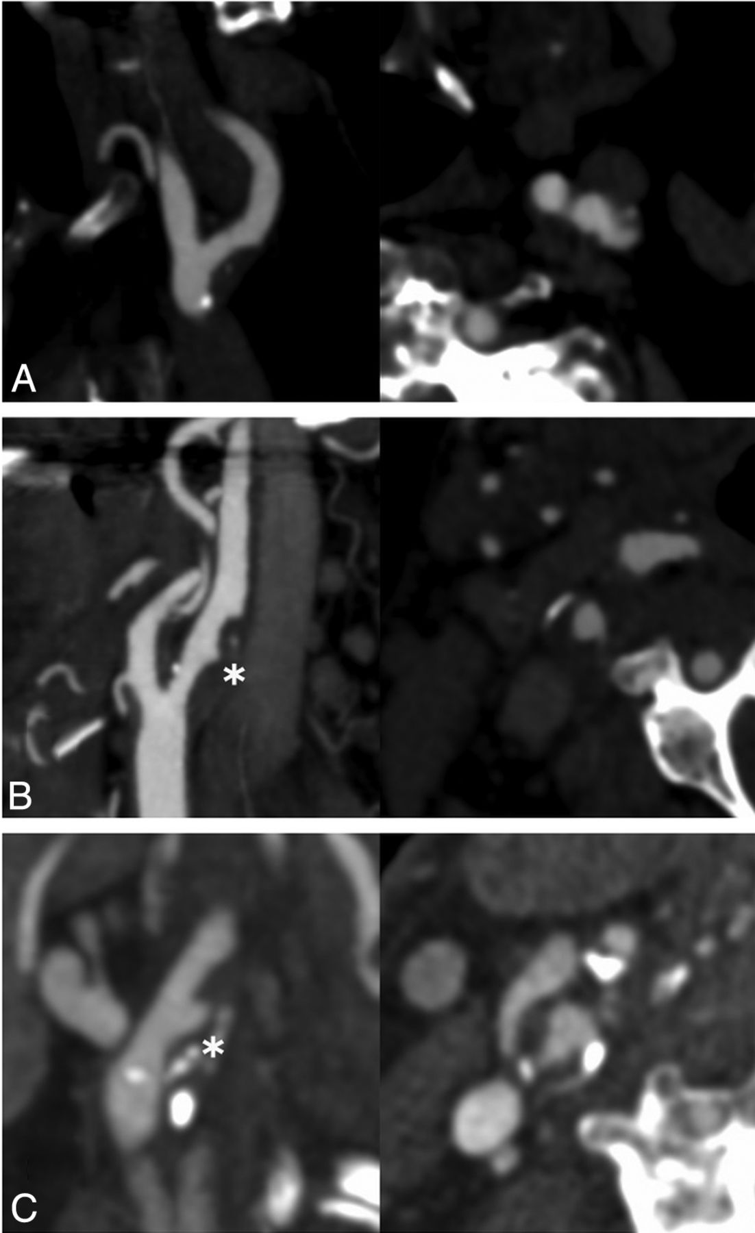
  • FIG 3.
    • Download figure
    • Open in new tab
    • Download powerpoint
    FIG 3.

    Sagittal oblique reformatted and axial CT angiographic images of exemplary cases of nonstenotic carotid disease with varying morphology in patients with ipsilateral ischemic stroke. A, Predominantly noncalcified plaque with an irregular surface. B, Carotid web. C, Predominantly noncalcified plaque with ulceration. D, Predominantly noncalcified plaque with hypodense plaque features.

Tables

  • Figures
    • View popup
    Table 1:

    Patient baseline characteristics for the entire patient sample (n = 367), patients with cardioembolic stroke (n = 226), and patients with cryptogenic stroke (n = 141)

    VariableEntire Patient Sample (n = 367)Cardioembolic Stroke (n = 226)Cryptogenic Stroke (n = 141)P Valuea
    Age (median) (IQR) (yr)71 (60–81)72 (62–82)69 (59–78).23
    Female sex (No.) (%)189/367 (51.5)116/226 (51.3)73/141 (51.8).51
    Baseline NIHSS (median) (IQR)16 (12–20)16 (12–20)17 (13–22).09
    Comorbidities (No.) (%)
     CAD107 (29.2)68 (30.1)39 (27.7).63
     Previous stroke46 (12.5)32 (14.2)14 (9.9).26
     Previous ICH7 (1.9)3 (1.3)4 (2.8).43
     Previous TIA24 (6.5)14 (6.2)10 (7.1).82
     Diabetes94 (25.6)54 (23.9)40 (28.4).39
     Hypertension267 (72.8)169 (74.8)98 (69.5).28
    Smoking (No.) (%)
     Current smoker69/333 (20.7)35/204 (17.2)34/129 (26.4).001
     Former smoker106/333 (31.8)80/204 (39.2)26/129 (24.5).001
    ASPECTS (median) (IQR)9 (8–9)9 (8–9)9 (8–9).88
    Occlusion site (No.) (%)(n = 330)(n = 210)(n = 120).43
     ICA (intracranial)47 (12.8)28 (12.4)19 (13.5)
     M1235 (64.0)147 (65.0)88 (62.4)
     M265 (17.7)42 (18.6)23 (16.3)
     Other20 (5.5)9 (4.0)11 (7.8)
    • Note:—CAD indicates coronary artery disease; ICH, intracerebral hemorrhage

    • ↵a Derived from the Wilcoxon rank sum test (continuous variables) or Fisher exact test (categoric variables).

    • View popup
    Table 2:

    Ipsilateral and contralateral nonstenotic carotid plaque in patients with cryptogenic and cardioembolic stroke

    Overall Ischemic Strokes (n = 367 Patients/734 Carotid Arteries)
    Cardioembolic strokeb (n = 226 patients/452 carotid arteries)
     Ipsilateral strokeYesNo (contralateral/unaffected side)
     Nonstenotic carotid plaque (No.) (%)35 (16.7)23 (9.5)
     No nonstenotic carotid plaque (No.) (%)175 (83.3)219 (90.5)
     Total (No.) (%)a210 (100)242 (100)
    Cryptogenic strokeb (n = 141 patients/282 carotid arteries)
     Ipsilateral strokeYesNo (contralateral/unaffected side)
     Nonstenotic carotid plaque (No.) (%)31 (23.9)24 (15.8)
     No nonstenotic carotid plaque (No.) (%)99 (76.2)128 (84.2)
     Total (No.) (%)a130 (100)152 (100)
    • ↵a Note that 16 patients in the cardioembolic group and 11 patients in the cryptogenic group had bilateral and/or posterior circulation strokes that were not confined to the ipsilateral ICA vascular territory. These strokes were, therefore, classified neither as right-sided nor left-sided strokes.

    • ↵b Two-sided P value (derived from the Fisher exact test) = .025 for cardioembolic strokes and .099 for cryptogenic stroke.

    • View popup
    Table 3:

    Plaque features with significant association with ipsilateral strokes in patients with cryptogenic and cardioembolic strokes

    Plaque FeatureuOR (95%CI)aOR (95%CI)a
    Cardioembolic stroke (n = 226 patients/452 carotid arteries)
     Predominantly calcified plaque0.75 (0.52–1.09)
     Irregularity1.50 (0.81–2.79)
     Ulceration1.94 (0.46–8.23)
     Hypodense plaque1.17 (0.77–1.76)
     Maximum plaque thickness1.11 (0.95–1.30)
     Donut sign–b
     Stenosis degree (31%–50% vs 0%–30%)1.05 (0.77–1.43)
     Webc3.49 (0.36–33.83)
     Unremarkable carotid artery (absence of any of the above–mentioned features)0.96 (0.66–1.40)
    Cryptogenic stroke (n = 141 patients/282 carotid arteries)
     Predominantly calcified plaque0.94 (0.58–1.51)
     Irregularity2.32 (1.15–4.70)2.50 (1.22–5.14)
     Ulceration1.47 (0.39–5.59)
     Hypodense plaque1.78 (1.09–2.92)1.89 (1.14–3.14)
     Maximum plaque thickness1.24 (1.03–1.50)1.29 (1.05–1.57)
     Donut signc1.17 (0.07–18.90)
     Stenosis degree (31%–50% vs. 0%–30%)1.31 (0.89–1.96)
     Webc1.77 (0.29–10.77)
     Unremarkable carotid artery (absence of any of the above-mentioned features)0.75 (0.46–1.21)
    • Note:—uOR indicates unadjusted OR.

    • ↵a With adjustment for age and sex and patient as a cluster variable. Multivariable analysis was performed for variables only when a significant association was found in univariable analysis.

    • ↵b Omitted because of collinearity (all patients with a donut sign had ipsilateral strokes).

    • ↵c Not a plaque feature but included in the definition of nonstenotic carotid disease.

PreviousNext
Back to top

In this issue

American Journal of Neuroradiology: 42 (9)
American Journal of Neuroradiology
Vol. 42, Issue 9
1 Sep 2021
  • Table of Contents
  • Index by author
  • Complete Issue (PDF)
Advertisement
Print
Download PDF
Email Article

Thank you for your interest in spreading the word on American Journal of Neuroradiology.

NOTE: We only request your email address so that the person you are recommending the page to knows that you wanted them to see it, and that it is not junk mail. We do not capture any email address.

Enter multiple addresses on separate lines or separate them with commas.
Nonstenotic Carotid Plaques in Ischemic Stroke: Analysis of the STRATIS Registry
(Your Name) has sent you a message from American Journal of Neuroradiology
(Your Name) thought you would like to see the American Journal of Neuroradiology web site.
CAPTCHA
This question is for testing whether or not you are a human visitor and to prevent automated spam submissions.
Cite this article
N. Singh, J. Ospel, A. Mayank, M. Marko, O.O. Zaidat, N.H. Mueller-Kronast, D.S. Liebeskind, M. Goyal, on behalf of the STRATIS Investigators
Nonstenotic Carotid Plaques in Ischemic Stroke: Analysis of the STRATIS Registry
American Journal of Neuroradiology Sep 2021, 42 (9) 1645-1652; DOI: 10.3174/ajnr.A7218

Citation Manager Formats

  • BibTeX
  • Bookends
  • EasyBib
  • EndNote (tagged)
  • EndNote 8 (xml)
  • Medlars
  • Mendeley
  • Papers
  • RefWorks Tagged
  • Ref Manager
  • RIS
  • Zotero
0 Responses
Respond to this article
Share
Bookmark this article
Nonstenotic Carotid Plaques in Ischemic Stroke: Analysis of the STRATIS Registry
N. Singh, J. Ospel, A. Mayank, M. Marko, O.O. Zaidat, N.H. Mueller-Kronast, D.S. Liebeskind, M. Goyal, on behalf of the STRATIS Investigators
American Journal of Neuroradiology Sep 2021, 42 (9) 1645-1652; DOI: 10.3174/ajnr.A7218
del.icio.us logo Twitter logo Facebook logo Mendeley logo
  • Tweet Widget
  • Facebook Like
  • Google Plus One
Purchase

Jump to section

  • Article
    • Abstract
    • ABBREVIATIONS:
    • MATERIALS AND METHODS
    • RESULTS
    • DISCUSSION
    • CONCLUSIONS
    • Footnotes
    • References
  • Figures & Data
  • Info & Metrics
  • Responses
  • References
  • PDF

Related Articles

  • No related articles found.
  • PubMed
  • Google Scholar

Cited By...

  • Symptomatic non-stenotic carotid disease: current challenges and opportunities for diagnosis and treatment
  • MR Imaging of Carotid Artery Atherosclerosis: Updated Evidence on High-Risk Plaque Features and Emerging Trends
  • Symptomatic non-stenotic carotid disease: current challenges and opportunities for diagnosis and treatment
  • Nonstenotic Carotid Plaques and Embolic Stroke of Undetermined Source: A Multimodality Review
  • Crossref
  • Google Scholar

This article has not yet been cited by articles in journals that are participating in Crossref Cited-by Linking.

More in this TOC Section

  • Proximal Vertebral Artery Variants and Embryology
  • High-Risk Plaque Features in Carotid MRI
  • Nonstenotic Carotid Plaques and Stroke Review
Show more Extracranial Vascular

Similar Articles

Advertisement

Indexed Content

  • Current Issue
  • Accepted Manuscripts
  • Article Preview
  • Past Issues
  • Editorials
  • Editor's Choice
  • Fellows' Journal Club
  • Letters to the Editor
  • Video Articles

Cases

  • Case Collection
  • Archive - Case of the Week
  • Archive - Case of the Month
  • Archive - Classic Case

More from AJNR

  • Trainee Corner
  • Imaging Protocols
  • MRI Safety Corner
  • Book Reviews

Multimedia

  • AJNR Podcasts
  • AJNR Scantastics

Resources

  • Turnaround Time
  • Submit a Manuscript
  • Submit a Video Article
  • Submit an eLetter to the Editor/Response
  • Manuscript Submission Guidelines
  • Statistical Tips
  • Fast Publishing of Accepted Manuscripts
  • Graphical Abstract Preparation
  • Imaging Protocol Submission
  • Evidence-Based Medicine Level Guide
  • Publishing Checklists
  • Author Policies
  • Become a Reviewer/Academy of Reviewers
  • News and Updates

About Us

  • About AJNR
  • Editorial Board
  • Editorial Board Alumni
  • Alerts
  • Permissions
  • Not an AJNR Subscriber? Join Now
  • Advertise with Us
  • Librarian Resources
  • Feedback
  • Terms and Conditions
  • AJNR Editorial Board Alumni

American Society of Neuroradiology

  • Not an ASNR Member? Join Now

© 2025 by the American Society of Neuroradiology All rights, including for text and data mining, AI training, and similar technologies, are reserved.
Print ISSN: 0195-6108 Online ISSN: 1936-959X

Powered by HighWire