Advertisement

Main menu

  • Home
  • Content
    • Current Issue
    • Accepted Manuscripts
    • Article Preview
    • Past Issue Archive
    • Video Articles
    • AJNR Case Collection
    • Case of the Week Archive
    • Case of the Month Archive
    • Classic Case Archive
  • Special Collections
    • AJNR Awards
    • Low-Field MRI
    • Alzheimer Disease
    • ASNR Foundation Special Collection
    • Photon-Counting CT
    • View All
  • Multimedia
    • AJNR Podcasts
    • AJNR SCANtastic
    • Trainee Corner
    • MRI Safety Corner
    • Imaging Protocols
  • For Authors
    • Submit a Manuscript
    • Submit a Video Article
    • Submit an eLetter to the Editor/Response
    • Manuscript Submission Guidelines
    • Statistical Tips
    • Fast Publishing of Accepted Manuscripts
    • Graphical Abstract Preparation
    • Imaging Protocol Submission
    • Author Policies
  • About Us
    • About AJNR
    • Editorial Board
    • Editorial Board Alumni
  • More
    • Become a Reviewer/Academy of Reviewers
    • Subscribers
    • Permissions
    • Alerts
    • Feedback
    • Advertisers
    • ASNR Home

User menu

  • Alerts
  • Log in

Search

  • Advanced search
American Journal of Neuroradiology

American Journal of Neuroradiology

ASHNR American Society of Functional Neuroradiology ASHNR American Society of Pediatric Neuroradiology ASSR
  • Alerts
  • Log in

Advanced Search

  • Home
  • Content
    • Current Issue
    • Accepted Manuscripts
    • Article Preview
    • Past Issue Archive
    • Video Articles
    • AJNR Case Collection
    • Case of the Week Archive
    • Case of the Month Archive
    • Classic Case Archive
  • Special Collections
    • AJNR Awards
    • Low-Field MRI
    • Alzheimer Disease
    • ASNR Foundation Special Collection
    • Photon-Counting CT
    • View All
  • Multimedia
    • AJNR Podcasts
    • AJNR SCANtastic
    • Trainee Corner
    • MRI Safety Corner
    • Imaging Protocols
  • For Authors
    • Submit a Manuscript
    • Submit a Video Article
    • Submit an eLetter to the Editor/Response
    • Manuscript Submission Guidelines
    • Statistical Tips
    • Fast Publishing of Accepted Manuscripts
    • Graphical Abstract Preparation
    • Imaging Protocol Submission
    • Author Policies
  • About Us
    • About AJNR
    • Editorial Board
    • Editorial Board Alumni
  • More
    • Become a Reviewer/Academy of Reviewers
    • Subscribers
    • Permissions
    • Alerts
    • Feedback
    • Advertisers
    • ASNR Home
  • Follow AJNR on Twitter
  • Visit AJNR on Facebook
  • Follow AJNR on Instagram
  • Join AJNR on LinkedIn
  • RSS Feeds

AJNR Awards, New Junior Editors, and more. Read the latest AJNR updates

Review ArticleAdult Brain
Open Access

Radiology-Pathology Correlations of Intracranial Clots: Current Theories, Clinical Applications, and Future Directions

J.C. Benson, D.F. Kallmes, A.S. Larson and W. Brinjikji
American Journal of Neuroradiology September 2021, 42 (9) 1558-1565; DOI: https://doi.org/10.3174/ajnr.A7249
J.C. Benson
aFrom the Department of Neuroradiology, Mayo Clinic, Rochester, Minnesota
  • Find this author on Google Scholar
  • Find this author on PubMed
  • Search for this author on this site
  • ORCID record for J.C. Benson
D.F. Kallmes
aFrom the Department of Neuroradiology, Mayo Clinic, Rochester, Minnesota
  • Find this author on Google Scholar
  • Find this author on PubMed
  • Search for this author on this site
  • ORCID record for D.F. Kallmes
A.S. Larson
aFrom the Department of Neuroradiology, Mayo Clinic, Rochester, Minnesota
  • Find this author on Google Scholar
  • Find this author on PubMed
  • Search for this author on this site
  • ORCID record for A.S. Larson
W. Brinjikji
aFrom the Department of Neuroradiology, Mayo Clinic, Rochester, Minnesota
  • Find this author on Google Scholar
  • Find this author on PubMed
  • Search for this author on this site
  • ORCID record for W. Brinjikji
  • Article
  • Figures & Data
  • Info & Metrics
  • Responses
  • References
  • PDF
Loading

Article Figures & Data

Figures

  • Tables
  • Fig 1.
    • Download figure
    • Open in new tab
    • Download powerpoint
    Fig 1.

    Histologic examples of various clot types. RBC-rich (A), fibrin-rich (B), platelet-rich (C) (all stained with Martius Scarlet Blue), and NET-rich (D) clots (immunohistochemistry using citrullinated H3 antibody) are shown, though standard definitions for these clot types have not yet been created.
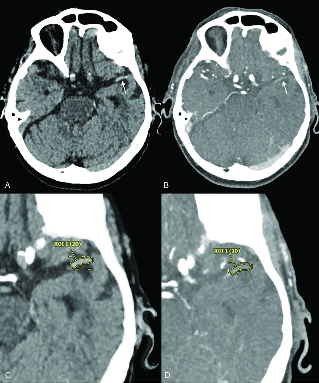
  • Fig 2.
    • Download figure
    • Open in new tab
    • Download powerpoint
    Fig 2.

    Measurement of clot perviousness on CT using TAI. NCCT (A) demonstrates a hyperdense vessel sign in the distal left M1 (arrow), with faint attenuation in this region also seen on CTA (arrow, B). Comparison of intraclot attenuation between the NCCT (C) and CTA (D) yields a 23% increase in Hounsfield units following contrast administration.

  • Fig 3.
    • Download figure
    • Open in new tab
    • Download powerpoint
    Fig 3.

    Intracranial clot evaluation on NCCT. Both the subjective presence of a hyperdense MCA sign (arrow in A) and objective measurement of intraclot attenuation (B) are predictive the histologic composition of a clot and responsiveness to treatment. Relative attenuation, in which the involved artery is compared with the contralateral side, may also be used (not shown).

Tables

  • Figures
  • Summary of the most commonly used methods for determining clot histology and predicted treatment response on NCCT, CTA, and MR imaginga

    Measurement MethodHistologic CorrelationFavorable Therapeutic Response
    NCCTHU↑ HU = ↑ RBC, ↓ fibrin/platelets↑ HU
    CTATAIConflicting data↑ TAI
    MR imagingSVSSVS = ↑ RBC, ↓ fibrin/platelets+ SVS
    • Note:—HU indicates Hounsfield unit.

    • ↵a Up and down arrows denote greater or lesser values (HU or TAI) and relative histologic composition. The plus sign denotes the presence of an imaging sign (ie, SVS).

PreviousNext
Back to top

In this issue

American Journal of Neuroradiology: 42 (9)
American Journal of Neuroradiology
Vol. 42, Issue 9
1 Sep 2021
  • Table of Contents
  • Index by author
  • Complete Issue (PDF)
Advertisement
Print
Download PDF
Email Article

Thank you for your interest in spreading the word on American Journal of Neuroradiology.

NOTE: We only request your email address so that the person you are recommending the page to knows that you wanted them to see it, and that it is not junk mail. We do not capture any email address.

Enter multiple addresses on separate lines or separate them with commas.
Radiology-Pathology Correlations of Intracranial Clots: Current Theories, Clinical Applications, and Future Directions
(Your Name) has sent you a message from American Journal of Neuroradiology
(Your Name) thought you would like to see the American Journal of Neuroradiology web site.
CAPTCHA
This question is for testing whether or not you are a human visitor and to prevent automated spam submissions.
Cite this article
J.C. Benson, D.F. Kallmes, A.S. Larson, W. Brinjikji
Radiology-Pathology Correlations of Intracranial Clots: Current Theories, Clinical Applications, and Future Directions
American Journal of Neuroradiology Sep 2021, 42 (9) 1558-1565; DOI: 10.3174/ajnr.A7249

Citation Manager Formats

  • BibTeX
  • Bookends
  • EasyBib
  • EndNote (tagged)
  • EndNote 8 (xml)
  • Medlars
  • Mendeley
  • Papers
  • RefWorks Tagged
  • Ref Manager
  • RIS
  • Zotero
0 Responses
Respond to this article
Share
Bookmark this article
Radiology-Pathology Correlations of Intracranial Clots: Current Theories, Clinical Applications, and Future Directions
J.C. Benson, D.F. Kallmes, A.S. Larson, W. Brinjikji
American Journal of Neuroradiology Sep 2021, 42 (9) 1558-1565; DOI: 10.3174/ajnr.A7249
del.icio.us logo Twitter logo Facebook logo Mendeley logo
  • Tweet Widget
  • Facebook Like
  • Google Plus One
Purchase

Jump to section

  • Article
    • Abstract
    • ABBREVIATIONS:
    • CONCLUSIONS
    • Footnotes
    • References
  • Figures & Data
  • Info & Metrics
  • Responses
  • References
  • PDF

Related Articles

  • No related articles found.
  • PubMed
  • Google Scholar

Cited By...

  • Multimodal CT imaging of ischemic stroke thrombi identifies scale-invariant radiomic features that reflect clot biology
  • Thrombus imaging characteristics within acute ischemic stroke: similarities and interdependence
  • Crossref (13)
  • Google Scholar

This article has been cited by the following articles in journals that are participating in Crossref Cited-by Linking.

  • Impact of vessel tortuosity and radiological thrombus characteristics on the choice of first-line thrombectomy strategy: Results from the ESCAPE-NA1 trial
    Fouzi Bala, Petra Cimflova, Nishita Singh, Jianhai Zhang, Manon Kappelhof, Beom Joon Kim, Mohamed Najm, Rotem Golan, Ibukun Elebute, Faysal Benali, Nerea Arrarte Terreros, Henk Marquering, Charles Majoie, Mohammed Almekhlafi, Mayank Goyal, Michael D Hill, Wu Qiu, Bijoy K Menon
    European Stroke Journal 2023 8 3
  • A nomogram for predicting thrombus composition in stroke patients with large vessel occlusion: combination of thrombus density and perviousness with clinical features
    Chendong Wang, Yu Hang, Yuezhou Cao, Linbo Zhao, Jincheng Jiao, Mingfang Li, Xiaoquan Xu, Shanshan Lu, Lei Jiang, Qianghui Liu, Haibin Shi, Sheng Liu, Zhenyu Jia
    Neuroradiology 2023 65 2
  • Predictive value of clot imaging in acute ischemic stroke: A systematic review of artificial intelligence and conventional studies
    Daniela Dumitriu LaGrange, Jeremy Hofmeister, Andrea Rosi, Maria Isabel Vargas, Isabel Wanke, Paolo Machi, Karl-Olof Lövblad
    Neuroscience Informatics 2023 3 1
  • Multimodal CT imaging of ischemic stroke thrombi identifies scale-invariant radiomic features that reflect clot biology
    Briana A Santo, Seyyed Mostafa Mousavi Janbeh Sarayi, Andrew D McCall, Andre Monteiro, Brianna Donnelly, Adnan H Siddiqui, Vincent M Tutino
    Journal of NeuroInterventional Surgery 2023 15 12
  • Thrombus imaging characteristics within acute ischemic stroke: similarities and interdependence
    Nerea Arrarte Terreros, Agnetha AE Bruggeman, Manon Kappelhof, Manon L Tolhuisen, Josje Brouwer, Jan W Hoving, Praneeta R Konduri, Katinka R van Kranendonk, Bruna G Dutra, Heitor CBR Alves, Diederik WJ Dippel, Wim H van Zwam, Ludo FM Beenen, Lonneke SF Yo, Ed van Bavel, Charles BLM Majoie, Henk A Marquering
    Journal of NeuroInterventional Surgery 2023 15 e1
  • Mechanical thrombectomy using the Nimbus stent-retriever – initial experiences in a single-center observational study
    Jan Boriesosdick, Arwed Elias Michael, Jan-Robert Kröger, Julius Henning Niehoff, Saher Saeed, Marc Pflug, Peter Schellinger, Volker Maus, Jan Borggrefe, Christoph Mönninghoff
    Interventional Neuroradiology 2024 30 3
  • Radiomics features on computed tomography reflect thrombus histological age prior to endovascular treatment of acute ischemic stroke
    Chendong Wang, Tao Li, Zhenyu Jia, Kai Qiu, Runhao Jiang, Yu Hang, Heng Ni, Yuezhou Cao, Linbo Zhao, Mingfang Li, Jincheng Jiao, Haibin Shi, Jiulou Zhang, Sheng Liu
    Journal of Stroke and Cerebrovascular Diseases 2023 32 11
  • Clinical Significance of Stiffness during Endoscopic Surgery for Intracerebral Hemorrhage: A Retrospective Study
    Kengo KISHIDA, Daisuke MARUYAMA, Saki KOTANI, Nobukuni MURAKAMI, Naoya HASHIMOTO
    Neurologia medico-chirurgica 2023 63 12
  • What is a Challenging Clot?
    Johanna M. Ospel, Mahmood Mirza, Frédéric Clarençon, Adnan Siddiqui, Karen Doyle, Arturo Consoli, Maxim Mokin, Teresa Ullberg, Osama Zaidat, Romain Bourcier, Zsolt Kulcsar, Matthew J. Gounis, David S. Liebeskind, Jens Fiehler, Ana Paula Narata, Marc Ribo, Tudor Jovin, Nobuyuki Sakai, Ansaar Rai, Ray McCarthy, Franziska Dorn, Tommy Andersson, Charles B. L. M. Majoie, Ricardo Hanel, Ashutosh Jadhav, Christian Riedel, Angel Chamorro, Waleed Brinjikji, Vincent Costalat, Simon F. DeMeyer, Raul G. Nogueira, Christophe Cognard, Joan Montaner, Thomas W. Leung, Carlos Molina, Heleen van Beusekom, Antoni Davalos, John Weisel, Rene Chapot, Markus Möhlenbruch, Patrick Brouwer
    Clinical Neuroradiology 2023 33 4
  • MRI characterization of in vitro clots at 3T and 7T: A technical note
    Daniela Dumitriu LaGrange, Lijing Xin, François Lazeyras, Karen M. Doyle, Isabel Wanke, Karl-Olof Lövblad
    Journal of Neuroradiology 2024 51 1

More in this TOC Section

  • Diagnostic Neuroradiology of Monoclonal Antibodies
  • Clinical Outcomes After Chiari I Decompression
  • Segmentation of Brain Metastases with BLAST
Show more ADULT BRAIN

Similar Articles

Advertisement

Indexed Content

  • Current Issue
  • Accepted Manuscripts
  • Article Preview
  • Past Issues
  • Editorials
  • Editor's Choice
  • Fellows' Journal Club
  • Letters to the Editor
  • Video Articles

Cases

  • Case Collection
  • Archive - Case of the Week
  • Archive - Case of the Month
  • Archive - Classic Case

More from AJNR

  • Trainee Corner
  • Imaging Protocols
  • MRI Safety Corner
  • Book Reviews

Multimedia

  • AJNR Podcasts
  • AJNR Scantastics

Resources

  • Turnaround Time
  • Submit a Manuscript
  • Submit a Video Article
  • Submit an eLetter to the Editor/Response
  • Manuscript Submission Guidelines
  • Statistical Tips
  • Fast Publishing of Accepted Manuscripts
  • Graphical Abstract Preparation
  • Imaging Protocol Submission
  • Evidence-Based Medicine Level Guide
  • Publishing Checklists
  • Author Policies
  • Become a Reviewer/Academy of Reviewers
  • News and Updates

About Us

  • About AJNR
  • Editorial Board
  • Editorial Board Alumni
  • Alerts
  • Permissions
  • Not an AJNR Subscriber? Join Now
  • Advertise with Us
  • Librarian Resources
  • Feedback
  • Terms and Conditions
  • AJNR Editorial Board Alumni

American Society of Neuroradiology

  • Not an ASNR Member? Join Now

© 2025 by the American Society of Neuroradiology All rights, including for text and data mining, AI training, and similar technologies, are reserved.
Print ISSN: 0195-6108 Online ISSN: 1936-959X

Powered by HighWire