Advertisement

Main menu

  • Home
  • Content
    • Current Issue
    • Accepted Manuscripts
    • Article Preview
    • Past Issue Archive
    • Video Articles
    • AJNR Case Collection
    • Case of the Week Archive
    • Case of the Month Archive
    • Classic Case Archive
  • Special Collections
    • Advancing NeuroMRI with High-Relaxivity Contrast Agents
    • AJNR Awards
    • Low-Field MRI
    • Alzheimer Disease
    • ASNR Foundation Special Collection
    • Photon-Counting CT
    • View All
  • Multimedia
    • AJNR Podcasts
    • AJNR SCANtastic
    • Trainee Corner
    • MRI Safety Corner
    • Imaging Protocols
  • For Authors
    • Submit a Manuscript
    • Submit a Video Article
    • Submit an eLetter to the Editor/Response
    • Manuscript Submission Guidelines
    • Statistical Tips
    • Fast Publishing of Accepted Manuscripts
    • Graphical Abstract Preparation
    • Imaging Protocol Submission
    • Author Policies
  • About Us
    • About AJNR
    • Editorial Board
    • Editorial Board Alumni
  • More
    • Become a Reviewer/Academy of Reviewers
    • Subscribers
    • Permissions
    • Alerts
    • Feedback
    • Advertisers
    • ASNR Home

User menu

  • Alerts
  • Log in

Search

  • Advanced search
American Journal of Neuroradiology

American Journal of Neuroradiology

ASHNR American Society of Functional Neuroradiology ASHNR American Society of Pediatric Neuroradiology ASSR
  • Alerts
  • Log in

Advanced Search

  • Home
  • Content
    • Current Issue
    • Accepted Manuscripts
    • Article Preview
    • Past Issue Archive
    • Video Articles
    • AJNR Case Collection
    • Case of the Week Archive
    • Case of the Month Archive
    • Classic Case Archive
  • Special Collections
    • Advancing NeuroMRI with High-Relaxivity Contrast Agents
    • AJNR Awards
    • Low-Field MRI
    • Alzheimer Disease
    • ASNR Foundation Special Collection
    • Photon-Counting CT
    • View All
  • Multimedia
    • AJNR Podcasts
    • AJNR SCANtastic
    • Trainee Corner
    • MRI Safety Corner
    • Imaging Protocols
  • For Authors
    • Submit a Manuscript
    • Submit a Video Article
    • Submit an eLetter to the Editor/Response
    • Manuscript Submission Guidelines
    • Statistical Tips
    • Fast Publishing of Accepted Manuscripts
    • Graphical Abstract Preparation
    • Imaging Protocol Submission
    • Author Policies
  • About Us
    • About AJNR
    • Editorial Board
    • Editorial Board Alumni
  • More
    • Become a Reviewer/Academy of Reviewers
    • Subscribers
    • Permissions
    • Alerts
    • Feedback
    • Advertisers
    • ASNR Home
  • Follow AJNR on Twitter
  • Visit AJNR on Facebook
  • Follow AJNR on Instagram
  • Join AJNR on LinkedIn
  • RSS Feeds

AJNR Awards, New Junior Editors, and more. Read the latest AJNR updates


Improved Turnaround Times | Median time to first decision: 12 days

Research ArticleAdult Brain

Comparison of Measurement Techniques and Response Criteria for MR Imaging Follow-up in Adult Primary Central Nervous System Lymphoma

K. Massicotte-Tisluck, D. Vanderweyen, J.-F. Vendrell, D. Fortin and G. Gahide
American Journal of Neuroradiology August 2021, 42 (8) 1415-1420; DOI: https://doi.org/10.3174/ajnr.A7132
K. Massicotte-Tisluck
aFrom the Département d'Imagerie Médicale, (K.M.-T., D.V., G.G.), Centre Hospitalier Universitaire de Sherbrooke, Sherbrooke, Québec, Canada
cFaculté de Médecine et des Sciences de la Santé (K.M.-T., D.V.), Université de Sherbrooke, Sherbrooke, Québec, Canada
  • Find this author on Google Scholar
  • Find this author on PubMed
  • Search for this author on this site
  • ORCID record for K. Massicotte-Tisluck
D. Vanderweyen
aFrom the Département d'Imagerie Médicale, (K.M.-T., D.V., G.G.), Centre Hospitalier Universitaire de Sherbrooke, Sherbrooke, Québec, Canada
cFaculté de Médecine et des Sciences de la Santé (K.M.-T., D.V.), Université de Sherbrooke, Sherbrooke, Québec, Canada
  • Find this author on Google Scholar
  • Find this author on PubMed
  • Search for this author on this site
  • ORCID record for D. Vanderweyen
J.-F. Vendrell
  • Find this author on Google Scholar
  • Find this author on PubMed
  • Search for this author on this site
  • ORCID record for J.-F. Vendrell
D. Fortin
bCentre de Recherche du Centre Hospitalier Universitaire de Sherbrooke, Etienne Le Bel (D.F., G.G.), Université de Sherbrooke, Sherbrooke, Québec, Canada
dService de Neurochirurgie, Département de Chirurgie (D.F.), Centre Hospitalier Universitaire de Sherbrooke, Sherbrooke, Québec, Canada.
  • Find this author on Google Scholar
  • Find this author on PubMed
  • Search for this author on this site
  • ORCID record for D. Fortin
G. Gahide
aFrom the Département d'Imagerie Médicale, (K.M.-T., D.V., G.G.), Centre Hospitalier Universitaire de Sherbrooke, Sherbrooke, Québec, Canada
dService de Neurochirurgie, Département de Chirurgie (D.F.), Centre Hospitalier Universitaire de Sherbrooke, Sherbrooke, Québec, Canada.
  • Find this author on Google Scholar
  • Find this author on PubMed
  • Search for this author on this site
  • ORCID record for G. Gahide
  • Article
  • Figures & Data
  • Supplemental
  • Info & Metrics
  • Responses
  • References
  • PDF
Loading

Article Figures & Data

Figures

  • Tables
  • FIG 1.
    • Download figure
    • Open in new tab
    • Download powerpoint
    FIG 1.

    3D measurement example on postcontrast 3D T1 MPRAGE. The blue line represents the longest 1D. The orange line is the longest length perpendicular to the blue line; their product represents 2D. The green line is the longest length perpendicular to the blue and orange lines; those 3 measures are used to calculate the volume for 3D.

  • FIG 2.
    • Download figure
    • Open in new tab
    • Download powerpoint
    FIG 2.

    Postcontrast 3D T1 MPRAGE in a 56-year-old woman treated for a left temporal PCNSL. Comparison of the 3D measurements performed by the 2 readers (reader 1, A and B; reader 2, C and D).
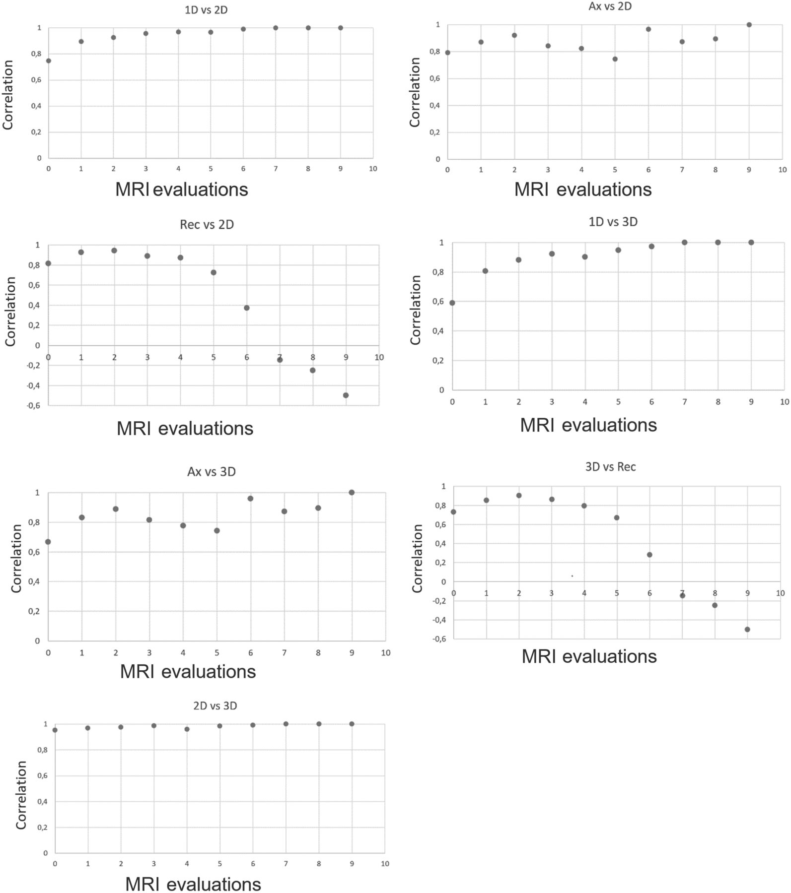
  • FIG 3.
    • Download figure
    • Open in new tab
    • Download powerpoint
    FIG 3.

    Correlation of raw measurements calculated with the Spearman correlation after we applied cubic root on volume calculated with the 3D method and square root on surface area calculated with the 2D methods (exclusion of zero values). Rec indicates RECIST; Ax, axial.

Tables

  • Figures
    • View popup
    Table 1:

    Included patient characteristics (N = 40)

    Patient CharacteristicsValue (SD)
    Mean age (yr)61.5 (11.1)
    Age range (yr)29–81
    Male/female ratio23:17
    Pathology
        Primary CNS large B-cell lymphoma40
        Prior WBRT5
        Median follow-up (yr)a3.01
    Total baseline MR imaging33
        Total baseline CT7
    Total follow-up MR imaging254
        Total follow-up CT10
    Mean number of MR images per patient7.60 (3.49)
    Mean number of lesions on baseline MR image2.33 (1.14)
    Disease location
        Superficial16
        Deep15
        Mixed9
    Particular disease distribution
        Midline crossing lesion10
        Multifocal, bilateral lesions16
    Induction responsea22
    Relapse after inductiona12
    • Note:—WBRT indicates whole brain radiotherapy; SD, standard deviation.

    • ↵a One patient was lost to follow-up, and 5 stopped their treatment for palliative care and were not included in this analysis.

    • View popup
    Table 2:

    Intraobserver and interobserver correlations calculated with the ICC for each measurement method in raw units (95% CI)

    Measurement MethodIntraobserver Correlation (95% CI)Interobserver Correlation (95% CI)
    Longest 1D0.993 (0.985–0.997)0.967 (0.947–0.979)
    2D0.997 (0.994–0.998)0.982 (0.969–0.989)
    3D0.993 (0.985–0.997)0.992 (0.986–0.995)
    • View popup
    Table 3:

    Correlation between raw measurements with the Spearman rank correlation coefficient

    Measurement MethodSpearman Rank Correlation Coefficient (95% CI)
    Longest 1D vs axial 1D0.977
    Longest 1D vs RECIST0.930
    Longest 1D vs 2D0.965
    Longest 1D vs 3D0.935
    Axial 1D vs RECIST0.915
    Axial 1D vs 2D0.963
    Axial 1D vs 3D0.942
    RECIST vs 2D0.924
    RECIST vs 3D0.898
    2D vs 3D0.990
    • View popup
    Table 4:

    Spearman rank correlation coefficients of the lesion variation in size compared with baseline MR imagesa

    Measurement MethodSpearman Correlation Coefficient (95% CI)
    Longest 1D vs axial 1D0.890
    Longest 1D vs RECIST0.898
    Longest 1D vs 2D0.962
    Longest 1D vs 3D0.918
    Axial 1D vs RECIST0.796
    Axial 1D vs 2D0.872
    Axial 1D vs 3D0.831
    RECIST vs 2D0.847
    RECIST vs 3D0.794
    2D vs 3D0.980
    • ↵a Cubic roots and square roots were applied for 3D and 2D measurements, respectively.

PreviousNext
Back to top

In this issue

American Journal of Neuroradiology: 42 (8)
American Journal of Neuroradiology
Vol. 42, Issue 8
1 Aug 2021
  • Table of Contents
  • Index by author
  • Complete Issue (PDF)
Advertisement
Print
Download PDF
Email Article

Thank you for your interest in spreading the word on American Journal of Neuroradiology.

NOTE: We only request your email address so that the person you are recommending the page to knows that you wanted them to see it, and that it is not junk mail. We do not capture any email address.

Enter multiple addresses on separate lines or separate them with commas.
Comparison of Measurement Techniques and Response Criteria for MR Imaging Follow-up in Adult Primary Central Nervous System Lymphoma
(Your Name) has sent you a message from American Journal of Neuroradiology
(Your Name) thought you would like to see the American Journal of Neuroradiology web site.
CAPTCHA
This question is for testing whether or not you are a human visitor and to prevent automated spam submissions.
Cite this article
K. Massicotte-Tisluck, D. Vanderweyen, J.-F. Vendrell, D. Fortin, G. Gahide
Comparison of Measurement Techniques and Response Criteria for MR Imaging Follow-up in Adult Primary Central Nervous System Lymphoma
American Journal of Neuroradiology Aug 2021, 42 (8) 1415-1420; DOI: 10.3174/ajnr.A7132

Citation Manager Formats

  • BibTeX
  • Bookends
  • EasyBib
  • EndNote (tagged)
  • EndNote 8 (xml)
  • Medlars
  • Mendeley
  • Papers
  • RefWorks Tagged
  • Ref Manager
  • RIS
  • Zotero
0 Responses
Respond to this article
Share
Bookmark this article
Comparison of Measurement Techniques and Response Criteria for MR Imaging Follow-up in Adult Primary Central Nervous System Lymphoma
K. Massicotte-Tisluck, D. Vanderweyen, J.-F. Vendrell, D. Fortin, G. Gahide
American Journal of Neuroradiology Aug 2021, 42 (8) 1415-1420; DOI: 10.3174/ajnr.A7132
del.icio.us logo Twitter logo Facebook logo Mendeley logo
  • Tweet Widget
  • Facebook Like
  • Google Plus One
Purchase

Jump to section

  • Article
    • Abstract
    • ABBREVIATIONS:
    • MATERIALS AND METHODS
    • RESULTS
    • DISCUSSION
    • CONCLUSIONS
    • ACKNOWLEDGMENTS
    • Footnotes
    • References
  • Figures & Data
  • Supplemental
  • Info & Metrics
  • Responses
  • References
  • PDF

Related Articles

  • PubMed
  • Google Scholar

Cited By...

  • No citing articles found.
  • Crossref
  • Google Scholar

This article has not yet been cited by articles in journals that are participating in Crossref Cited-by Linking.

More in this TOC Section

  • Diagnostic Neuroradiology of Monoclonal Antibodies
  • Clinical Outcomes After Chiari I Decompression
  • Segmentation of Brain Metastases with BLAST
Show more Adult Brain

Similar Articles

Advertisement

Indexed Content

  • Current Issue
  • Accepted Manuscripts
  • Article Preview
  • Past Issues
  • Editorials
  • Editor's Choice
  • Fellows' Journal Club
  • Letters to the Editor
  • Video Articles

Cases

  • Case Collection
  • Archive - Case of the Week
  • Archive - Case of the Month
  • Archive - Classic Case

More from AJNR

  • Trainee Corner
  • Imaging Protocols
  • MRI Safety Corner
  • Book Reviews

Multimedia

  • AJNR Podcasts
  • AJNR Scantastics

Resources

  • Turnaround Time
  • Submit a Manuscript
  • Submit a Video Article
  • Submit an eLetter to the Editor/Response
  • Manuscript Submission Guidelines
  • Statistical Tips
  • Fast Publishing of Accepted Manuscripts
  • Graphical Abstract Preparation
  • Imaging Protocol Submission
  • Evidence-Based Medicine Level Guide
  • Publishing Checklists
  • Author Policies
  • Become a Reviewer/Academy of Reviewers
  • News and Updates

About Us

  • About AJNR
  • Editorial Board
  • Editorial Board Alumni
  • Alerts
  • Permissions
  • Not an AJNR Subscriber? Join Now
  • Advertise with Us
  • Librarian Resources
  • Feedback
  • Terms and Conditions
  • AJNR Editorial Board Alumni

American Society of Neuroradiology

  • Not an ASNR Member? Join Now

© 2025 by the American Society of Neuroradiology All rights, including for text and data mining, AI training, and similar technologies, are reserved.
Print ISSN: 0195-6108 Online ISSN: 1936-959X

Powered by HighWire