Advertisement

Main menu

  • Home
  • Content
    • Current Issue
    • Accepted Manuscripts
    • Article Preview
    • Past Issue Archive
    • Video Articles
    • AJNR Case Collection
    • Case of the Week Archive
    • Case of the Month Archive
    • Classic Case Archive
  • Special Collections
    • Advancing NeuroMRI with High-Relaxivity Contrast Agents
    • AJNR Awards
    • Low-Field MRI
    • Alzheimer Disease
    • ASNR Foundation Special Collection
    • Photon-Counting CT
    • View All
  • Multimedia
    • AJNR Podcasts
    • AJNR SCANtastic
    • Trainee Corner
    • MRI Safety Corner
    • Imaging Protocols
  • For Authors
    • Submit a Manuscript
    • Submit a Video Article
    • Submit an eLetter to the Editor/Response
    • Manuscript Submission Guidelines
    • Statistical Tips
    • Fast Publishing of Accepted Manuscripts
    • Graphical Abstract Preparation
    • Imaging Protocol Submission
    • Author Policies
  • About Us
    • About AJNR
    • Editorial Board
    • Editorial Board Alumni
  • More
    • Become a Reviewer/Academy of Reviewers
    • Subscribers
    • Permissions
    • Alerts
    • Feedback
    • Advertisers
    • ASNR Home

User menu

  • Alerts
  • Log in

Search

  • Advanced search
American Journal of Neuroradiology

American Journal of Neuroradiology

ASHNR American Society of Functional Neuroradiology ASHNR American Society of Pediatric Neuroradiology ASSR
  • Alerts
  • Log in

Advanced Search

  • Home
  • Content
    • Current Issue
    • Accepted Manuscripts
    • Article Preview
    • Past Issue Archive
    • Video Articles
    • AJNR Case Collection
    • Case of the Week Archive
    • Case of the Month Archive
    • Classic Case Archive
  • Special Collections
    • Advancing NeuroMRI with High-Relaxivity Contrast Agents
    • AJNR Awards
    • Low-Field MRI
    • Alzheimer Disease
    • ASNR Foundation Special Collection
    • Photon-Counting CT
    • View All
  • Multimedia
    • AJNR Podcasts
    • AJNR SCANtastic
    • Trainee Corner
    • MRI Safety Corner
    • Imaging Protocols
  • For Authors
    • Submit a Manuscript
    • Submit a Video Article
    • Submit an eLetter to the Editor/Response
    • Manuscript Submission Guidelines
    • Statistical Tips
    • Fast Publishing of Accepted Manuscripts
    • Graphical Abstract Preparation
    • Imaging Protocol Submission
    • Author Policies
  • About Us
    • About AJNR
    • Editorial Board
    • Editorial Board Alumni
  • More
    • Become a Reviewer/Academy of Reviewers
    • Subscribers
    • Permissions
    • Alerts
    • Feedback
    • Advertisers
    • ASNR Home
  • Follow AJNR on Twitter
  • Visit AJNR on Facebook
  • Follow AJNR on Instagram
  • Join AJNR on LinkedIn
  • RSS Feeds

AJNR Awards, New Junior Editors, and more. Read the latest AJNR updates


Improved Turnaround Times | Median time to first decision: 12 days

Research ArticleAdult Brain
Open Access

Diagnostic Accuracy of Screening Arterial Spin-Labeling MRI Using Hadamard Encoding for the Detection of Reduced CBF in Adult Patients with Ischemic Moyamoya Disease

K. Setta, T. Matsuda, M. Sasaki, T. Chiba, S. Fujiwara, M. Kobayashi, K. Yoshida, Y. Kubo, M. Suzuki, K. Yoshioka and K. Ogasawara
American Journal of Neuroradiology August 2021, 42 (8) 1403-1409; DOI: https://doi.org/10.3174/ajnr.A7167
K. Setta
aFrom the Department of Neurosurgery (K.S., T.C., S.F., M.K., K. Yoshida, Y. Kubo, K.O.), Institute for Biomedical Sciences, Iwate Medical University School of Medicine, Yahaba-cho, Japan
  • Find this author on Google Scholar
  • Find this author on PubMed
  • Search for this author on this site
  • ORCID record for K. Setta
T. Matsuda
cDivision of Ultrahigh Field MRI, Institute for Biomedical Sciences (T.M., M. Sasaki), Iwate Medical University School of Medicine, Yahaba-cho, Japan
  • Find this author on Google Scholar
  • Find this author on PubMed
  • Search for this author on this site
  • ORCID record for T. Matsuda
M. Sasaki
bDepartment of Radiology (M. Suzuki, K. Yoshioka) Institute for Biomedical Sciences (TM, MS), Iwate Medical University School of Medicine, Yahaba-cho, Japan
cDivision of Ultrahigh Field MRI, Institute for Biomedical Sciences (T.M., M. Sasaki), Iwate Medical University School of Medicine, Yahaba-cho, Japan
  • Find this author on Google Scholar
  • Find this author on PubMed
  • Search for this author on this site
  • ORCID record for M. Sasaki
T. Chiba
aFrom the Department of Neurosurgery (K.S., T.C., S.F., M.K., K. Yoshida, Y. Kubo, K.O.), Institute for Biomedical Sciences, Iwate Medical University School of Medicine, Yahaba-cho, Japan
  • Find this author on Google Scholar
  • Find this author on PubMed
  • Search for this author on this site
  • ORCID record for T. Chiba
S. Fujiwara
aFrom the Department of Neurosurgery (K.S., T.C., S.F., M.K., K. Yoshida, Y. Kubo, K.O.), Institute for Biomedical Sciences, Iwate Medical University School of Medicine, Yahaba-cho, Japan
  • Find this author on Google Scholar
  • Find this author on PubMed
  • Search for this author on this site
  • ORCID record for S. Fujiwara
M. Kobayashi
aFrom the Department of Neurosurgery (K.S., T.C., S.F., M.K., K. Yoshida, Y. Kubo, K.O.), Institute for Biomedical Sciences, Iwate Medical University School of Medicine, Yahaba-cho, Japan
  • Find this author on Google Scholar
  • Find this author on PubMed
  • Search for this author on this site
  • ORCID record for M. Kobayashi
K. Yoshida
aFrom the Department of Neurosurgery (K.S., T.C., S.F., M.K., K. Yoshida, Y. Kubo, K.O.), Institute for Biomedical Sciences, Iwate Medical University School of Medicine, Yahaba-cho, Japan
bDepartment of Radiology (M. Suzuki, K. Yoshioka) Institute for Biomedical Sciences (TM, MS), Iwate Medical University School of Medicine, Yahaba-cho, Japan
  • Find this author on Google Scholar
  • Find this author on PubMed
  • Search for this author on this site
  • ORCID record for K. Yoshida
Y. Kubo
aFrom the Department of Neurosurgery (K.S., T.C., S.F., M.K., K. Yoshida, Y. Kubo, K.O.), Institute for Biomedical Sciences, Iwate Medical University School of Medicine, Yahaba-cho, Japan
  • Find this author on Google Scholar
  • Find this author on PubMed
  • Search for this author on this site
  • ORCID record for Y. Kubo
M. Suzuki
  • Find this author on Google Scholar
  • Find this author on PubMed
  • Search for this author on this site
  • ORCID record for M. Suzuki
K. Yoshioka
aFrom the Department of Neurosurgery (K.S., T.C., S.F., M.K., K. Yoshida, Y. Kubo, K.O.), Institute for Biomedical Sciences, Iwate Medical University School of Medicine, Yahaba-cho, Japan
bDepartment of Radiology (M. Suzuki, K. Yoshioka) Institute for Biomedical Sciences (TM, MS), Iwate Medical University School of Medicine, Yahaba-cho, Japan
  • Find this author on Google Scholar
  • Find this author on PubMed
  • Search for this author on this site
  • ORCID record for K. Yoshioka
K. Ogasawara
aFrom the Department of Neurosurgery (K.S., T.C., S.F., M.K., K. Yoshida, Y. Kubo, K.O.), Institute for Biomedical Sciences, Iwate Medical University School of Medicine, Yahaba-cho, Japan
  • Find this author on Google Scholar
  • Find this author on PubMed
  • Search for this author on this site
  • ORCID record for K. Ogasawara
  • Article
  • Figures & Data
  • Supplemental
  • Info & Metrics
  • Responses
  • References
  • PDF
Loading

Article Figures & Data

Figures

  • FIG 1.
    • Download figure
    • Open in new tab
    • Download powerpoint
    FIG 1.

    Comparisons (upper graphs) and Bland-Altman plots (lower graphs) of SPECT-CBF and sASL-CBF ratios and sASL-CBF (left graphs) or hASL-CBF ratios (right graphs) for each MCA ROI in each patient. In the upper graphs, each horizontal line denotes the cutoff point for indicating a reduced SPECT-CBF ratio (0.686) and each vertical line denotes the cutoff point lying closest to the upper left corner of the receiver operating characteristic curve for detecting a reduced SPECT-CBF ratio (0.502 for sASL-CBF, 0.801 for hASL-CBF).

  • FIG 2.
    • Download figure
    • Open in new tab
    • Download powerpoint
    FIG 2.

    Receiver operating characteristic curves used to compare the accuracy of sASL-CBF and hASL-CBF ratios for detecting a reduced SPECT-CBF ratio. Pair-wise comparison analysis shows a significantly greater area under the receiver operating characteristic curve for the hASL-CBF (dotted line) than for the sASL-CBF ratio (solid line).
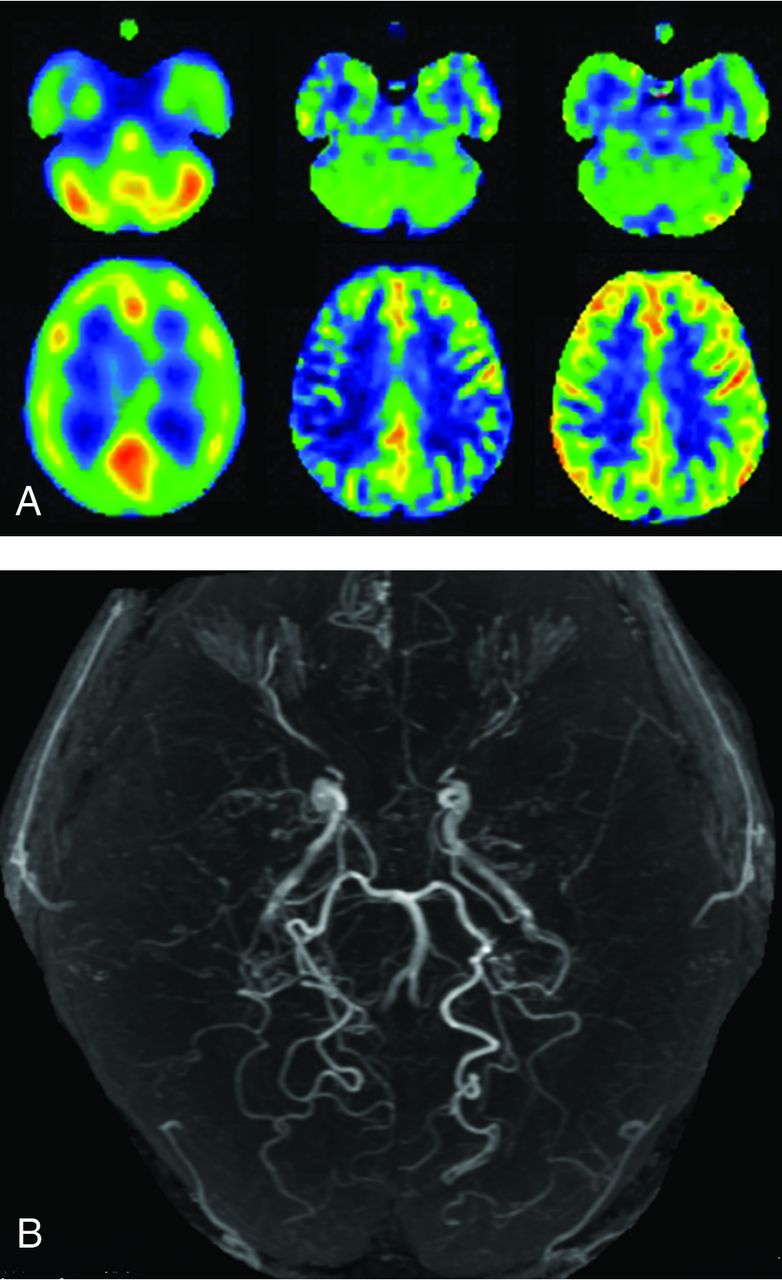
  • FIG 3.
    • Download figure
    • Open in new tab
    • Download powerpoint
    FIG 3.

    MRA (B), brain perfusion SPECT images (A, left), and sASL (A, middle) and hASL (A, right) MR images of a 43-year-old male patient with transient ischemic attacks complicated by right-sided hemiparesis. MRA shows stage 3 Moyamoya disease. On all 3 SPECT and ASL images, the CBF in the left parietal region is severely reduced compared with that in the left cerebellar hemisphere.

  • FIG 4.
    • Download figure
    • Open in new tab
    • Download powerpoint
    FIG 4.

    MRA (B), brain perfusion SPECT images (A, left), and sALS (A, middle) and hASL (A, right) MR images of a 34-year-old female patient with transient ischemic attacks complicated by left-sided hemiparesis. MRA shows stage 3 Moyamoya disease. On perfusion SPECT images, the CBF in the right MCA territory is slightly reduced compared with that in the right cerebellar hemisphere. The CBF on sASL MR images is severely reduced in the right central, parietal, and angular regions, whereas the CBF on hASL MR imaging is not reduced relative to the right cerebellar hemisphere.

PreviousNext
Back to top

In this issue

American Journal of Neuroradiology: 42 (8)
American Journal of Neuroradiology
Vol. 42, Issue 8
1 Aug 2021
  • Table of Contents
  • Index by author
  • Complete Issue (PDF)
Advertisement
Print
Download PDF
Email Article

Thank you for your interest in spreading the word on American Journal of Neuroradiology.

NOTE: We only request your email address so that the person you are recommending the page to knows that you wanted them to see it, and that it is not junk mail. We do not capture any email address.

Enter multiple addresses on separate lines or separate them with commas.
Diagnostic Accuracy of Screening Arterial Spin-Labeling MRI Using Hadamard Encoding for the Detection of Reduced CBF in Adult Patients with Ischemic Moyamoya Disease
(Your Name) has sent you a message from American Journal of Neuroradiology
(Your Name) thought you would like to see the American Journal of Neuroradiology web site.
CAPTCHA
This question is for testing whether or not you are a human visitor and to prevent automated spam submissions.
Cite this article
K. Setta, T. Matsuda, M. Sasaki, T. Chiba, S. Fujiwara, M. Kobayashi, K. Yoshida, Y. Kubo, M. Suzuki, K. Yoshioka, K. Ogasawara
Diagnostic Accuracy of Screening Arterial Spin-Labeling MRI Using Hadamard Encoding for the Detection of Reduced CBF in Adult Patients with Ischemic Moyamoya Disease
American Journal of Neuroradiology Aug 2021, 42 (8) 1403-1409; DOI: 10.3174/ajnr.A7167

Citation Manager Formats

  • BibTeX
  • Bookends
  • EasyBib
  • EndNote (tagged)
  • EndNote 8 (xml)
  • Medlars
  • Mendeley
  • Papers
  • RefWorks Tagged
  • Ref Manager
  • RIS
  • Zotero
0 Responses
Respond to this article
Share
Bookmark this article
Diagnostic Accuracy of Screening Arterial Spin-Labeling MRI Using Hadamard Encoding for the Detection of Reduced CBF in Adult Patients with Ischemic Moyamoya Disease
K. Setta, T. Matsuda, M. Sasaki, T. Chiba, S. Fujiwara, M. Kobayashi, K. Yoshida, Y. Kubo, M. Suzuki, K. Yoshioka, K. Ogasawara
American Journal of Neuroradiology Aug 2021, 42 (8) 1403-1409; DOI: 10.3174/ajnr.A7167
del.icio.us logo Twitter logo Facebook logo Mendeley logo
  • Tweet Widget
  • Facebook Like
  • Google Plus One
Purchase

Jump to section

  • Article
    • Abstract
    • ABBREVIATIONS:
    • MATERIALS AND METHODS
    • RESULTS
    • DISCUSSION
    • CONCLUSIONS
    • Footnotes
    • References
  • Figures & Data
  • Supplemental
  • Info & Metrics
  • Responses
  • References
  • PDF

Related Articles

  • PubMed
  • Google Scholar

Cited By...

  • No citing articles found.
  • Crossref
  • Google Scholar

This article has not yet been cited by articles in journals that are participating in Crossref Cited-by Linking.

More in this TOC Section

Adult Brain

  • Diagnostic Neuroradiology of Monoclonal Antibodies
  • Clinical Outcomes After Chiari I Decompression
  • Segmentation of Brain Metastases with BLAST
Show more Adult Brain

Functional

  • Kurtosis and Epileptogenic Tubers: A Pilot Study
  • Glutaric Aciduria Type 1: DK vs. Conventional MRI
  • WM Neuroaxonal Loss in Type 1 Diabetes
Show more Functional

Similar Articles

Advertisement

Indexed Content

  • Current Issue
  • Accepted Manuscripts
  • Article Preview
  • Past Issues
  • Editorials
  • Editor's Choice
  • Fellows' Journal Club
  • Letters to the Editor
  • Video Articles

Cases

  • Case Collection
  • Archive - Case of the Week
  • Archive - Case of the Month
  • Archive - Classic Case

More from AJNR

  • Trainee Corner
  • Imaging Protocols
  • MRI Safety Corner
  • Book Reviews

Multimedia

  • AJNR Podcasts
  • AJNR Scantastics

Resources

  • Turnaround Time
  • Submit a Manuscript
  • Submit a Video Article
  • Submit an eLetter to the Editor/Response
  • Manuscript Submission Guidelines
  • Statistical Tips
  • Fast Publishing of Accepted Manuscripts
  • Graphical Abstract Preparation
  • Imaging Protocol Submission
  • Evidence-Based Medicine Level Guide
  • Publishing Checklists
  • Author Policies
  • Become a Reviewer/Academy of Reviewers
  • News and Updates

About Us

  • About AJNR
  • Editorial Board
  • Editorial Board Alumni
  • Alerts
  • Permissions
  • Not an AJNR Subscriber? Join Now
  • Advertise with Us
  • Librarian Resources
  • Feedback
  • Terms and Conditions
  • AJNR Editorial Board Alumni

American Society of Neuroradiology

  • Not an ASNR Member? Join Now

© 2025 by the American Society of Neuroradiology All rights, including for text and data mining, AI training, and similar technologies, are reserved.
Print ISSN: 0195-6108 Online ISSN: 1936-959X

Powered by HighWire