
Article Figures & Data
Figures
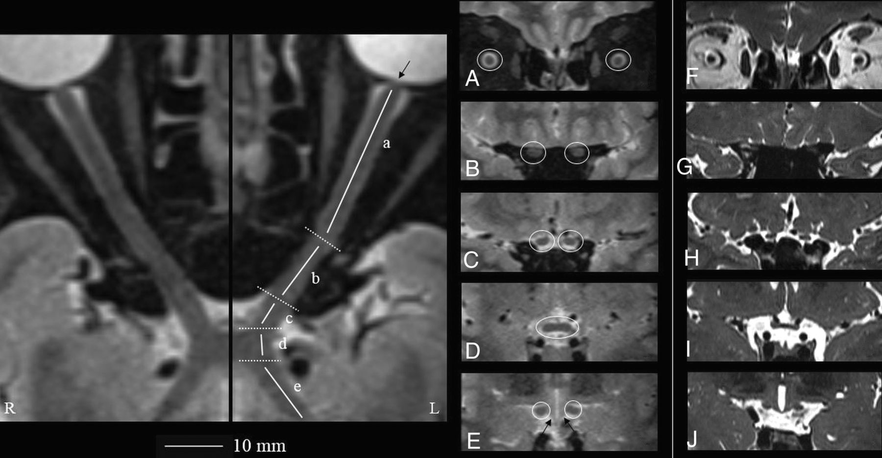
- FIG 1.
Geometrically unbiased, high-resolution representation of the aVP segment anatomy by 3D-T2-STIR-ZOOMit (healthy control No. 9001).The left panel shows curved reconstructions along the true long axis of the right and left aVPs. The thin arrow points to the porus opticus. Coronal-oblique reconstruction images corresponding to the iOrb (A, from the eye bulb to the optic canal), iCanal (B, in the optic canal), and iCran (C, from the optic canal to the chiasm) segments and OC (D) and OT (E) segments highlighted in white circles are presented. By comparison, the right panel (F–J, different healthy volunteer) shows corresponding coronal-oblique images obtained with a 3D-ZOOMit standard sequence without the STIR preparation pulse. Note substantial contrast resolution gain with 3D-T2-STIR-ZOOMit, particularly at interfaces between different signal tissues and between the OT and adjacent hypothalamus (thin arrows in E).
- FIG 2.
Flow chart showing the numbers of participants and healthy participants with MS included in the study who were assessed and evaluated.
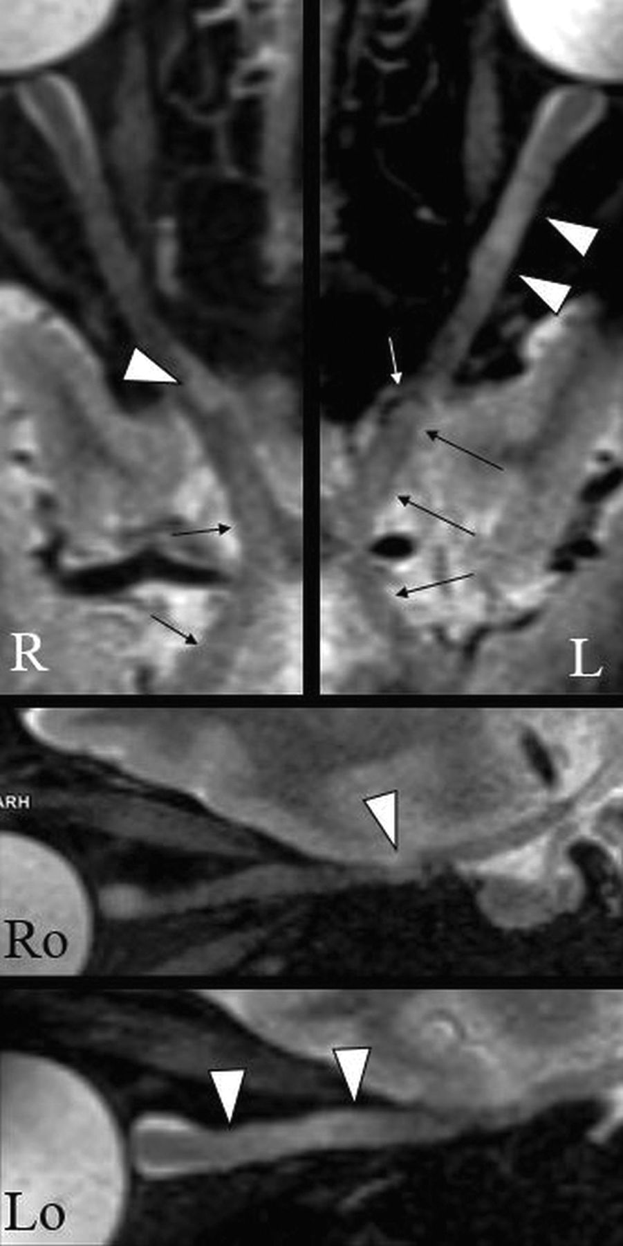
- FIG 3.
An example of the DL appearance on 3D-T2-STIR-ZOOMit images in a patient with MS (No. 0034). The upper panel shows curved reconstructions along the true long axis of the right and left aVPs. Focal signal hyperintensities indicate DLs with partial (right) and complete (left) involvement of the optic nerve cross-section (arrowheads). There are areas of diffuse signal hyperintensity with no definite boundaries along the aVP (thin arrows), possibly related to diffuse demyelination and/or degeneration. The white arrow indicates the ophthalmic artery causing nerve impression. Ro and Lo images present sagittal-oblique reconstructions of R and L.
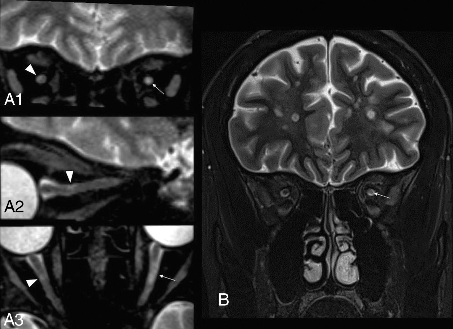
- FIG 4.
Comparison of extra cranial aVP detection in a participant with MS with previous unilateral AON on the left side (patient with MS No. 0038). A1–3, A very small, asymptomatic DL partially involving the superolateral aspect of the right iOrb segment (arrowheads on coronal, sagittal-oblique, and axial-oblique images) was missed on the 2D-T2-STIR image (B). A larger L iOrb lesion was instead detected by both sequences (thin arrows in A1, A3 and B), possibly representing chronic changes related to the previous AON.
- FIG 5.
Intracranial aVP detection of asymptomatic DL in a participant with MS without a previous AON history (patient with MS No. 0020) on 3D-T2-STIR-ZOOMit images. A1, and B1, Bilateral DLs partially affecting the OTs and impairing tissue contrast (arrowheads) with the adjacent hypothalamus ventrolateral aspects (asterisks). C1, Further depiction of DLs with partial involvement of the OC (arrowheads). All these lesions were not detectable on 2D-T2-STIR (middle panel, B2, C2). The right panel (A3, B3, C3) provides the reference normal anatomy (empty arrowheads) from a healthy control (No. 9005). Because of the axial plane appearance of the OTs and hypothalamus ventrolateral aspects resembling lobster antennas, we propose the “lobster antenna” sign to indicate loss of normal tissue contrast between the OT and hypothalamus. Thin arrows in A1 and A3 indicate the mammillary bodies, corresponding to the lobster's eyes in the artwork (A2). Courtesy of Dr. Andrea Diociasi.
- FIG 6.
Proportion of participants with MS with at least 1 identified DL (at any site and any side) by previous AON history (pON–/pON+) and MR imaging technique (2D-T2-STIR/3D-T2-STIR-ZOOMit).


Tables
Parameter 3D-T2-STIR-ZOOMit 2D-T2-STIR Mode 3D 2D Acquisition plane Transversal Coronal TR/TE (ms) 240/119 4880/101 Magnetization preparation Non-selective STIR Non-selective STIR TI (ms) 220 220 FOV read/FOV phase 80 mm × 100% 180 mm × 100% Base resolution/phase/section 128/100%/100% 320/100%/– Acquired voxel size (X, Y, Z) (mm) 0.6, 0.6, 0.6 0.6, 0.6, 2.5 Interpolated voxel size (X, Y, Z) (mm) 0.3, 0.3, 0.6 Flip angle Variable 140° Turbo factor 46 35 Averages 1.4 1 Acceleration factor (GRAPPA) 2 None Scan duration (min/sec) 8/54 2/57 Note:—GRAPPA, generalized autocalibrating partially parallel acquisition; STIR, short tau inversion recovery.
- Table 2:
Confusion matrix illustrating DL detection rates of 3D-T2-STIR-ZOOMit and 2D-T2-STIR per each aVP segment in all patients with MSa
3D-T2-STIR-ZOOMit iOrb– iOrb+ iCanal– iCanal+ iCran– iCran+ OC– OC+ OT– OT+ 2D-T2-STIR – 67 (69.8) 9 (9.4) 68 (70.8) 8 (8.3) 87 (90.6) 7 (7.3) 79 (82.3) 16 (16.7) 86 (89.6) 10 (10.4) + 0 (0) 20 (20.8) 4 (4.2) 16 (16.7) 0 (0) 2 (2.1) 0 (0) 1 (1.0) 0 (0) 0 (0) ↵a Data are number of DLs (n = 96, for each aVP segment) with percentages in parentheses and refer to 96 observations (left and right sides together in 48 patients) in each aVP segment. Absence of a lesion is indicated with –; presence, with +”.


