Advertisement

Main menu

  • Home
  • Content
    • Current Issue
    • Accepted Manuscripts
    • Article Preview
    • Past Issue Archive
    • Video Articles
    • AJNR Case Collection
    • Case of the Week Archive
    • Case of the Month Archive
    • Classic Case Archive
  • Special Collections
    • Advancing NeuroMRI with High-Relaxivity Contrast Agents
    • AJNR Awards
    • Low-Field MRI
    • Alzheimer Disease
    • ASNR Foundation Special Collection
    • Photon-Counting CT
    • View All
  • Multimedia
    • AJNR Podcasts
    • AJNR SCANtastic
    • Trainee Corner
    • MRI Safety Corner
    • Imaging Protocols
  • For Authors
    • Submit a Manuscript
    • Submit a Video Article
    • Submit an eLetter to the Editor/Response
    • Manuscript Submission Guidelines
    • Statistical Tips
    • Fast Publishing of Accepted Manuscripts
    • Graphical Abstract Preparation
    • Imaging Protocol Submission
    • Author Policies
  • About Us
    • About AJNR
    • Editorial Board
    • Editorial Board Alumni
  • More
    • Become a Reviewer/Academy of Reviewers
    • Subscribers
    • Permissions
    • Alerts
    • Feedback
    • Advertisers
    • ASNR Home

User menu

  • Alerts
  • Log in

Search

  • Advanced search
American Journal of Neuroradiology

American Journal of Neuroradiology

ASHNR American Society of Functional Neuroradiology ASHNR American Society of Pediatric Neuroradiology ASSR
  • Alerts
  • Log in

Advanced Search

  • Home
  • Content
    • Current Issue
    • Accepted Manuscripts
    • Article Preview
    • Past Issue Archive
    • Video Articles
    • AJNR Case Collection
    • Case of the Week Archive
    • Case of the Month Archive
    • Classic Case Archive
  • Special Collections
    • Advancing NeuroMRI with High-Relaxivity Contrast Agents
    • AJNR Awards
    • Low-Field MRI
    • Alzheimer Disease
    • ASNR Foundation Special Collection
    • Photon-Counting CT
    • View All
  • Multimedia
    • AJNR Podcasts
    • AJNR SCANtastic
    • Trainee Corner
    • MRI Safety Corner
    • Imaging Protocols
  • For Authors
    • Submit a Manuscript
    • Submit a Video Article
    • Submit an eLetter to the Editor/Response
    • Manuscript Submission Guidelines
    • Statistical Tips
    • Fast Publishing of Accepted Manuscripts
    • Graphical Abstract Preparation
    • Imaging Protocol Submission
    • Author Policies
  • About Us
    • About AJNR
    • Editorial Board
    • Editorial Board Alumni
  • More
    • Become a Reviewer/Academy of Reviewers
    • Subscribers
    • Permissions
    • Alerts
    • Feedback
    • Advertisers
    • ASNR Home
  • Follow AJNR on Twitter
  • Visit AJNR on Facebook
  • Follow AJNR on Instagram
  • Join AJNR on LinkedIn
  • RSS Feeds

AJNR Awards, New Junior Editors, and more. Read the latest AJNR updates


Improved Turnaround Times | Median time to first decision: 12 days

Research ArticlePediatric Neuroimaging
Open Access

Radiomics of Pediatric Low-Grade Gliomas: Toward a Pretherapeutic Differentiation of BRAF-Mutated and BRAF-Fused Tumors

M.W. Wagner, N. Hainc, F. Khalvati, K. Namdar, L. Figueiredo, M. Sheng, S. Laughlin, M.M. Shroff, E. Bouffet, U. Tabori, C. Hawkins, K.W. Yeom and B.B. Ertl-Wagner
American Journal of Neuroradiology April 2021, 42 (4) 759-765; DOI: https://doi.org/10.3174/ajnr.A6998
M.W. Wagner
aFrom the Departments of Diagnostic Imaging (M.W.W., N.H., F.K., K.N., M.S., S.L., M.M.S., B.B.E.-W.)
  • Find this author on Google Scholar
  • Find this author on PubMed
  • Search for this author on this site
  • ORCID record for M.W. Wagner
N. Hainc
aFrom the Departments of Diagnostic Imaging (M.W.W., N.H., F.K., K.N., M.S., S.L., M.M.S., B.B.E.-W.)
dDepartment of Neuroradiology (N.H.), Zurich University Hospital, University of Zurich, Zurich, Switzerland
  • Find this author on Google Scholar
  • Find this author on PubMed
  • Search for this author on this site
  • ORCID record for N. Hainc
F. Khalvati
aFrom the Departments of Diagnostic Imaging (M.W.W., N.H., F.K., K.N., M.S., S.L., M.M.S., B.B.E.-W.)
  • Find this author on Google Scholar
  • Find this author on PubMed
  • Search for this author on this site
  • ORCID record for F. Khalvati
K. Namdar
aFrom the Departments of Diagnostic Imaging (M.W.W., N.H., F.K., K.N., M.S., S.L., M.M.S., B.B.E.-W.)
  • Find this author on Google Scholar
  • Find this author on PubMed
  • Search for this author on this site
  • ORCID record for K. Namdar
L. Figueiredo
bDivision of Neuroradiology, Neurooncology (L.F., E.B., U.T.)
  • Find this author on Google Scholar
  • Find this author on PubMed
  • Search for this author on this site
  • ORCID record for L. Figueiredo
M. Sheng
aFrom the Departments of Diagnostic Imaging (M.W.W., N.H., F.K., K.N., M.S., S.L., M.M.S., B.B.E.-W.)
  • Find this author on Google Scholar
  • Find this author on PubMed
  • Search for this author on this site
  • ORCID record for M. Sheng
S. Laughlin
aFrom the Departments of Diagnostic Imaging (M.W.W., N.H., F.K., K.N., M.S., S.L., M.M.S., B.B.E.-W.)
  • Find this author on Google Scholar
  • Find this author on PubMed
  • Search for this author on this site
  • ORCID record for S. Laughlin
M.M. Shroff
aFrom the Departments of Diagnostic Imaging (M.W.W., N.H., F.K., K.N., M.S., S.L., M.M.S., B.B.E.-W.)
  • Find this author on Google Scholar
  • Find this author on PubMed
  • Search for this author on this site
  • ORCID record for M.M. Shroff
E. Bouffet
bDivision of Neuroradiology, Neurooncology (L.F., E.B., U.T.)
  • Find this author on Google Scholar
  • Find this author on PubMed
  • Search for this author on this site
  • ORCID record for E. Bouffet
U. Tabori
bDivision of Neuroradiology, Neurooncology (L.F., E.B., U.T.)
  • Find this author on Google Scholar
  • Find this author on PubMed
  • Search for this author on this site
  • ORCID record for U. Tabori
C. Hawkins
cPaediatric Laboratory Medicine (C.H.), Division of Pathology, The Hospital for Sick Children and Department of Medical Imaging, University of Toronto, Ontario, Canada
  • Find this author on Google Scholar
  • Find this author on PubMed
  • Search for this author on this site
  • ORCID record for C. Hawkins
K.W. Yeom
eDepartment of Radiology (K.W.Y.), Stanford University School of Medicine, Lucile Packard Children’s Hospital, Palo Alto, California
  • Find this author on Google Scholar
  • Find this author on PubMed
  • Search for this author on this site
  • ORCID record for K.W. Yeom
B.B. Ertl-Wagner
aFrom the Departments of Diagnostic Imaging (M.W.W., N.H., F.K., K.N., M.S., S.L., M.M.S., B.B.E.-W.)
  • Find this author on Google Scholar
  • Find this author on PubMed
  • Search for this author on this site
  • ORCID record for B.B. Ertl-Wagner
  • Article
  • Figures & Data
  • Supplemental
  • Info & Metrics
  • Responses
  • References
  • PDF
Loading

Article Figures & Data

Figures

  • Tables
  • FIG 1.
    • Download figure
    • Open in new tab
    • Download powerpoint
    FIG 1.

    Receiver operating characteristic curve with a 4-fold cross-validation scheme to predict BRAF status using radiomics of FLAIR MR images. Std. dev. indicates standard deviation.
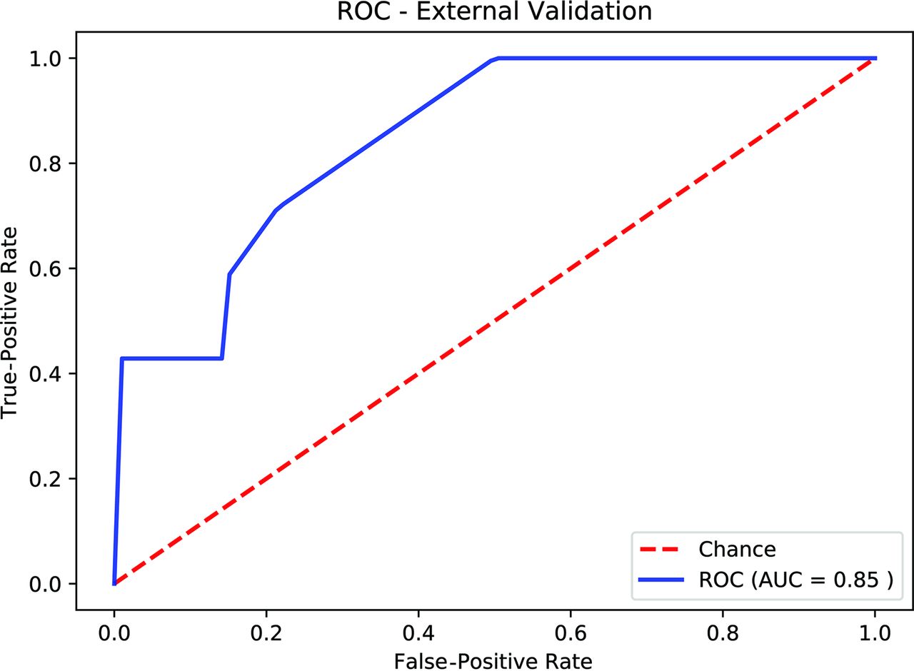
  • FIG 2.
    • Download figure
    • Open in new tab
    • Download powerpoint
    FIG 2.

    Receiver operating characteristic curve of the external validation using the optimal hyperparameters obtained by 4-fold cross-validation.

  • FIG 3.
    • Download figure
    • Open in new tab
    • Download powerpoint
    FIG 3.

    Axial FLAIR images of pLGG. A, A 7-year-old boy. Infratentorial, BRAF V600E-mutated JPA. B, A 12-year-old boy. Supratentorial intraventricular, BRAF-fused ganglioma. C, A 7-year-old boy. Left temporal BRAF V600E-mutated dysembryoplastic neuroepithelial tumor. D, An 8-year-old boy. Right temporal BRAF V600E-mutated pleomorphic xanthoastrocytoma.

Tables

  • Figures
    • View popup
    Table 1:

    Patient demographics

    Institutional Cohort
    TorontoStanford
    No. of patients9421
    Age (mean) (yr)9.48.37
    Male sex (No.) (%)45 (48)12 (57)
    Histologic diagnosis (No.)
     JPA5412
     GG147
     LGA11
     PMA42
     PXA5
     DNET2
     DA2
     GC1
     ODG1
    Molecular subgroup (No.) (%)
     BRAF fusion62 (66)14 (66)
     BRAF mutation32 (34)7 (34)
    FLAIR availability (No.)9421
    Supratentorial (No.) (%)43 (46)6 (28)
    Transtentorial (No.) (%)01 (5)
    Infratentorial (No.) (%)51 (54)14 (67)
    • Note:—GG indicates ganglioglioma; LGA, low-grade astrocytoma; PMA, pilomyxoid astrocytoma; PXA, pleomorphic xanthoastrocytoma; DNET, dysembryoplastic neuroepithelial tumor; DA, diffuse astrocytoma; GC, gangliocytoma; ODG, oligodendroglioma.

    • View popup
    Table 2:

    Performance of radiomic features

    No. of FoldsNo. of TreesAUC (SD) (95% CI)Mean Sensitivity (95% CI)Mean Specificity (95% CI)Mean PPV (95% CI)Mean NPV (95% CI)Top 10 Predictive Features on the External Dataset
    4250.75 (SD, 0.12) (0.62–0.89)0.72 (0.60–0.84)0.86 (0.76–0.95)0.73 (0.60–0.87)0.85 (0.80–0.91)(585, 374, 761, 22, 17, 560, 344, 258, 148, 108)
    • Note:—SD indicates Standard Deviation; PPV, Positive Predictive Value; NPV; Negative Predictive Value.

    • View popup
    Table 3:

    Predictive radiomic featuresa

    No.SourceFeature CategoryFeature
    5853D wavelet transformGray-level difference matrixSmall dependence low gray-level emphasis
    3743D wavelet transformGray-level size zone matrixZone percentage
    7613D wavelet transformGray-level difference matrixDependence entropy
    22OriginalGray-level difference matrixDependence nonuniformity normalized
    17OriginalGray-level difference matrixDependence entropy
    5603D wavelet transformGray-level size zone matrixZone percentage
    3443D wavelet transformHistogramEntropy
    2583D wavelet transformGray-level run-lengthGray-level variance
    1483D wavelet transformHistogramUniformity
    1083D wavelet transformGray-level difference matrixGray-level variance
    • ↵a Radiomic features are ranked from top to bottom according to their importance.

    • View popup
    Table 4:

    Discriminative clinical factorsa

    VariableP ValueOdds Ratio (95% CI)
    Age.041.14 (1.008–1.30)
    Location<.00118.80 (4.96–94.6)
    Sex.96
    • ↵a Older age and supratentorial location of tumor are significant predictors of BRAF V600E mutation. Sex is not a predictor.

PreviousNext
Back to top

In this issue

American Journal of Neuroradiology: 42 (4)
American Journal of Neuroradiology
Vol. 42, Issue 4
1 Apr 2021
  • Table of Contents
  • Index by author
  • Complete Issue (PDF)
Advertisement
Print
Download PDF
Email Article

Thank you for your interest in spreading the word on American Journal of Neuroradiology.

NOTE: We only request your email address so that the person you are recommending the page to knows that you wanted them to see it, and that it is not junk mail. We do not capture any email address.

Enter multiple addresses on separate lines or separate them with commas.
Radiomics of Pediatric Low-Grade Gliomas: Toward a Pretherapeutic Differentiation of BRAF-Mutated and BRAF-Fused Tumors
(Your Name) has sent you a message from American Journal of Neuroradiology
(Your Name) thought you would like to see the American Journal of Neuroradiology web site.
CAPTCHA
This question is for testing whether or not you are a human visitor and to prevent automated spam submissions.
Cite this article
M.W. Wagner, N. Hainc, F. Khalvati, K. Namdar, L. Figueiredo, M. Sheng, S. Laughlin, M.M. Shroff, E. Bouffet, U. Tabori, C. Hawkins, K.W. Yeom, B.B. Ertl-Wagner
Radiomics of Pediatric Low-Grade Gliomas: Toward a Pretherapeutic Differentiation of BRAF-Mutated and BRAF-Fused Tumors
American Journal of Neuroradiology Apr 2021, 42 (4) 759-765; DOI: 10.3174/ajnr.A6998

Citation Manager Formats

  • BibTeX
  • Bookends
  • EasyBib
  • EndNote (tagged)
  • EndNote 8 (xml)
  • Medlars
  • Mendeley
  • Papers
  • RefWorks Tagged
  • Ref Manager
  • RIS
  • Zotero
0 Responses
Respond to this article
Share
Bookmark this article
Radiomics of Pediatric Low-Grade Gliomas: Toward a Pretherapeutic Differentiation of BRAF-Mutated and BRAF-Fused Tumors
M.W. Wagner, N. Hainc, F. Khalvati, K. Namdar, L. Figueiredo, M. Sheng, S. Laughlin, M.M. Shroff, E. Bouffet, U. Tabori, C. Hawkins, K.W. Yeom, B.B. Ertl-Wagner
American Journal of Neuroradiology Apr 2021, 42 (4) 759-765; DOI: 10.3174/ajnr.A6998
del.icio.us logo Twitter logo Facebook logo Mendeley logo
  • Tweet Widget
  • Facebook Like
  • Google Plus One
Purchase

Jump to section

  • Article
    • Abstract
    • ABBREVIATIONS:
    • MATERIALS AND METHODS
    • RESULTS
    • DISCUSSION
    • CONCLUSIONS
    • Footnotes
    • References
  • Figures & Data
  • Supplemental
  • Info & Metrics
  • Responses
  • References
  • PDF

Related Articles

  • PubMed
  • Google Scholar

Cited By...

  • Imaging Clusters of Pediatric Low-Grade Glioma Are Associated with Distinct Molecular Characteristics
  • Segmentation-Free Pretherapeutic Assessment of BRAF-Status in Pediatric Low-Grade Gliomas
  • Multiparametric MRI Along with Machine Learning Informs on Molecular Underpinnings, Prognosis, and Treatment Response in Pediatric Low-Grade Glioma
  • Imaging Clusters of Pediatric Low-Grade Glioma are Associated with Distinct Molecular Characteristics
  • Identification of Multiclass Pediatric Low-Grade Neuroepithelial Tumor Molecular Subtype with ADC MR Imaging and Machine Learning
  • Artificial Intelligence in the Future Landscape of Pediatric Neuroradiology: Opportunities and Challenges
  • Juvenile pilocytic astrocytoma in a child with a prior normal neonatal brain MRI
  • Early prognostication of overall survival for pediatric diffuse midline gliomas using MRI radiomics and machine learning: a two-center study
  • Systematic Literature Review of Machine Learning Algorithms Using Pretherapy Radiologic Imaging for Glioma Molecular Subtype Prediction
  • Utilizing the Amide Proton Transfer Technique to Characterize Diffuse Gliomas Based on the WHO 2021 Classification of CNS Tumors
  • MR Imaging Characteristics and ADC Histogram Metrics for Differentiating Molecular Subgroups of Pediatric Low-Grade Gliomas
  • Evolving Role and Translation of Radiomics and Radiogenomics in Adult and Pediatric Neuro-Oncology
  • ADC Histogram Analysis of Pediatric Low-Grade Glioma Treated with Selumetinib: A Report from the Pediatric Brain Tumor Consortium
  • Crossref
  • Google Scholar

This article has not yet been cited by articles in journals that are participating in Crossref Cited-by Linking.

More in this TOC Section

  • Pediatric Glioma:Imaging Clusters,Molecular Traits
  • Clinical SVR of Fetal Brain MRI
  • FRACTURE MR in Congenital Vertebral Anomalies
Show more PEDIATRIC NEUROIMAGING

Similar Articles

Advertisement

Indexed Content

  • Current Issue
  • Accepted Manuscripts
  • Article Preview
  • Past Issues
  • Editorials
  • Editor's Choice
  • Fellows' Journal Club
  • Letters to the Editor
  • Video Articles

Cases

  • Case Collection
  • Archive - Case of the Week
  • Archive - Case of the Month
  • Archive - Classic Case

More from AJNR

  • Trainee Corner
  • Imaging Protocols
  • MRI Safety Corner
  • Book Reviews

Multimedia

  • AJNR Podcasts
  • AJNR Scantastics

Resources

  • Turnaround Time
  • Submit a Manuscript
  • Submit a Video Article
  • Submit an eLetter to the Editor/Response
  • Manuscript Submission Guidelines
  • Statistical Tips
  • Fast Publishing of Accepted Manuscripts
  • Graphical Abstract Preparation
  • Imaging Protocol Submission
  • Evidence-Based Medicine Level Guide
  • Publishing Checklists
  • Author Policies
  • Become a Reviewer/Academy of Reviewers
  • News and Updates

About Us

  • About AJNR
  • Editorial Board
  • Editorial Board Alumni
  • Alerts
  • Permissions
  • Not an AJNR Subscriber? Join Now
  • Advertise with Us
  • Librarian Resources
  • Feedback
  • Terms and Conditions
  • AJNR Editorial Board Alumni

American Society of Neuroradiology

  • Not an ASNR Member? Join Now

© 2025 by the American Society of Neuroradiology All rights, including for text and data mining, AI training, and similar technologies, are reserved.
Print ISSN: 0195-6108 Online ISSN: 1936-959X

Powered by HighWire