Advertisement

Main menu

  • Home
  • Content
    • Current Issue
    • Accepted Manuscripts
    • Article Preview
    • Past Issue Archive
    • Video Articles
    • AJNR Case Collection
    • Case of the Week Archive
    • Case of the Month Archive
    • Classic Case Archive
  • Special Collections
    • AJNR Awards
    • Low-Field MRI
    • Alzheimer Disease
    • ASNR Foundation Special Collection
    • Photon-Counting CT
    • View All
  • Multimedia
    • AJNR Podcasts
    • AJNR SCANtastic
    • Trainee Corner
    • MRI Safety Corner
    • Imaging Protocols
  • For Authors
    • Submit a Manuscript
    • Submit a Video Article
    • Submit an eLetter to the Editor/Response
    • Manuscript Submission Guidelines
    • Statistical Tips
    • Fast Publishing of Accepted Manuscripts
    • Graphical Abstract Preparation
    • Imaging Protocol Submission
    • Author Policies
  • About Us
    • About AJNR
    • Editorial Board
    • Editorial Board Alumni
  • More
    • Become a Reviewer/Academy of Reviewers
    • Subscribers
    • Permissions
    • Alerts
    • Feedback
    • Advertisers
    • ASNR Home

User menu

  • Alerts
  • Log in

Search

  • Advanced search
American Journal of Neuroradiology

American Journal of Neuroradiology

ASHNR American Society of Functional Neuroradiology ASHNR American Society of Pediatric Neuroradiology ASSR
  • Alerts
  • Log in

Advanced Search

  • Home
  • Content
    • Current Issue
    • Accepted Manuscripts
    • Article Preview
    • Past Issue Archive
    • Video Articles
    • AJNR Case Collection
    • Case of the Week Archive
    • Case of the Month Archive
    • Classic Case Archive
  • Special Collections
    • AJNR Awards
    • Low-Field MRI
    • Alzheimer Disease
    • ASNR Foundation Special Collection
    • Photon-Counting CT
    • View All
  • Multimedia
    • AJNR Podcasts
    • AJNR SCANtastic
    • Trainee Corner
    • MRI Safety Corner
    • Imaging Protocols
  • For Authors
    • Submit a Manuscript
    • Submit a Video Article
    • Submit an eLetter to the Editor/Response
    • Manuscript Submission Guidelines
    • Statistical Tips
    • Fast Publishing of Accepted Manuscripts
    • Graphical Abstract Preparation
    • Imaging Protocol Submission
    • Author Policies
  • About Us
    • About AJNR
    • Editorial Board
    • Editorial Board Alumni
  • More
    • Become a Reviewer/Academy of Reviewers
    • Subscribers
    • Permissions
    • Alerts
    • Feedback
    • Advertisers
    • ASNR Home
  • Follow AJNR on Twitter
  • Visit AJNR on Facebook
  • Follow AJNR on Instagram
  • Join AJNR on LinkedIn
  • RSS Feeds

AJNR Awards, New Junior Editors, and more. Read the latest AJNR updates

Research ArticleNeurointervention

CTA-Based Patient-Tailored Femoral or Radial Frontline Access Reduces the Rate of Catheterization Failure in Chronic Subdural Hematoma Embolization

E. Shotar, G. Pouliquen, K. Premat, A. Pouvelle, S. Mouyal, L. Meyblum, S. Lenck, V. Degos, S. Abi Jaoude, N. Sourour, B. Mathon and F. Clarençon
American Journal of Neuroradiology March 2021, 42 (3) 495-500; DOI: https://doi.org/10.3174/ajnr.A6951
E. Shotar
aFrom the Department of Neuroradiology (E.S., G.P., K.P., A.P., S.M., L.M., S.L., N.S., F.C.)
  • Find this author on Google Scholar
  • Find this author on PubMed
  • Search for this author on this site
  • ORCID record for E. Shotar
G. Pouliquen
aFrom the Department of Neuroradiology (E.S., G.P., K.P., A.P., S.M., L.M., S.L., N.S., F.C.)
  • Find this author on Google Scholar
  • Find this author on PubMed
  • Search for this author on this site
  • ORCID record for G. Pouliquen
K. Premat
aFrom the Department of Neuroradiology (E.S., G.P., K.P., A.P., S.M., L.M., S.L., N.S., F.C.)
dSorbonne Université (K.P., V.D., S.A.J., B.M., F.C.), Paris, France
  • Find this author on Google Scholar
  • Find this author on PubMed
  • Search for this author on this site
  • ORCID record for K. Premat
A. Pouvelle
aFrom the Department of Neuroradiology (E.S., G.P., K.P., A.P., S.M., L.M., S.L., N.S., F.C.)
  • Find this author on Google Scholar
  • Find this author on PubMed
  • Search for this author on this site
  • ORCID record for A. Pouvelle
S. Mouyal
aFrom the Department of Neuroradiology (E.S., G.P., K.P., A.P., S.M., L.M., S.L., N.S., F.C.)
  • Find this author on Google Scholar
  • Find this author on PubMed
  • Search for this author on this site
  • ORCID record for S. Mouyal
L. Meyblum
aFrom the Department of Neuroradiology (E.S., G.P., K.P., A.P., S.M., L.M., S.L., N.S., F.C.)
  • Find this author on Google Scholar
  • Find this author on PubMed
  • Search for this author on this site
  • ORCID record for L. Meyblum
S. Lenck
aFrom the Department of Neuroradiology (E.S., G.P., K.P., A.P., S.M., L.M., S.L., N.S., F.C.)
  • Find this author on Google Scholar
  • Find this author on PubMed
  • Search for this author on this site
  • ORCID record for S. Lenck
V. Degos
bNeurosurgical Anesthesiology and Critical Care (V.D.)
dSorbonne Université (K.P., V.D., S.A.J., B.M., F.C.), Paris, France
  • Find this author on Google Scholar
  • Find this author on PubMed
  • Search for this author on this site
  • ORCID record for V. Degos
S. Abi Jaoude
cNeurosurgery (S.A.J., B.M.), Pitié-Salpêtrière Hospital, Paris, France
dSorbonne Université (K.P., V.D., S.A.J., B.M., F.C.), Paris, France
  • Find this author on Google Scholar
  • Find this author on PubMed
  • Search for this author on this site
  • ORCID record for S. Abi Jaoude
N. Sourour
aFrom the Department of Neuroradiology (E.S., G.P., K.P., A.P., S.M., L.M., S.L., N.S., F.C.)
  • Find this author on Google Scholar
  • Find this author on PubMed
  • Search for this author on this site
  • ORCID record for N. Sourour
B. Mathon
cNeurosurgery (S.A.J., B.M.), Pitié-Salpêtrière Hospital, Paris, France
dSorbonne Université (K.P., V.D., S.A.J., B.M., F.C.), Paris, France
  • Find this author on Google Scholar
  • Find this author on PubMed
  • Search for this author on this site
  • ORCID record for B. Mathon
F. Clarençon
aFrom the Department of Neuroradiology (E.S., G.P., K.P., A.P., S.M., L.M., S.L., N.S., F.C.)
dSorbonne Université (K.P., V.D., S.A.J., B.M., F.C.), Paris, France
  • Find this author on Google Scholar
  • Find this author on PubMed
  • Search for this author on this site
  • ORCID record for F. Clarençon
  • Article
  • Figures & Data
  • Info & Metrics
  • Responses
  • References
  • PDF
Loading

Abstract

BACKGROUND AND PURPOSE: Chronic subdural hematoma embolization, an apparently simple procedure, can prove to be challenging because of the advanced age of the target population. The aim of this study was to compare 2 arterial-access strategies, femoral versus patient-tailored CTA-based frontline access selection, in chronic subdural hematoma embolization procedures.

MATERIALS AND METHODS: This was a monocentric retrospective study. From the March 15, 2018, to the February 14, 2019 (period 1), frontline femoral access was used. Between February 15, 2019, and March 30, 2020 (period 2), the choice of the frontline access, femoral or radial, was based on the CTA recommended as part of the preoperative work-up during both above-mentioned periods. The primary end point was the rate of catheterization failure. The secondary end points were the rate of access site conversion and fluoroscopy duration.

RESULTS: During the study period, 124 patients (with 143 chronic subdural hematomas) underwent an embolization procedure (mean age, 74 [SD, 13] years). Forty-eight chronic subdural hematomas (43 patients) were included during period 1 and were compared with 95 chronic subdural hematomas (81 patients) during period 2. During the first period, 5/48 (10%) chronic subdural hematoma embolizations were aborted due to failed catheterization, significantly more than during period 2 (1/95, 1%; P = .009). The rates of femoral-to-radial (P = .55) and total conversion (P = .86) did not differ between the 2 periods. No significant difference was found regarding the duration of fluoroscopy (P = .62).

CONCLUSIONS: A CTA-based patient-tailored choice of frontline arterial access reduces the rate of catheterization failure in chronic subdural hematoma embolization procedures.

ABBREVIATIONS:

CSDH
chronic subdural hematoma
MMA
middle meningeal artery

The annual incidence of chronic subdural hematomas (CSDHs), 14 to 20 per 100,000 individuals, means that the condition is one of the most frequently managed by neurosurgery departments.1,2 CSDHs are thought to be sentinel health events, akin to hip fractures, with important reduction in life expectancy for patients compared with age-matched controls.3 The condition is, moreover, associated with far-from-negligible rates of morbidity and mortality, around 11% and 4%, respectively.4

Standard management of symptomatic CSDHs includes surgical evacuation, mostly through twist drill or burr-hole craniostomy with closed-system drainage.4⇓-6 Recently, middle meningeal artery (MMA) embolization has emerged as a possible treatment of CSDHs.7,8 The procedure is simple in appearance but can prove to be challenging in a subset of patients because of tortuous vasculature. Indeed, CSDH is mostly a disease of the elderly with two-thirds of cases accounted for in patients older than 65 years of age.1 In the elderly, several factors, including peripheral vascular disease and vascular anatomy, can complicate or even preclude cervical vessel navigation by a traditional transfemoral approach.9,10

The transradial approach has recently emerged as an alternative to transfemoral access in interventional neuroradiology, with the stated aim of reducing access-related complications and patient discomfort.11 It has also been envisioned that radial access may facilitate anterior circulation navigation in some patients.10,11 The aim of this study was to compare 2 arterial-access strategies, frontline femoral versus patient-tailored frontline access selection (femoral or radial), based on a preoperative CTA, in CSDH embolization procedures.

MATERIALS and METHODS

Study Design

This was a monocentric retrospective study performed in a tertiary care and teaching hospital. Starting from March 15, 2018, CSDH has been proposed as a treatment of CSDH, initially as an adjunct to surgery and more recently as a possible sole treatment in a minority of patients. The most common indication for CSDH embolization at our institution is a CSDH recurrence or a CSDH with an independent recurrence risk factor, including antiplatelet therapy, anticoagulation therapy, hepatopathy, or alcohol abuse, for instance.12 Eligible patients were excluded if they refused the embolization procedure or were denied embolization by the attending physician because they presented in a moribund state, had a contraindication to the embolization procedure like severe renal dysfunction, or had a life expectancy of <6 months. A pre-embolization aortic arch and supra-aortic trunk CTA was recommended to rule out anatomic contraindications. The choice of the MMA embolization technique was left to the discretion of the attending interventional neuroradiologist.

From March 15, 2018, to February 14, 2019 (period 1), a femoral access was systematically used as a frontline strategy for CSDH embolization, in line with wider institutional habits. On the basis of this initial experience, from February 15, 2019, to March 30, 2020 (period 2), the choice of the arterial frontline access, femoral or radial, was patient-tailored and based on the available CTA. The choice of frontline access was left to the discretion of the attending interventional neuroradiologist. Radial frontline access was, nevertheless, strongly recommended for right carotid catheterization in case of a type III aortic arch with proximal common carotid artery tortuosity and for left carotid navigation in case of a bovine arch configuration.10,11 For radial access (up to 6F sheaths), micropuncture under sonographic guidance was preferred, without prior testing of collateral circulation. Intra-arterial injection of verapamil (3 mg) through the sheath was performed. Given the context of intracranial hemorrhage, radial access in this specific setting was usually performed without anticoagulation. Postprocedure, patent hemostasis for 2 hours was recommended. No left radial or distal radial access was used in this series.

Demographic and clinical data were retrieved by retrospective review of patient medical records. Imaging data were analyzed on the local PACS.

End Points

The primary end point was the rate of catheterization failure leading to procedure abortion. The secondary end points were rate of access site conversion and fluoroscopy duration. Because some procedures were performed in a biplane angiosuite while others were performed in a monoplane angiosuite, only the fluoroscopy duration of the anterior-posterior plane was considered for biplane procedures.

Statistical Analysis

Data are expressed as a percentage for binary variables and as mean [SD] for continuous variables. Probability values are provided uncorrected. Probability values < .05 were considered significant. A χ2 test was used to compare frequencies, and comparison of means was performed using a Student t test. Statistical analyses were performed using MedCalc, Version 19.2 (MedCalc Software).

Ethical Statement

The institutional review board approved this study (Comité d’Ethique pour la Recherche en Imagerie Médicale CRM-2003–063) and the need for signed patient consent was waived.

Data-Sharing Statement

Raw data are available on reasonable request from the corresponding author.

RESULTS

Population Characteristics

One hundred thirty-eight patients were referred for MMA embolization during the study period (Fig 1). Six patients were deemed unfit to undergo embolization, and another 6 patients refused to undergo the procedure. Two patients were excluded on the basis of the result of the CTA because of extensive supra-aortic trunk atheroma precluding safe endovascular navigation. A total of 124 patients underwent embolization procedures of 143 target MMAs (105 unilateral and 19 bilateral CSDHs). The mean age of the study population was 74 [SD, 13] years, and most of the patients (94; 76%) were men. There was no difference in terms of demographics, clinical and radiologic presentation of the CSDHs, management strategy, and choice of anesthesia technique between patients managed during periods 1 and 2 (Table 1).

FIG 1.
  • Download figure
  • Open in new tab
  • Download powerpoint
FIG 1.

Flow chart. E° indicates embolization.

View this table:
  • View inline
  • View popup
Table 1:

Patient characteristicsa

Embolization Procedures

During period 1, all 48 CSDH embolizations were performed via a frontline femoral access. In 1 instance (2%), a femoral-to-radial conversion was deemed necessary during the procedure (Fig 2, illustrative example C). During period 2, a frontline femoral access was chosen for 57/95 (60%) CSDH embolizations. Of these, a femoral-to-radial conversion was deemed necessary during the procedure in 1/57 (2%) instances (Fig 2, illustrative example D). Conversely, a frontline radial access was chosen for 38/95 (40%) cases with 3/38 (8%) radial-to-femoral per-procedure conversions. Figure 2 shows a series of anatomic configurations having led to difficult endovascular navigation or catheterization failure.

FIG 2.
  • Download figure
  • Open in new tab
  • Download powerpoint
FIG 2.

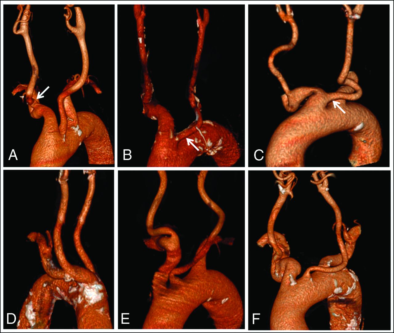
Illustrative examples of challenging and failed catheterization cases. Manually-segmented surface-rendering 3D reconstructions of CTAs of aortic arches of patients included during periods 1 (A–C) and 2 (D–F). A, Anterior view of a case of failed right carotid catheterization by femoral access due to a proximal kinking of the right common carotid artery (right arrow) in a type III aortic arch. B, Anterior view in a case of failed left carotid catheterization by femoral access due to a bovine arch configuration (right arrow), which, in retrospect, would have been an ideal candidate for radial access. C, Anterior view in a case of failed left carotid catheterization by femoral access converted secondarily to transradial catheterization in relation to a bovine arch (right arrow). D, Posterior view of a case of failed right carotid catheterization by a frontline femoral access most probably due to the angulation between the brachiocephalic trunk and the aortic arch, with subsequent successful radial access conversion. E, Anterior view of a bilateral CSDH in which a frontline radial access was chosen. Following right CSDH embolization by the transradial approach, failure to navigate the left carotid artery led to radial-to-femoral access conversion and left-sided successful embolization. F, Anterior view in a case of failed catheterization of the left carotid artery from a frontline femoral access after which the procedure was aborted.

The CSDH embolization procedure was aborted in 15/143 (10%) cases, in 6 instances because of agitation, in 6 cases because of failed catheterization, and in 3 cases because of a so-called “dangerous anastomosis,” including 2 cases in which the MMA originated from the ophthalmic artery. Notably, once the guiding catheter was successfully placed, microcatheter navigation in the external carotid artery and MMA was always achieved. A total of 128/143 (90%) MMAs were embolized. Embolization was performed with calibrated tris-acryl microspheres (300–500 µm in diameter) in 111/128 (87%) instances, with associated proximal MMA coiling in 33/111 (30%) cases; using an n-BCA liquid embolic agent in 6/128 (5%) cases; by proximal MMA coiling alone in 10/128 (8%) cases; and by a gelatin-based embolic agent in the remaining patient (1%). We registered 7 minor complications in 124 patients (6%): 1 partial seizure during the procedure, 1 reversible headache, 2 transient diplopias, 2 asymptomatic iatrogenic meningomeningeal fistulas treated by MMA coiling during the procedure, and 1 asymptomatic external carotid artery occlusion. The only major complication (1%) was a femoral artery occlusion at the access site, treated surgically. The patient died 6 weeks after the CSDH embolization procedure.

Outcome

During period one, 5/48 (10%) CSDH embolization procedures were aborted due to failed catheterization. In all cases of failed catheterization, only a femoral access was attempted (Fig 2, illustrative cases A and B). In comparison, during period 2, only 1/95 (1%) procedures was interrupted after a failed attempt to catheterize a left carotid artery by a femoral access (Fig 2, illustrative case F), significantly less than during period 1 (P = .009). During periods 1 and 2, all 5 femoral-to-radial and radial-to-femoral conversions led to successful CSDH embolizations (Fig 2, illustrative cases C, D, and E). In none of the 6 failed catheterizations was an access site conversion attempted. The mean age of patients with failed catheterization was 81 [SD, 9] years and tended to be higher than that in the general study population, but the difference did not reach statistical significance (P = .21).

The rates of femoral-to-radial (P = .55) and total conversion (P = .86) did not differ significantly between the 2 periods. No significant difference was found regarding the duration of fluoroscopy between the 2 periods (P = .62) (Table 2).

View this table:
  • View inline
  • View popup
Table 2:

Comparison of outcomesa

DISCUSSION

Main Results

This nonrandomized study shows that a CTA-based patient-tailored choice of frontline arterial femoral or radial access reduces the rate of catheterization failure in CSDH embolization procedures. Moreover, femoral-to-radial and radial-to-femoral per-procedure conversion should be considered when faced with challenging endovascular navigation because versatility appears to improve procedure outcomes.

MMA Embolization in the Treatment of CSDH

MMA embolization has been proposed as a potential treatment for CSDH.7,8 Enhanced understanding of CSDH physiopathology underlies the rationale for CSDH embolization.13 Morphologically, microvasculature alterations of the outer membrane of the CSDH include increased vessel density, capillary diameter, and the occurrence of large intercellular gaps between the endothelial cells.14,15 MMA embolization aims to occlude the so-called sinusoid neovessels of the CSDH outer membrane and subsequently reduce repeated microhemorrhage. Both curative embolization as a sole treatment7,16⇓-18 or combined management with embolization as an adjunct to surgical treatment have been proposed.7,19,20 A variety of embolic agents have been described in this context, mostly calibrated particles but also coils, n-BCA, or ethylene-vinyl alcohol copolymer liquid embolic agents.7,17,21⇓-23 Ban et al,7 in a relatively large study, compared 72 consecutive patients treated by CSDH embolization, either curative (27 patients) or as an adjunct to surgery (45 patients), with a historical control group of 469 patients managed conventionally either by an operation alone or conservatively. The treatment failure rate in the embolization group was found to be significantly reduced (1%) compared with historical controls (27.5%).

Challenging Catheterization and Radial Access

As stated above, CSDH is mostly a disease of the elderly.1 Indeed, the mean age of the study population described herein is >5 years higher than in patients included in real-life mechanical thrombectomy registries.24,25 Advanced age is associated with peripheral vascular disease, aortic arch elongation, and increased carotid artery tortuosity, all of which can increase technical difficulty or even preclude endovascular navigation by femoral access.9,10,26,27 The 2 largest CSDH embolization series to date have reported populations with an average age younger than 70 years,7,8 closer to the ages of patients undergoing mechanical thrombectomy than those reported herein. This may explain why technical difficulties related to CSDH embolization have not been highlighted before.

Radial access was described >3 decades ago.28 It has since become the access route of choice in percutaneous cardiac interventions, mainly due to demonstrated superiority in terms of patient satisfaction, cost effectiveness, and, more important, reduced local complication rates and even cardiac mortality.29,30 Transfemoral access exposes patients to a risk of retroperitoneal hematoma with potentially catastrophic consequences, especially in the setting of antiplatelet or anti-coagulant medication. Alternatively, hand ischemia is a potentially devastating complication of radial access, but in practice, radial artery occlusion remains essentially clinically silent.31 In the Minimizing Adverse Haemorrhagic Events by Transradial Access Site and Systemic Implementation of AngioX (MATRIX) trial, >4000 patients were randomized to transradial access and none presented with symptomatic hand ischemia.31,32

Interventional neuroradiology has lagged behind in adopting radial access, and several factors have been proposed to explain this hesitancy: Interventional neuroradiology training is dominated by femoral access, and there is a perceived difficulty in navigating the cerebrovasculature from the radial artery.11 Despite these potential impediments, several centers have successfully implemented radial-access strategies for diagnostic cerebral angiographies and neurointerventional therapeutic procedures.11,33⇓–35 Although definite evidence of the potential benefit of radial access in neurointerventional procedures is still lacking, several studies have pointed out reduced patient discomfort, reduced complication rates, and even enhanced technical feasibility in a subset of patients.10,11,33,34 The critical importance of both safety and technical feasibility in the specific context of CSDH embolization is underlined by the results of this study.

Both radial and femoral access strategies have specific complications and drawbacks, further highlighting the potential benefit of a tailored-access strategy. For instance, considering alternative access sites to transfemoral access has been recommended in patients with known prior surgery, stent placement, or occlusion of the femoral or iliac arteries or descending aorta.31 Alternatively, tortuosity and acute angulation of the left common carotid artery and internal carotid artery have been associated with radial approach failure.36

Study Limitations

The retrospective, monocentric, and nonrandomized design of this study are all potential sources of bias. In particular, it cannot be excluded that later-stage reduced rates of failure to catheterize are related to more aggressive endovascular navigation. Indeed, CSDH embolization is an emerging procedure for which the perceived utility may have increased across time during the study, therefore reducing the acceptability of procedural failure. However, the stability of fluoroscopy durations between the study periods contradicts this notion. Fluoroscopy duration as an outcome is, in itself, also subject to bias given that it does not distinguish the duration of endovascular navigation as opposed to embolization time. Also, criteria leading to the choice of a frontline radial or femoral access were largely based on operator preferences and eluded this study. Moreover, it could be that increased recourse to the radial route in itself, rather than the patient-tailored strategy of frontline access, reduced catheterization failure. Indeed, an increased choice of radial access as a frontline option and the propensity to switch from femoral to radial access in case of challenging anatomies may be thought of as mutually reinforcing. As stated earlier, in none of the 6 failed catheterizations was an access site conversion attempted, stressing the fact that improved catheterization rates may be a marker of increased versatility. This may be especially true in the general context of the modification of access strategies in interventional neuroradiology underway.

Also, the learning curve effect cannot be excluded to explain reduced later-stage catheterization failure. Experience gained during period 1 could have led to better operator performance during period 2. This is unlikely, however, for femoral access, given that challenging anatomies have become common with the advent of mechanical thrombectomy, and catheterization failure is now exceptional in this context, albeit with dedicated catheters with enhanced navigability. Also, given the potentially debilitating consequences of stroke, physicians may be less reluctant to perform more aggressive endovascular maneuvers to reach the target vessel in this setting. Finally, as illustrated in this study population, agitation is also a frequent cause of procedural abortion with the patient under local anesthesia or even conscious sedation. General anesthesia should be considered in a subset of patients to increase procedural success rates.

CONCLUSIONS

A CTA-based patient-tailored choice of frontline arterial access reduces the rate of catheterization failure in CSDH embolization procedures.

Footnotes

  • Disclosures: Eimad Shotar—UNRELATED: Grants/Grants Pending: Principal Investigator of the EMPROTECT randomized trial.* Nader Sourour—UNRELATED: Consultancy: Medtronic, Balt. Frédéric Clarençon—UNRELATED: Board Membership: ArteDrone; Payment for Development of Educational Presentations: Balt, Penumbra, Guerbet. *Money paid to the institution.

References

  1. 1.↵
    1. Kudo H,
    2. Kuwamura K,
    3. Izawa I, et al
    . Chronic subdural hematoma in elderly people: present status on Awaji Island and epidemiological prospect. Neurol Med Chir (Tokyo) 1992;32:207–09 doi:10.2176/nmc.32.207 pmid:1378564
    CrossRefPubMed
  2. 2.↵
    1. Iorio-Morin C,
    2. Touchette C,
    3. Lévesque M, et al
    . Chronic subdural hematoma: toward a new management paradigm for an increasingly complex population. J Neurotrauma 2018;35:1882–85 doi:10.1089/neu.2018.5872 pmid:30074869
    CrossRefPubMed
  3. 3.↵
    1. Dumont TM,
    2. Rughani AI,
    3. Goeckes T, et al
    . Chronic subdural hematoma: a sentinel health event. World Neurosurg 2013;80:889–92 doi:10.1016/j.wneu.2012.06.026 pmid:22722034
    CrossRefPubMed
  4. 4.↵
    1. Almenawer SA,
    2. Farrokhyar F,
    3. Hong C, et al
    . Chronic subdural hematoma management: a systematic review and meta-analysis of 34,829 patients. Ann Surg 2014;259:449–57 doi:10.1097/SLA.0000000000000255 pmid:24096761
    CrossRefPubMed
  5. 5.↵
    1. Liu W,
    2. Bakker NA,
    3. Groen RJ
    . Chronic subdural hematoma: a systematic review and meta-analysis of surgical procedures. J Neurosurg 2014;121:665–73 doi:10.3171/2014.5.JNS132715 pmid:24995782
    CrossRefPubMed
  6. 6.↵
    1. Santarius T,
    2. Kirkpatrick PJ,
    3. Ganesan D, et al
    . Use of drains versus no drains after burr-hole evacuation of chronic subdural haematoma: a randomised controlled trial. Lancet 2009;374:1067–73 doi:10.1016/S0140-6736(09)61115-6 pmid:9782872
    CrossRefPubMed
  7. 7.↵
    1. Ban SP,
    2. Hwang G,
    3. Byoun HS, et al
    . Middle meningeal artery embolization for chronic subdural hematoma. Radiology 2018;286:992–99 doi:10.1148/radiol.2017170053 pmid:29019449
    CrossRefPubMed
  8. 8.↵
    1. Link TW,
    2. Boddu S,
    3. Paine SM, et al
    . Middle meningeal artery embolization for chronic subdural hematoma: a series of 60 cases. Neurosurgery 2019;85:801–07 doi:10.1093/neuros/nyy521 pmid:30418606
    CrossRefPubMed
  9. 9.↵
    1. Lam RC,
    2. Lin SC,
    3. DeRubertis B, et al
    . The impact of increasing age on anatomic factors affecting carotid angioplasty and stenting. J Vasc Surg 2007;45:875–80 doi:10.1016/j.jvs.2006.12.059 pmid:17466784
    CrossRefPubMed
  10. 10.↵
    1. Sur S,
    2. Snelling B,
    3. Khandelwal P, et al
    . Transradial approach for mechanical thrombectomy in anterior circulation large-vessel occlusion. Neurosurg Focus 2017;42:E13 doi:10.3171/2017.1.FOCUS16525 pmid:28366055
    CrossRefPubMed
  11. 11.↵
    1. Snelling BM,
    2. Sur S,
    3. Shah SS, et al
    . Transradial cerebral angiography: techniques and outcomes. J Neurointerv Surg 2018;10:874–81 doi:10.1136/neurintsurg-2017-013584 pmid:29311120
    Abstract/FREE Full Text
  12. 12.↵
    1. Shotar E,
    2. Meyblum L,
    3. Premat K, et al
    . Middle meningeal artery embolization reduces the post-operative recurrence rate of at-risk chronic subdural hematoma. J Neurointerv Surg 2020;12:1209–13 doi:10.1136/neurintsurg-2020-016048 pmid:32439812
    Abstract/FREE Full Text
  13. 13.↵
    1. Pouvelle A,
    2. Pouliquen G,
    3. Premat K, et al
    . Larger middle meningeal arteries on computed tomography angiography in patients with chronic subdural hematomas as compared to matched controls. J Neurotrauma 2020 Jul 10. [Epub ahead of print] ]doi:10.1089/neu.2020.7168 pmid:32546051
    CrossRefPubMed
  14. 14.↵
    1. Friede RL,
    2. Schachenmayr W
    . The origin of subdural neomembranes, II: fine structural of neomembranes. Am J Pathol 1978;92:69–84 pmid:686149
    PubMed
  15. 15.↵
    1. Yamashima T,
    2. Yamamoto S,
    3. Friede RL
    . The role of endothelial gap junctions in the enlargement of chronic subdural hematomas. J Neurosurg 1983;59:298–303 doi:10.3171/jns.1983.59.2.0298 pmid:6864298
    CrossRefPubMed
  16. 16.↵
    1. Mandai S,
    2. Sakurai M,
    3. Matsumoto Y
    . Middle meningeal artery embolization for refractory chronic subdural hematoma: case report. J Neurosurg 2000;93:686–88 doi:10.3171/jns.2000.93.4.0686 pmid:11014549
    CrossRefPubMed
  17. 17.↵
    1. Ishihara H,
    2. Ishihara S,
    3. Kohyama S, et al
    . Experience in endovascular treatment of recurrent chronic subdural hematoma. Interv Neuroradiol 2007;13(Suppl 1):141–44 doi:10.1177/15910199070130S121 pmid:20566092
    CrossRefPubMed
  18. 18.↵
    1. Tempaku A,
    2. Yamauchi S,
    3. Ikeda H, et al
    . Usefulness of interventional embolization of the middle meningeal artery for recurrent chronic subdural hematoma: five cases and a review of the literature. Interv Neuroradiol 2015;21:366–71 doi:10.1177/1591019915583224 pmid:26015518
    CrossRefPubMed
  19. 19.↵
    1. Nakagawa I,
    2. Park HS,
    3. Kotsugi M, et al
    . Enhanced hematoma membrane on DynaCT images during middle meningeal artery embolization for persistently recurrent chronic subdural hematoma. World Neurosurg 2019;126:e473–79 doi:10.1016/j.wneu.2019.02.074 pmid:30825631
    CrossRefPubMed
  20. 20.↵
    1. Hashimoto T,
    2. Ohashi T,
    3. Watanabe D, et al
    . Usefulness of embolization of the middle meningeal artery for refractory chronic subdural hematomas. Surg Neurol Int 2013;4:104 doi:10.4103/2152-7806.116679 pmid:24032079
    CrossRefPubMed
  21. 21.↵
    1. Fiorella D,
    2. Arthur AS
    . Middle meningeal artery embolization for the management of chronic subdural hematoma. J Neurointerv Surg 2019;11:912–15 doi:10.1136/neurintsurg-2019-014730 pmid:30798265
    Abstract/FREE Full Text
  22. 22.↵
    1. Kang J,
    2. Whang K,
    3. Hong SK, et al
    . Middle meningeal artery embolization in recurrent chronic subdural hematoma combined with arachnoid cyst. Korean J Neurotrauma 2015;11:187–90 doi:10.13004/kjnt.2015.11.2.187 pmid:27169092
    CrossRefPubMed
  23. 23.↵
    1. Matsumoto H,
    2. Hanayama H,
    3. Okada T, et al
    . Which surgical procedure is effective for refractory chronic subdural hematoma? Analysis of our surgical procedures and literature review. J Clin Neurosci 2018;49:40–47 doi:10.1016/j.jocn.2017.11.009 pmid:29274740
    CrossRefPubMed
  24. 24.↵
    1. Boisseau W,
    2. Fahed R,
    3. Lapergue B
    , et al. ETIS Investigators. Predictors of parenchymal hematoma after mechanical thrombectomy: a multicenter study. Stroke 2019;50:2364–70 doi:10.1161/STROKEAHA.118.024512 pmid:31670928
    CrossRefPubMed
  25. 25.↵
    1. Zaidat OO,
    2. Castonguay AC,
    3. Nogueira RG, et al
    . TREVO stent-retriever mechanical thrombectomy for acute ischemic stroke secondary to large vessel occlusion registry. J Neurointerv Surg 2018;10:516–24 doi:10.1136/neurintsurg-2017-013328 pmid:28963367
    Abstract/FREE Full Text
  26. 26.↵
    1. Faggioli G,
    2. Ferri M,
    3. Gargiulo M, et al
    . Measurement and impact of proximal and distal tortuosity in carotid stenting procedures. J Vasc Surg 2007;46:1119–24 doi:10.1016/j.jvs.2007.08.027 pmid:18154988
    CrossRefPubMed
  27. 27.↵
    1. Mendiz OA,
    2. Sampaolesi AH,
    3. Londero HF, et al
    . Initial experience with transradial access for carotid artery stenting. Vasc Endovascular Surg 2011;45:499–503 doi:10.1177/1538574411405547 pmid:21844492
    CrossRefPubMed
  28. 28.↵
    1. Campeau L
    . Percutaneous radial artery approach for coronary angiography. Cathet Cardiovasc Diagn 1989;16:3–7 doi:10.1002/ccd.1810160103 pmid:2912567
    CrossRefPubMed
  29. 29.↵
    1. Romagnoli E,
    2. Biondi-Zoccai G,
    3. Sciahbasi A, et al
    . Radial versus femoral randomized investigation in ST-segment elevation acute coronary syndrome: the RIFLE-STEACS (Radial Versus Femoral Randomized Investigation in ST-Elevation Acute Coronary Syndrome) study. J Am Coll Cardiol 2012;60:2481–89 doi:10.1016/j.jacc.2012.06.017 pmid:22858390
    FREE Full Text
  30. 30.↵
    1. Jolly SS,
    2. Yusuf S,
    3. Cairns J, et al
    . Radial versus femoral access for coronary angiography and intervention in patients with acute coronary syndromes (RIVAL): a randomised, parallel group, multicentre trial. Lancet 2011;377:1409–20 doi:10.1016/S0140-6736(11)60404-2 pmid:21470671
    CrossRefPubMed
  31. 31.↵
    1. Starke RM,
    2. Snelling B,
    3. Al-Mufti F, et al
    ; Society of NeuroInterventional Surgery. Transarterial and transvenous access for neurointerventional surgery: report of the SNIS Standards and Guidelines Committee. J Neurointerv Surg 2020;12:733–41 doi:10.1136/neurintsurg-2019-015573 pmid:31818970
    Abstract/FREE Full Text
  32. 32.↵
    1. Valgimigli M,
    2. Frigoli E,
    3. Leonardi S, et al
    ; MATRIX Investigators. Radial versus femoral access and bivalirudin versus unfractionated heparin in invasively managed patients with acute coronary syndrome (MATRIX): final 1-year results of a multicentre, randomised controlled trial. Lancet 2018;392:835–48 doi:10.1016/S0140-6736(18)31714-8 pmid:30153988
    CrossRefPubMed
  33. 33.↵
    1. Stone JG,
    2. Zussman BM,
    3. Tonetti DA, et al
    . Transradial versus transfemoral approaches for diagnostic cerebral angiography: a prospective, single-center, non-inferiority comparative effectiveness study. J Neurointerv Surg 2020;12:993–98 doi:10.1136/neurintsurg-2019-015642 pmid:31974282
    Abstract/FREE Full Text
  34. 34.↵
    1. Catapano JS,
    2. Fredrickson VL,
    3. Fujii T, et al
    . Complications of femoral versus radial access in neuroendovascular procedures with propensity adjustment. J Neurointerv Surg 2020;12:611–15 doi:10.1136/neurintsurg-2019-015569 pmid:31843764
    Abstract/FREE Full Text
  35. 35.↵
    1. Khanna O,
    2. Sweid A,
    3. Mouchtouris N, et al
    . Radial artery catheterization for neuroendovascular procedures. Stroke 2019;50:2587–90 doi:10.1161/STROKEAHA.119.025811 pmid:31311466
    CrossRefPubMed
  36. 36.↵
    1. Waqas M,
    2. Vakharia K,
    3. Dossani RH, et al
    . Transradial access for flow diversion of intracranial aneurysms: case series. Interv Neuroradiol 2000 July 5. [Epub ahead of print] doi:10.1177/1591019920938961 pmid:32623930
    CrossRefPubMed
  • Received July 5, 2020.
  • Accepted after revision October 12, 2020.
  • © 2021 by American Journal of Neuroradiology
PreviousNext
Back to top

In this issue

American Journal of Neuroradiology: 42 (3)
American Journal of Neuroradiology
Vol. 42, Issue 3
1 Mar 2021
  • Table of Contents
  • Index by author
  • Complete Issue (PDF)
Advertisement
Print
Download PDF
Email Article

Thank you for your interest in spreading the word on American Journal of Neuroradiology.

NOTE: We only request your email address so that the person you are recommending the page to knows that you wanted them to see it, and that it is not junk mail. We do not capture any email address.

Enter multiple addresses on separate lines or separate them with commas.
CTA-Based Patient-Tailored Femoral or Radial Frontline Access Reduces the Rate of Catheterization Failure in Chronic Subdural Hematoma Embolization
(Your Name) has sent you a message from American Journal of Neuroradiology
(Your Name) thought you would like to see the American Journal of Neuroradiology web site.
CAPTCHA
This question is for testing whether or not you are a human visitor and to prevent automated spam submissions.
Cite this article
E. Shotar, G. Pouliquen, K. Premat, A. Pouvelle, S. Mouyal, L. Meyblum, S. Lenck, V. Degos, S. Abi Jaoude, N. Sourour, B. Mathon, F. Clarençon
CTA-Based Patient-Tailored Femoral or Radial Frontline Access Reduces the Rate of Catheterization Failure in Chronic Subdural Hematoma Embolization
American Journal of Neuroradiology Mar 2021, 42 (3) 495-500; DOI: 10.3174/ajnr.A6951

Citation Manager Formats

  • BibTeX
  • Bookends
  • EasyBib
  • EndNote (tagged)
  • EndNote 8 (xml)
  • Medlars
  • Mendeley
  • Papers
  • RefWorks Tagged
  • Ref Manager
  • RIS
  • Zotero
0 Responses
Respond to this article
Share
Bookmark this article
CTA-Based Patient-Tailored Femoral or Radial Frontline Access Reduces the Rate of Catheterization Failure in Chronic Subdural Hematoma Embolization
E. Shotar, G. Pouliquen, K. Premat, A. Pouvelle, S. Mouyal, L. Meyblum, S. Lenck, V. Degos, S. Abi Jaoude, N. Sourour, B. Mathon, F. Clarençon
American Journal of Neuroradiology Mar 2021, 42 (3) 495-500; DOI: 10.3174/ajnr.A6951
del.icio.us logo Twitter logo Facebook logo Mendeley logo
  • Tweet Widget
  • Facebook Like
  • Google Plus One
Purchase

Jump to section

  • Article
    • Abstract
    • ABBREVIATIONS:
    • MATERIALS and METHODS
    • RESULTS
    • DISCUSSION
    • CONCLUSIONS
    • Footnotes
    • References
  • Figures & Data
  • Info & Metrics
  • Responses
  • References
  • PDF

Related Articles

  • No related articles found.
  • PubMed
  • Google Scholar

Cited By...

  • Femoral versus radial access for middle meningeal artery embolization for chronic subdural hematomas: multicenter propensity score matched study
  • Evolution of Radial Access in Neurointerventional Surgery
  • Crossref (5)
  • Google Scholar

This article has been cited by the following articles in journals that are participating in Crossref Cited-by Linking.

  • Transradial versus transfemoral access for acute stroke endovascular thrombectomy: a 4-year experience in a high-volume center
    Roger Barranco-Pons, Isabel Rodríguez Caamaño, Anna Nuñez Guillen, Oscar Sabino Chirife, Helena Quesada, Pere Cardona
    Neuroradiology 2022 64 5
  • Femoral versus radial access for middle meningeal artery embolization for chronic subdural hematomas: multicenter propensity score matched study
    Mohamed M Salem, Georgios S Sioutas, Avi Gajjar, Jane Khalife, Okkes Kuybu, Kate T Carroll, Alex Nguyen Hoang, Ammad A Baig, Mira Salih, Cordell Baker, Gustavo M Cortez, Zack Abecassis, Juan Francisco Ruiz Rodriguez, Jason M Davies, C Michael Cawley, Howard Riina, Alejandro M Spiotta, Alexander Khalessi, Brian M Howard, Ricardo A Hanel, Omar Tanweer, Daniel Tonetti, Adnan H Siddiqui, Michael Lang, Elad I Levy, Christopher S Ogilvy, Visish M Srinivasan, Peter Kan, Bradley A Gross, Brian Jankowitz, Michael R Levitt, Ajith J Thomas, Ramesh Grandhi, Jan Karl Burkhardt
    Journal of NeuroInterventional Surgery 2024
  • Evolution of Radial Access in Neurointerventional Surgery
    D. Dornbos
    American Journal of Neuroradiology 2021 42 4
  • Forearm access for carotid artery stenting using the dual protection of flow reversal and distal filter: Trans-forearm dual protection technique
    Yoshiki Hanaoka, Jun-ichi Koyama, Yuki Kubota, Takuya Nakamura, Satoshi Kitamura, Daisuke Yamazaki, Tetsuyoshi Horiuchi
    Interventional Neuroradiology 2024
  • Embolisation bei chronischem subduralem Hämatom: Welcher arterielle Zugang?
    Neuroradiologie Scan 2021 11 04

More in this TOC Section

  • Optimizing Voxel Size in 3D Rotational Angiography
  • Neuroform Atlas Stent for Intracranial Aneurysms
  • Neurologic Complication in Transradial Angiography
Show more NEUROINTERVENTION

Similar Articles

Advertisement

Indexed Content

  • Current Issue
  • Accepted Manuscripts
  • Article Preview
  • Past Issues
  • Editorials
  • Editor's Choice
  • Fellows' Journal Club
  • Letters to the Editor
  • Video Articles

Cases

  • Case Collection
  • Archive - Case of the Week
  • Archive - Case of the Month
  • Archive - Classic Case

More from AJNR

  • Trainee Corner
  • Imaging Protocols
  • MRI Safety Corner

Multimedia

  • AJNR Podcasts
  • AJNR Scantastics

Resources

  • Turnaround Time
  • Submit a Manuscript
  • Submit a Video Article
  • Submit an eLetter to the Editor/Response
  • Manuscript Submission Guidelines
  • Statistical Tips
  • Fast Publishing of Accepted Manuscripts
  • Graphical Abstract Preparation
  • Imaging Protocol Submission
  • Evidence-Based Medicine Level Guide
  • Publishing Checklists
  • Author Policies
  • Become a Reviewer/Academy of Reviewers
  • News and Updates

About Us

  • About AJNR
  • Editorial Board
  • Editorial Board Alumni
  • Alerts
  • Permissions
  • Not an AJNR Subscriber? Join Now
  • Advertise with Us
  • Librarian Resources
  • Feedback
  • Terms and Conditions
  • AJNR Editorial Board Alumni

American Society of Neuroradiology

  • Not an ASNR Member? Join Now

© 2025 by the American Society of Neuroradiology All rights, including for text and data mining, AI training, and similar technologies, are reserved.
Print ISSN: 0195-6108 Online ISSN: 1936-959X

Powered by HighWire