
Article Figures & Data
Figures
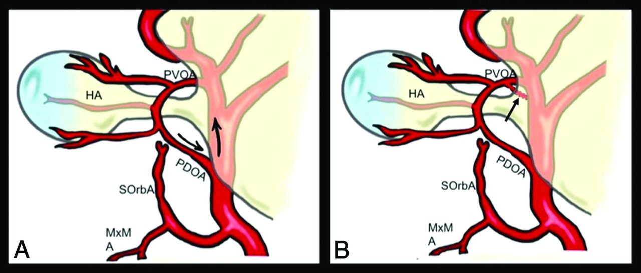
- FIG 1.
Theories of Padget and Lasjauinias et al about OA origin migration. When the embryo is about 18 mm, the OA reaches its definitive origin on the supraclinoid ICA. This phenomenon is explained by Padget13 by the cranial elongation of the ICA during this stage with the consequent movement of the PDOA origin (black arrows in A). On the other hand, Lasjaunias et al11 hypothesized the presence of an intradural anastomosis between the PVOA and the primitive ICA (black arrow in B) in correspondence with the future origin with successive regression of the original stem. HA indicates hyaloid artery; M×M, maxillo-mandibulary artery; SOrbA, supraorbital artery.
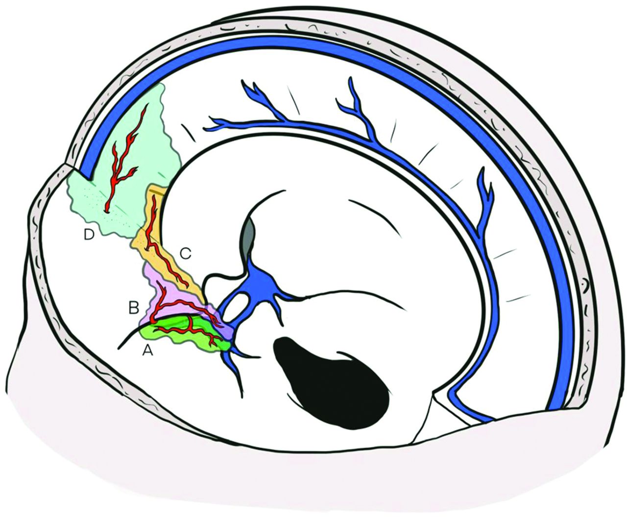
- FIG 2.
Dural territories of OA branches. A, Territory of the deep recurrent ophthalmic artery (green), which exits from the medial part of the superior orbital fissure and supplies the dura of the lateral wall of the cavernous sinus. B, Dural territory of the superficial recurrent ophthalmic artery (pink), which passes through the lateral part of the superior orbital fissure to reach the dura over the anterior clinoid process and the cavernous sinus roof. C, The posterior ethmoidal artery (orange) passes through the posterior ethmoidal canal to reach the dura of the planum sphenoidale, the posterior cribriform plate, and the anterior clinoid process. D, The anterior ethmoidal artery (light blue) passes through the anterior ethmoidal canal, and its meningeal territory consists of the anterior part of the cribriform plate, the medial part of the orbital roof, and the anterior third of the falx cerebri.
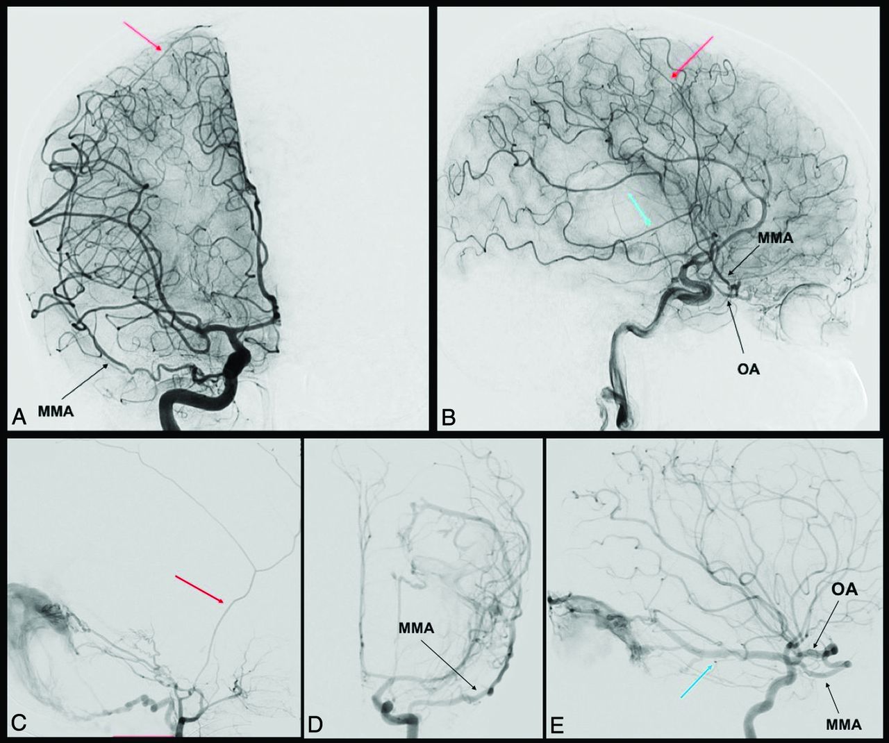
- FIG 3.
MMA origin from the OA. Anterior-posterior and lateral view angiograms (A and B) show a rare case of complete MMA origin from the OA. The OA, through the superficial recurrent OA, gives rise to the MMA, which passes through the lateral part of the superior orbital fissure and gives off its anterior (red arrow) and posterior divisions (blue arrow). In the angiograms C, D, and E, a rare case of partial origin of the MMA from the OA is shown. The angiograms D and E show a left ICA injection in frontal and lateral views, where the posterior branch of the MMA (blue arrow) originates from the OA and feeds a tentorial arteriovenous fistula. After the ECA injection (C), only the anterior branch of the MMA is enhanced (red arrow). Reproduced from Bonasia et al.27
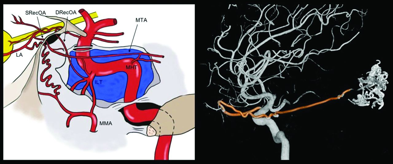
- FIG 4.
Marginal tentorial artery origin and course. The marginal tentorial artery, also called artery of the free margin of the tentorium or artery of Bernasconi and Cassinari, may have different origins, which are shown in the graphic representation. It can arise from the lacrimal artery (LA) within the orbit, through the superficial recurrent ophthalmic artery (SRecOA), from the inferolateral trunk (ILT), and from the meningohypophyseal trunk (MHT). The artery courses posterolaterally along the free margin of the tentorium. Note a 3D-DSA reconstruction of a rare case of MTA (highlighted in red) origin from the OA. The MTA exits the orbit through the superior orbital fissure (SOF) and is directed posteriorly to feed an arteriovenous malformation. DRecOA indicatesdeep recurrent ophthalmic artery .
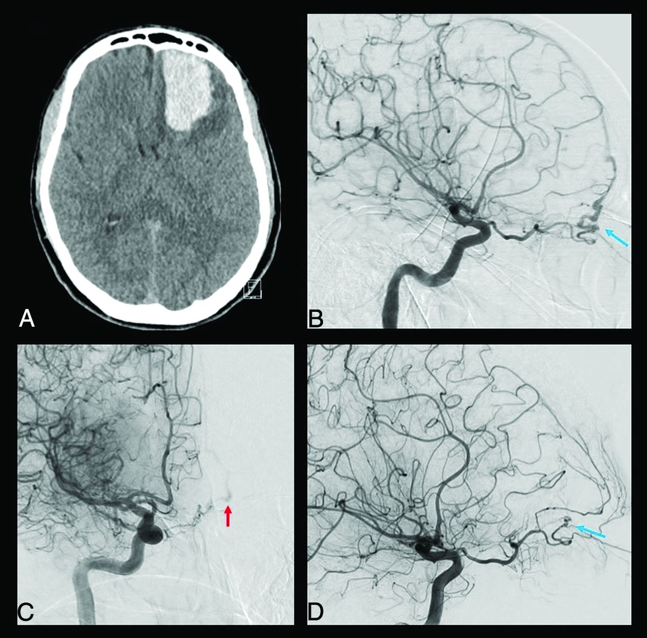
- FIG 5.
Clinical case of a ruptured cribriform plate dAVF. A 49-year-old man was admitted for sudden onset of unusual headache with nausea and vomiting. The CT scan performed in the emergency department (A) shows a left frontal basis intraparenchymal hematoma. The DSA highlighted a cribriform plate dAVF with major feeders represented by the left anterior ethmoidal artery from the left OA (blue arrow in B). The right ICA injection also showed a contribution from the contralateral OA through its ethmoidal branches (red arrow in C). The venous drainage was represented by a single cortical vein directed into the superior sagittal sinus (type III according to the Cognard-Lariboisière classification26). The patient successfully underwent left supraorbital craniotomy and clipping of the dAVF (D), with no enhancement of the dAVF on the postoperative DSA (blue arrow) and complete clinical recovery.
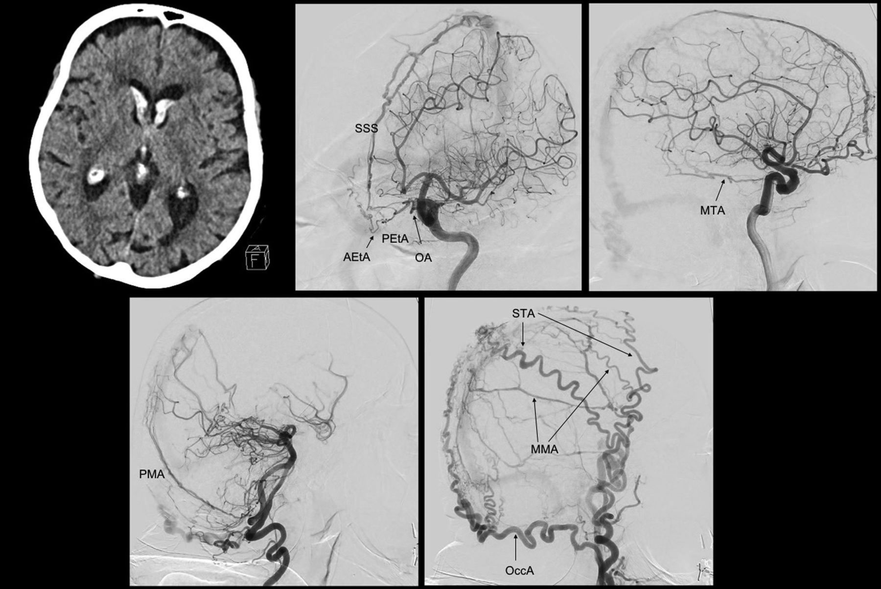
- FIG 6.
Clinical case of a dAVF fed by multiple OA dural branches. An 89-year-old woman, previously having undergone an operation for a pituitary adenoma, was admitted for unusual headache associated with vomiting. The CT scan shows an intraventricular hemorrhage with mild hydrocephalus. The diagnostic DSA shows a complex dAVF (Cognard-Lariboisière grade IIa+b26) supplied by the OA through the anterior and posterior ethmoidal arteries (AEtA, PEtA), with both direct and indirect shunts with the superior sagittal sinus (SSS). Another point of shunt with the SSS is reached by the MTA and the posterior meningeal artery (PMA) and the MMA. Also, other branches from the ECA contribute to the shunt, like the occipital artery (OccA) and the superficial temporal artery (STA). Because of the patient’s age, the complexity of the dAVF, the high risk associated with every option of treatment, and the absence of alteration of consciousness, we managed the dAVF conservatively.


Tables
OA Branches Origin from the OA Foramen Dural Territory Possible Anastomosis Clinical Consequences in Case of Embolism Deep recurrent OA First segment Superior orbital fissure Superior orbital fissure (lateral part), sphenoid wing Inferolateral trunk (ICA) Cerebrovascular accident Superficial recurrent OA Second segment Superior orbital fissure Anterior clinoid processLesser sphenoid wingMiddle fossa (anteromedial portion) Posterior ethmoidal arteryMMA (anterior division)Medial tentorial artery (ICA) Cerebrovascular accident, loss of vision Anterior ethmoidal artery Third segment Anterior ethmoidal canal Anterior convexity (anterior meningeal artery)Anterior cranial fossa (medial third)Anterior falx cerebri (anterior falcine artery) Contralateral anterior ethmoidal arteryBilateral MMAsPosterior ethmoidal arteryOlfactory branch (ACA) Posterior ethmoidal artery Third segment Posterior ethmoidal canal Anterior cranial fossa (medial third)Anterior clinoid processChiasmatic groove Contralateral posterior ethmoidal arteryAnterior ethmoidal arteryMMA (anterior division) Note:—ACA indicates anterior cerebral artery.
Type Vascular Anatomy Foramen Spinosum I Complete OA origin of the MMA Absent II Partial OA origin of the MMAAnterior division from the OAPosterior division from the IMA Reduced in size III OA origin of the accessory meningeal artery Normal Note:—IMA indicates internal maxillary artery.


Jump to section
Related Articles
Cited By...
- No citing articles found.