Advertisement

Main menu

  • Home
  • Content
    • Current Issue
    • Accepted Manuscripts
    • Article Preview
    • Past Issue Archive
    • Video Articles
    • AJNR Case Collection
    • Case of the Week Archive
    • Case of the Month Archive
    • Classic Case Archive
  • Special Collections
    • AJNR Awards
    • Low-Field MRI
    • Alzheimer Disease
    • ASNR Foundation Special Collection
    • Photon-Counting CT
    • View All
  • Multimedia
    • AJNR Podcasts
    • AJNR SCANtastic
    • Trainee Corner
    • MRI Safety Corner
    • Imaging Protocols
  • For Authors
    • Submit a Manuscript
    • Submit a Video Article
    • Submit an eLetter to the Editor/Response
    • Manuscript Submission Guidelines
    • Statistical Tips
    • Fast Publishing of Accepted Manuscripts
    • Graphical Abstract Preparation
    • Imaging Protocol Submission
    • Author Policies
  • About Us
    • About AJNR
    • Editorial Board
    • Editorial Board Alumni
  • More
    • Become a Reviewer/Academy of Reviewers
    • Subscribers
    • Permissions
    • Alerts
    • Feedback
    • Advertisers
    • ASNR Home

User menu

  • Alerts
  • Log in

Search

  • Advanced search
American Journal of Neuroradiology

American Journal of Neuroradiology

ASHNR American Society of Functional Neuroradiology ASHNR American Society of Pediatric Neuroradiology ASSR
  • Alerts
  • Log in

Advanced Search

  • Home
  • Content
    • Current Issue
    • Accepted Manuscripts
    • Article Preview
    • Past Issue Archive
    • Video Articles
    • AJNR Case Collection
    • Case of the Week Archive
    • Case of the Month Archive
    • Classic Case Archive
  • Special Collections
    • AJNR Awards
    • Low-Field MRI
    • Alzheimer Disease
    • ASNR Foundation Special Collection
    • Photon-Counting CT
    • View All
  • Multimedia
    • AJNR Podcasts
    • AJNR SCANtastic
    • Trainee Corner
    • MRI Safety Corner
    • Imaging Protocols
  • For Authors
    • Submit a Manuscript
    • Submit a Video Article
    • Submit an eLetter to the Editor/Response
    • Manuscript Submission Guidelines
    • Statistical Tips
    • Fast Publishing of Accepted Manuscripts
    • Graphical Abstract Preparation
    • Imaging Protocol Submission
    • Author Policies
  • About Us
    • About AJNR
    • Editorial Board
    • Editorial Board Alumni
  • More
    • Become a Reviewer/Academy of Reviewers
    • Subscribers
    • Permissions
    • Alerts
    • Feedback
    • Advertisers
    • ASNR Home
  • Follow AJNR on Twitter
  • Visit AJNR on Facebook
  • Follow AJNR on Instagram
  • Join AJNR on LinkedIn
  • RSS Feeds

AJNR Awards, New Junior Editors, and more. Read the latest AJNR updates

Research ArticlePediatrics

Persistent Trigeminal Artery: A Novel Imaging Finding in CHARGE Syndrome

A. Siddiqui, P. Touska, D. Josifova and S.E.J. Connor
American Journal of Neuroradiology October 2021, 42 (10) 1898-1903; DOI: https://doi.org/10.3174/ajnr.A7240
A. Siddiqui
aFrom the Department of Radiology (A.S., P.T., S.E.J.C.), Guy's and St. Thomas' NHS Foundation Trust, London, UK
bDepartment of Neuroradiology (A.S., S.E.J.C.), King's College Hospital NHS Foundation Trust, London, UK
  • Find this author on Google Scholar
  • Find this author on PubMed
  • Search for this author on this site
  • ORCID record for A. Siddiqui
P. Touska
aFrom the Department of Radiology (A.S., P.T., S.E.J.C.), Guy's and St. Thomas' NHS Foundation Trust, London, UK
  • Find this author on Google Scholar
  • Find this author on PubMed
  • Search for this author on this site
  • ORCID record for P. Touska
D. Josifova
cDepartment of Genetics (D.J.), Guy's and St. Thomas' NHS Foundation Trust, London, UK
  • Find this author on Google Scholar
  • Find this author on PubMed
  • Search for this author on this site
  • ORCID record for D. Josifova
S.E.J. Connor
aFrom the Department of Radiology (A.S., P.T., S.E.J.C.), Guy's and St. Thomas' NHS Foundation Trust, London, UK
bDepartment of Neuroradiology (A.S., S.E.J.C.), King's College Hospital NHS Foundation Trust, London, UK
dSchool of Biomedical Engineering and Imaging Sciences (S.E.J.C.), St. Thomas' Hospital, King's College, London, UK
  • Find this author on Google Scholar
  • Find this author on PubMed
  • Search for this author on this site
  • ORCID record for S.E.J. Connor
  • Article
  • Figures & Data
  • Info & Metrics
  • Responses
  • References
  • PDF
Loading

Article Figures & Data

Figures

  • Tables
  • FIG 1.
    • Download figure
    • Open in new tab
    • Download powerpoint
    FIG 1.

    PTA in CHARGE syndrome, medial variant. Axial T2WI (A and B), sagittal T2WI (C), axial CT (D), and MRA transverse (E) and lateral (F) views in a neonate demonstrate a PTA (long arrows) connecting the intracavernous ICA to the basilar artery. Note the bilateral ocular colobomas (arrowheads), absent semicircular canals, and clival cleft (arrow in D).
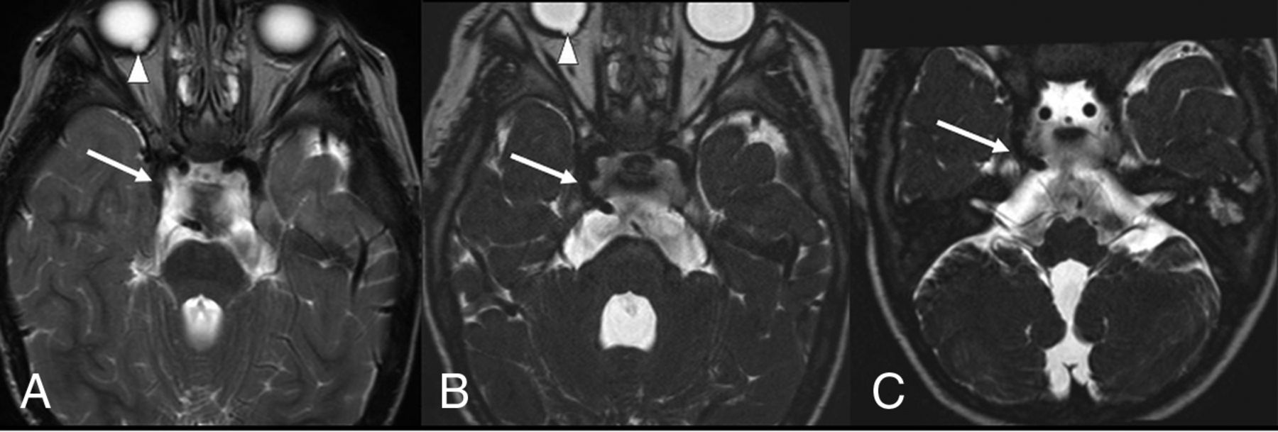
  • FIG 2.
    • Download figure
    • Open in new tab
    • Download powerpoint
    FIG 2.

    PTA in CHARGE syndrome, lateral variant. Standard 5-mm brain axial T2WI (A) and thin-section submillimetric T2WI (B and C) show a lateral-type PTA (long arrows), bilateral coloboma (arrowheads), and aplasia of the semicircular canals. The PTA is less clearly evident on the standard brain T2WI (A) compared with the thin-section images (B and C). Note the hypoplastic basilar artery below the PTA.

Tables

  • Figures
  • Prevalence of PTA versus other CHARGE clinical criteria and of the PTA in cases in which the clinical criterion was absent

    Clinical CriterionPrevalence in Literature12,25Prevalence in Our Study (95% CIs)Prevalence of PTA When the Clinical Criterion Was Absent
    PTA56%, 14/25 (95% CI, 0.35–0.76)
    Coloboma75%–90%64%, 16/25 (95% CI, 0.42–0.82)55%, 5/9 (95% CI, 0.21–0.86)
    Choanal atresia35%–65%40%, 10/25 (95% CI, 0.21–0.61)53%, 8/15 (95% CI, 0.27–0.79)
    CHARGE ear95%–100%100%, 25/25 (95% CI, 0.86–1)
    Cranial nerve dysfunction40–>95%100%, 25/25 (95% CI, 0.86–1)
    Cardiovascular anomalies50%–85%72%, 18/25 (95% CI, 0.51–0.88)57%, 4/7 (95% CI, 0.18–0.9)
    Gonadal/genitourinary anomalies50%–70%40%, 10/25 (95% CI, 0.21–0.61)67%, 10/15 (95% CI, 0.21–0.61)
    Clefting, orofacial/larynx15%–20%32%, 8/25 (95% CI, 0.15–0.53)65%, 11/17 (95% CI, 0.38–0.86)
    Growth deficiency/developmental delay70–>90%100%, 25/25 (95% CI, 0.86–1)
    Tracheoesophageal fistula/anomalies15%–20%8%, 2/25 (95% CI, 0.01–0.26)56%, 13/23 (95% CI, 0.34–0.77)
PreviousNext
Back to top

In this issue

American Journal of Neuroradiology: 42 (10)
American Journal of Neuroradiology
Vol. 42, Issue 10
1 Oct 2021
  • Table of Contents
  • Index by author
  • Complete Issue (PDF)
Advertisement
Print
Download PDF
Email Article

Thank you for your interest in spreading the word on American Journal of Neuroradiology.

NOTE: We only request your email address so that the person you are recommending the page to knows that you wanted them to see it, and that it is not junk mail. We do not capture any email address.

Enter multiple addresses on separate lines or separate them with commas.
Persistent Trigeminal Artery: A Novel Imaging Finding in CHARGE Syndrome
(Your Name) has sent you a message from American Journal of Neuroradiology
(Your Name) thought you would like to see the American Journal of Neuroradiology web site.
CAPTCHA
This question is for testing whether or not you are a human visitor and to prevent automated spam submissions.
Cite this article
A. Siddiqui, P. Touska, D. Josifova, S.E.J. Connor
Persistent Trigeminal Artery: A Novel Imaging Finding in CHARGE Syndrome
American Journal of Neuroradiology Oct 2021, 42 (10) 1898-1903; DOI: 10.3174/ajnr.A7240

Citation Manager Formats

  • BibTeX
  • Bookends
  • EasyBib
  • EndNote (tagged)
  • EndNote 8 (xml)
  • Medlars
  • Mendeley
  • Papers
  • RefWorks Tagged
  • Ref Manager
  • RIS
  • Zotero
0 Responses
Respond to this article
Share
Bookmark this article
Persistent Trigeminal Artery: A Novel Imaging Finding in CHARGE Syndrome
A. Siddiqui, P. Touska, D. Josifova, S.E.J. Connor
American Journal of Neuroradiology Oct 2021, 42 (10) 1898-1903; DOI: 10.3174/ajnr.A7240
del.icio.us logo Twitter logo Facebook logo Mendeley logo
  • Tweet Widget
  • Facebook Like
  • Google Plus One
Purchase

Jump to section

  • Article
    • Abstract
    • ABBREVIATIONS:
    • MATERIALS AND METHODS
    • RESULTS
    • DISCUSSION
    • CONCLUSIONS
    • Footnotes
    • References
  • Figures & Data
  • Info & Metrics
  • Responses
  • References
  • PDF

Related Articles

  • No related articles found.
  • PubMed
  • Google Scholar

Cited By...

  • No citing articles found.
  • Crossref
  • Google Scholar

This article has not yet been cited by articles in journals that are participating in Crossref Cited-by Linking.

More in this TOC Section

  • SyMRI & MR Fingerprinting in Brainstem Myelination
  • Comparison of Image Quality and Radiation Dose in Pediatric Temporal Bone CT Using Photon-Counting Detector CT and Energy-Integrating Detector CT
  • Dual-Layer Detector CT for PEDS Image Quality
Show more Pediatrics

Similar Articles

Advertisement

Indexed Content

  • Current Issue
  • Accepted Manuscripts
  • Article Preview
  • Past Issues
  • Editorials
  • Editor's Choice
  • Fellows' Journal Club
  • Letters to the Editor
  • Video Articles

Cases

  • Case Collection
  • Archive - Case of the Week
  • Archive - Case of the Month
  • Archive - Classic Case

More from AJNR

  • Trainee Corner
  • Imaging Protocols
  • MRI Safety Corner
  • Book Reviews

Multimedia

  • AJNR Podcasts
  • AJNR Scantastics

Resources

  • Turnaround Time
  • Submit a Manuscript
  • Submit a Video Article
  • Submit an eLetter to the Editor/Response
  • Manuscript Submission Guidelines
  • Statistical Tips
  • Fast Publishing of Accepted Manuscripts
  • Graphical Abstract Preparation
  • Imaging Protocol Submission
  • Evidence-Based Medicine Level Guide
  • Publishing Checklists
  • Author Policies
  • Become a Reviewer/Academy of Reviewers
  • News and Updates

About Us

  • About AJNR
  • Editorial Board
  • Editorial Board Alumni
  • Alerts
  • Permissions
  • Not an AJNR Subscriber? Join Now
  • Advertise with Us
  • Librarian Resources
  • Feedback
  • Terms and Conditions
  • AJNR Editorial Board Alumni

American Society of Neuroradiology

  • Not an ASNR Member? Join Now

© 2025 by the American Society of Neuroradiology All rights, including for text and data mining, AI training, and similar technologies, are reserved.
Print ISSN: 0195-6108 Online ISSN: 1936-959X

Powered by HighWire