Advertisement

Main menu

  • Home
  • Content
    • Current Issue
    • Accepted Manuscripts
    • Article Preview
    • Past Issue Archive
    • Video Articles
    • AJNR Case Collection
    • Case of the Week Archive
    • Case of the Month Archive
    • Classic Case Archive
  • Special Collections
    • AJNR Awards
    • Low-Field MRI
    • Alzheimer Disease
    • ASNR Foundation Special Collection
    • Photon-Counting CT
    • View All
  • Multimedia
    • AJNR Podcasts
    • AJNR SCANtastic
    • Trainee Corner
    • MRI Safety Corner
    • Imaging Protocols
  • For Authors
    • Submit a Manuscript
    • Submit a Video Article
    • Submit an eLetter to the Editor/Response
    • Manuscript Submission Guidelines
    • Statistical Tips
    • Fast Publishing of Accepted Manuscripts
    • Graphical Abstract Preparation
    • Imaging Protocol Submission
    • Author Policies
  • About Us
    • About AJNR
    • Editorial Board
    • Editorial Board Alumni
  • More
    • Become a Reviewer/Academy of Reviewers
    • Subscribers
    • Permissions
    • Alerts
    • Feedback
    • Advertisers
    • ASNR Home

User menu

  • Alerts
  • Log in

Search

  • Advanced search
American Journal of Neuroradiology

American Journal of Neuroradiology

ASHNR American Society of Functional Neuroradiology ASHNR American Society of Pediatric Neuroradiology ASSR
  • Alerts
  • Log in

Advanced Search

  • Home
  • Content
    • Current Issue
    • Accepted Manuscripts
    • Article Preview
    • Past Issue Archive
    • Video Articles
    • AJNR Case Collection
    • Case of the Week Archive
    • Case of the Month Archive
    • Classic Case Archive
  • Special Collections
    • AJNR Awards
    • Low-Field MRI
    • Alzheimer Disease
    • ASNR Foundation Special Collection
    • Photon-Counting CT
    • View All
  • Multimedia
    • AJNR Podcasts
    • AJNR SCANtastic
    • Trainee Corner
    • MRI Safety Corner
    • Imaging Protocols
  • For Authors
    • Submit a Manuscript
    • Submit a Video Article
    • Submit an eLetter to the Editor/Response
    • Manuscript Submission Guidelines
    • Statistical Tips
    • Fast Publishing of Accepted Manuscripts
    • Graphical Abstract Preparation
    • Imaging Protocol Submission
    • Author Policies
  • About Us
    • About AJNR
    • Editorial Board
    • Editorial Board Alumni
  • More
    • Become a Reviewer/Academy of Reviewers
    • Subscribers
    • Permissions
    • Alerts
    • Feedback
    • Advertisers
    • ASNR Home
  • Follow AJNR on Twitter
  • Visit AJNR on Facebook
  • Follow AJNR on Instagram
  • Join AJNR on LinkedIn
  • RSS Feeds

AJNR Awards, New Junior Editors, and more. Read the latest AJNR updates

Research ArticleAdult Brain
Open Access

Noninvasive Determination of IDH and 1p19q Status of Lower-grade Gliomas Using MRI Radiomics: A Systematic Review

A.P. Bhandari, R. Liong, J. Koppen, S.V. Murthy and A. Lasocki
American Journal of Neuroradiology January 2021, 42 (1) 94-101; DOI: https://doi.org/10.3174/ajnr.A6875
A.P. Bhandari
aFrom the Department of Anatomy (A.P.B.)
cTownsville University Hospital (A.P.B., J.K.), Douglas, Queensland, Australia
  • Find this author on Google Scholar
  • Find this author on PubMed
  • Search for this author on this site
  • ORCID record for A.P. Bhandari
R. Liong
dDepartment of Medical Imaging Research Office (R.L.), Royal Brisbane and Women’s Hospital, Herston, Queensland, Australia
  • Find this author on Google Scholar
  • Find this author on PubMed
  • Search for this author on this site
  • ORCID record for R. Liong
J. Koppen
cTownsville University Hospital (A.P.B., J.K.), Douglas, Queensland, Australia
  • Find this author on Google Scholar
  • Find this author on PubMed
  • Search for this author on this site
  • ORCID record for J. Koppen
S.V. Murthy
bCollege of Medicine and Dentistry (S.V.M.), James Cook University, Townsville, Queensland, Australia
  • Find this author on Google Scholar
  • Find this author on PubMed
  • Search for this author on this site
  • ORCID record for S.V. Murthy
A. Lasocki
eDepartment of Cancer Imaging (A.L.), Peter MacCallum Cancer Centre, Melbourne, Victoria, Australia
fSir Peter MacCallum Department of Oncology (A.L.), The University of Melbourne, Melbourne, Victoria, Australia
  • Find this author on Google Scholar
  • Find this author on PubMed
  • Search for this author on this site
  • ORCID record for A. Lasocki
  • Article
  • Figures & Data
  • Supplemental
  • Info & Metrics
  • Responses
  • References
  • PDF
Loading

Article Figures & Data

Figures

  • Tables
  • FIG 1.
    • Download figure
    • Open in new tab
    • Download powerpoint
    FIG 1.

    IDH status forest plot of included studies with an AUC.
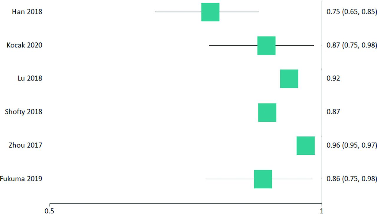
  • FIG 2.
    • Download figure
    • Open in new tab
    • Download powerpoint
    FIG 2.

    1p19q status forest plot of included studies with an AUC.

Tables

  • Figures
    • View popup
    Table 1:

    Derived aims and key findings of studies comparing IDH-mut and IDH wild-type LGG

    First Author and YearDerived AimKey Findings
    Fukuma 201922To integrate CNN deep learning features with conventional radiomic featuresConventional radiomic features: accuracy (mean ± 95% CI) = 71.7% ± 8.3%; AUC (± 95% CI) = 0.718 ± 0.139CNN features: accuracy = 69.6% ± 5.6%; AUC = 0.619 ± 0.132CNN and conventional radiomic features: accuracy = 73.1% ± 9.4%; AUC = 0.699 ± 0.145
    Gihr 202023To determine if intensity features relate to IDH statusEntropy, a second-order histogram parameter of the ADC volume was significant: IDH-mut versus IDH wild-type, mean ± SD = 5.5 ± 0.63 vs 4.75 ± 0.69; P = .0144
    Jakola 201824To determine if texture features can predict IDH status on FLAIRHomogeneity and volume could classify IDH status with an AUC = 0.940 (85% sensitivity, 100% specificity) using the generalized linear model
    Kim 202025To determine if DWI- and DSC perfusion-based image integration with standard imaging (T1WI postcontrast and FLAIR) can improve classificationIntegration increased the AUC (95% CI) = 0.747 (0.663–0.832); (53.6% sensitivity and 86.7% specificity) from 0.705 (0.613–0.796) (43.9% sensitivity and 88.8% specificity) compared with conventional MR imaging radiomics
    Li 201727To determine if integration of deep learning features into the radiogenomic pipeline improves classificationConventional radiomics produced an AUC = 0.85 (sensitivity of 82.9%, specificity of 73.5%)CNN deep learning–derived features plus conventional radiomic features with feature selection produced an AUC = 0.95 (sensitivity of 94.4%; specificity of 86.7%)
    Lu 201828To determine the best ML classifierLinear SVM classified IDH status with an AUC = 0.936 (sensitivity of 85.7%, specificity of 93.0%)
    Park 202029To determine if DTI improves classification when added to conventional radiomicsAddition of DTI radiomic features to conventional imaging radiomics increased the AUC (95% CI) = 0.900 (0.855–0.945) from 0.835 (0.773–0.896)
    Ren 201930To compare radiomic, VASARI, and radiomic plus VASARI features derived from FLAIR, ADC, eADC, and CBFRadiomics: AUC (95% CI) = 0.931 (0.842–1); sensitivity of 100%, specificity of 85.71%VASARI: AUC = 0.843 (sensitivity of 91.67%; specificity of 61.90%)Radiomics plus VASARI: AUC = 0.888 (0.786–0.989); sensitivity of 94.44% and specificity of 71.43%
    Yu 201732To classify using the improved genetic algorithm for feature selection and leave-one-out cross-validation method in WHO grade II LGGUsing the proposed method and the SVM ML classifier, an AUC = 0.71 (sensitivity = 56% and specificity = 74%) was achieved
    Zhou 201734To determine if VASARI annotations were superior to standard radiomic classification analysisIDH classification through texture features found an AUC (± 95% CI) = 0.79 ± 0.02; sensitivity 90%, specificity of 89%IDH classification through VASARI features, AUC = 0.73 ± 0.02; sensitivity of 69%, specificity of 69%
    Zhang 201833To classify by conventional radiomicsAUC = 0.830 (sensitivity = 82%, specificity = 92%) using SVM
    • Note:—eADC indicates exponential ADC.

    • View popup
    Table 2:

    Derived aims and key findings of studies examining 1p19q status of IDH-mut LGG

    First Author and YearDerived AimKey Findings
    Han 202035To determine if clinical and standard imaging factors improve classificationThe AUC (95% CI) = 0.753 (0.654–0.852) for clinical plus radiomic features versus AUC = 0.760 (0.663-0.857) for just radiomic features; radiomic features were superior to clinical features alone, AUC = 0.627 (0.551–0.703)
    Kocak 202026To determine the best ML classifierThe neural network produced the highest AUC (95% CI) = 0.869 (0.751–0.981); sensitivity of 87.5%, specificity of 75.8%
    Lu 201828To determine the best ML classifierClassification occurred with an AUC = 0.92 (sensitivity of 88.5%, specificity of 86.2%) using quadratic SVM
    Shofty 201831To determine the best ML classifierClassification occurred with an AUC = 0.87 (sensitivity of 92%, specificity of 83%) using ensemble bagged trees classifier
    Zhou 201734To determine if VASARI annotations were superior to standard radiomic analysis for classificationTexture features classified with an AUC (± 95% CI) = 0.96 ± 0.01; sensitivity of 90% ± 2%, specificity of 89% ± 2%VASARI features classified with an AUC = 0.78 ± 0.02; sensitivity of 72% ± 3%, specificity of 67% ±3%
    Fukuma 201922To determine if integration of CNN deep learning with radiomic features improved classificationConventional radiomic features (± 95% CI): accuracy = 59.0 ± 9.0%; AUC = 0.656 ± 0.113CNN features: accuracy = 84.0 ± 9.3%; AUC = 0.868 ± 0.099CNN and conventional radiomic features: accuracy = 79.8 ± 11.0%; AUC = 0.861 ± 0.116
PreviousNext
Back to top

In this issue

American Journal of Neuroradiology: 42 (1)
American Journal of Neuroradiology
Vol. 42, Issue 1
1 Jan 2021
  • Table of Contents
  • Index by author
  • Complete Issue (PDF)
Advertisement
Print
Download PDF
Email Article

Thank you for your interest in spreading the word on American Journal of Neuroradiology.

NOTE: We only request your email address so that the person you are recommending the page to knows that you wanted them to see it, and that it is not junk mail. We do not capture any email address.

Enter multiple addresses on separate lines or separate them with commas.
Noninvasive Determination of IDH and 1p19q Status of Lower-grade Gliomas Using MRI Radiomics: A Systematic Review
(Your Name) has sent you a message from American Journal of Neuroradiology
(Your Name) thought you would like to see the American Journal of Neuroradiology web site.
CAPTCHA
This question is for testing whether or not you are a human visitor and to prevent automated spam submissions.
Cite this article
A.P. Bhandari, R. Liong, J. Koppen, S.V. Murthy, A. Lasocki
Noninvasive Determination of IDH and 1p19q Status of Lower-grade Gliomas Using MRI Radiomics: A Systematic Review
American Journal of Neuroradiology Jan 2021, 42 (1) 94-101; DOI: 10.3174/ajnr.A6875

Citation Manager Formats

  • BibTeX
  • Bookends
  • EasyBib
  • EndNote (tagged)
  • EndNote 8 (xml)
  • Medlars
  • Mendeley
  • Papers
  • RefWorks Tagged
  • Ref Manager
  • RIS
  • Zotero
0 Responses
Respond to this article
Share
Bookmark this article
Noninvasive Determination of IDH and 1p19q Status of Lower-grade Gliomas Using MRI Radiomics: A Systematic Review
A.P. Bhandari, R. Liong, J. Koppen, S.V. Murthy, A. Lasocki
American Journal of Neuroradiology Jan 2021, 42 (1) 94-101; DOI: 10.3174/ajnr.A6875
del.icio.us logo Twitter logo Facebook logo Mendeley logo
  • Tweet Widget
  • Facebook Like
  • Google Plus One
Purchase

Jump to section

  • Article
    • Abstract
    • ABBREVIATIONS:
    • MATERIALS AND METHODS
    • RESULTS
    • DISCUSSION
    • CONCLUSIONS
    • Footnotes
    • References
  • Figures & Data
  • Supplemental
  • Info & Metrics
  • Responses
  • References
  • PDF

Related Articles

  • PubMed
  • Google Scholar

Cited By...

  • Automated Determination of the H3 K27-Altered Status in Spinal Cord Diffuse Midline Glioma by Radiomics Based on T2-Weighted MR Images
  • Radiogenomics Provides Insights into Gliomas Demonstrating Single-Arm 1p or 19q Deletion
  • Machine Learning in Differentiating Gliomas from Primary CNS Lymphomas: A Systematic Review, Reporting Quality, and Risk of Bias Assessment
  • Crossref (58)
  • Google Scholar

This article has been cited by the following articles in journals that are participating in Crossref Cited-by Linking.

  • Deep Learning With Radiomics for Disease Diagnosis and Treatment: Challenges and Potential
    Xingping Zhang, Yanchun Zhang, Guijuan Zhang, Xingting Qiu, Wenjun Tan, Xiaoxia Yin, Liefa Liao
    Frontiers in Oncology 2022 12
  • Systematic review of the radiomics quality score applications: an EuSoMII Radiomics Auditing Group Initiative
    Gaia Spadarella, Arnaldo Stanzione, Tugba Akinci D’Antonoli, Anna Andreychenko, Salvatore Claudio Fanni, Lorenzo Ugga, Elmar Kotter, Renato Cuocolo
    European Radiology 2022 33 3
  • MRI biomarkers in neuro-oncology
    Marion Smits
    Nature Reviews Neurology 2021 17 8
  • MRI-Based Radiomics and Radiogenomics in the Management of Low-Grade Gliomas: Evaluating the Evidence for a Paradigm Shift
    Ahmed Habib, Nicolina Jovanovich, Meagan Hoppe, Murat Ak, Priyadarshini Mamindla, Rivka R. Colen, Pascal O. Zinn
    Journal of Clinical Medicine 2021 10 7
  • Multicenter DSC–MRI-Based Radiomics Predict IDH Mutation in Gliomas
    Georgios C. Manikis, Georgios S. Ioannidis, Loizos Siakallis, Katerina Nikiforaki, Michael Iv, Diana Vozlic, Katarina Surlan-Popovic, Max Wintermark, Sotirios Bisdas, Kostas Marias
    Cancers 2021 13 16
  • Accuracy of Machine Learning Algorithms for the Classification of Molecular Features of Gliomas on MRI: A Systematic Literature Review and Meta-Analysis
    Evi J. van Kempen, Max Post, Manoj Mannil, Benno Kusters, Mark ter Laan, Frederick J. A. Meijer, Dylan J. H. A. Henssen
    Cancers 2021 13 11
  • Machine Learning-Based Multiparametric Magnetic Resonance Imaging Radiomics for Prediction of H3K27M Mutation in Midline Gliomas
    Sedat Giray Kandemirli, Burak Kocak, Shotaro Naganawa, Kerem Ozturk, Stephen S.F. Yip, Saurav Chopra, Luciano Rivetti, Amro Saad Aldine, Karra Jones, Zuzan Cayci, Toshio Moritani, Takashi Shawn Sato
    World Neurosurgery 2021 151
  • Multi-Modal Learning for Predicting the Genotype of Glioma
    Yiran Wei, Xi Chen, Lei Zhu, Lipei Zhang, Carola-Bibiane Schönlieb, Stephen Price, Chao Li
    IEEE Transactions on Medical Imaging 2023 42 11
  • Conventional MRI features can predict the molecular subtype of adult grade 2–3 intracranial diffuse gliomas
    Arian Lasocki, Michael E. Buckland, Katharine J. Drummond, Heng Wei, Jing Xie, Michael Christie, Andrew Neal, Frank Gaillard
    Neuroradiology 2022 64 12
  • Development and external validation of a non-invasive molecular status predictor of chromosome 1p/19q co-deletion based on MRI radiomics analysis of Low Grade Glioma patients
    Roberto Casale, Elizaveta Lavrova, Sebastian Sanduleanu, Henry C. Woodruff, Philippe Lambin
    European Journal of Radiology 2021 139

More in this TOC Section

Adult Brain

  • Diagnostic Neuroradiology of Monoclonal Antibodies
  • Clinical Outcomes After Chiari I Decompression
  • Segmentation of Brain Metastases with BLAST
Show more Adult Brain

Functional

  • Kurtosis and Epileptogenic Tubers: A Pilot Study
  • Glutaric Aciduria Type 1: DK vs. Conventional MRI
  • Multiparametric MRI in PEDS Pontine Glioma
Show more Functional

Similar Articles

Advertisement

Indexed Content

  • Current Issue
  • Accepted Manuscripts
  • Article Preview
  • Past Issues
  • Editorials
  • Editor's Choice
  • Fellows' Journal Club
  • Letters to the Editor
  • Video Articles

Cases

  • Case Collection
  • Archive - Case of the Week
  • Archive - Case of the Month
  • Archive - Classic Case

More from AJNR

  • Trainee Corner
  • Imaging Protocols
  • MRI Safety Corner
  • Book Reviews

Multimedia

  • AJNR Podcasts
  • AJNR Scantastics

Resources

  • Turnaround Time
  • Submit a Manuscript
  • Submit a Video Article
  • Submit an eLetter to the Editor/Response
  • Manuscript Submission Guidelines
  • Statistical Tips
  • Fast Publishing of Accepted Manuscripts
  • Graphical Abstract Preparation
  • Imaging Protocol Submission
  • Evidence-Based Medicine Level Guide
  • Publishing Checklists
  • Author Policies
  • Become a Reviewer/Academy of Reviewers
  • News and Updates

About Us

  • About AJNR
  • Editorial Board
  • Editorial Board Alumni
  • Alerts
  • Permissions
  • Not an AJNR Subscriber? Join Now
  • Advertise with Us
  • Librarian Resources
  • Feedback
  • Terms and Conditions
  • AJNR Editorial Board Alumni

American Society of Neuroradiology

  • Not an ASNR Member? Join Now

© 2025 by the American Society of Neuroradiology All rights, including for text and data mining, AI training, and similar technologies, are reserved.
Print ISSN: 0195-6108 Online ISSN: 1936-959X

Powered by HighWire