
Article Figures & Data
Figures
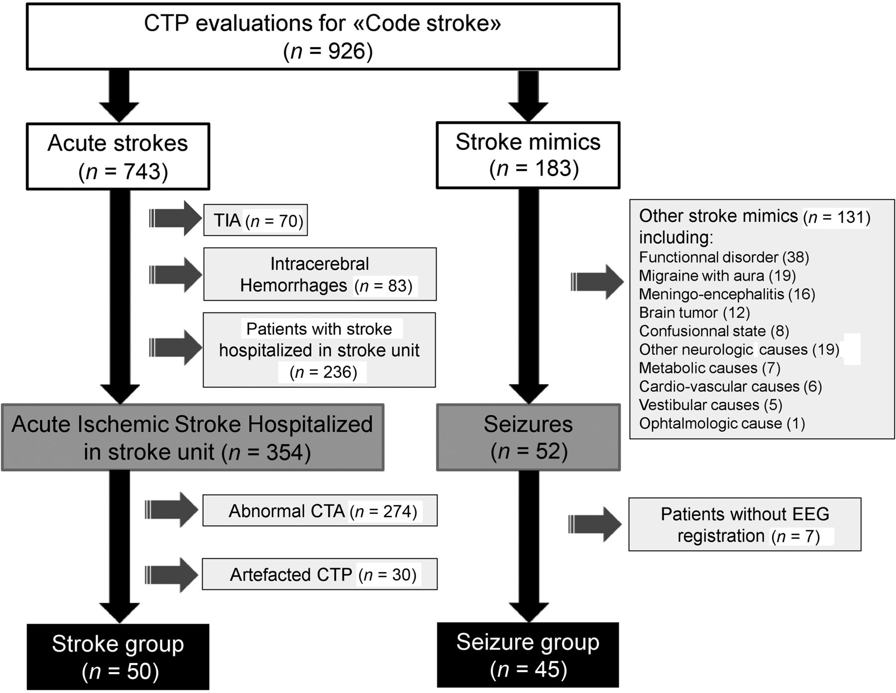
- FIG 1.
Flow chart.
- FIG 2.
CTP images showing examples of hyperperfusion respecting (A) or not (B) a vascular territory in the seizure group. A, An 84-year-old woman presenting with Wernicke aphasia and complete right hemianopsia: left fronto-parieto-occipital hyperperfusion, which may correspond with the posterior cortical branch of left middle cerebral artery. B, An 80-year-old woman presenting with mutism, right hemiplegia, and left forced gaze deviation: left parieto-occipital hyperperfusion not respecting a vascular territory (the left posterior cerebral artery was exclusively from the basilar artery).
- FIG 3.
A 92-year-old patient presenting with aphasia and right central facial palsy: focal hyperperfusion on CTP in favor of immediate poststroke “luxury perfusion.” A, Focal decrease of the Tmax parameter in the left prefrontal area in favor of relative hyperperfusion. B, Focal increase of the CBV parameter in the same area. C, Correlation with follow-up MR imaging performed 26 hours after clinical presentation identifying a bifocal acute ischemic stroke.
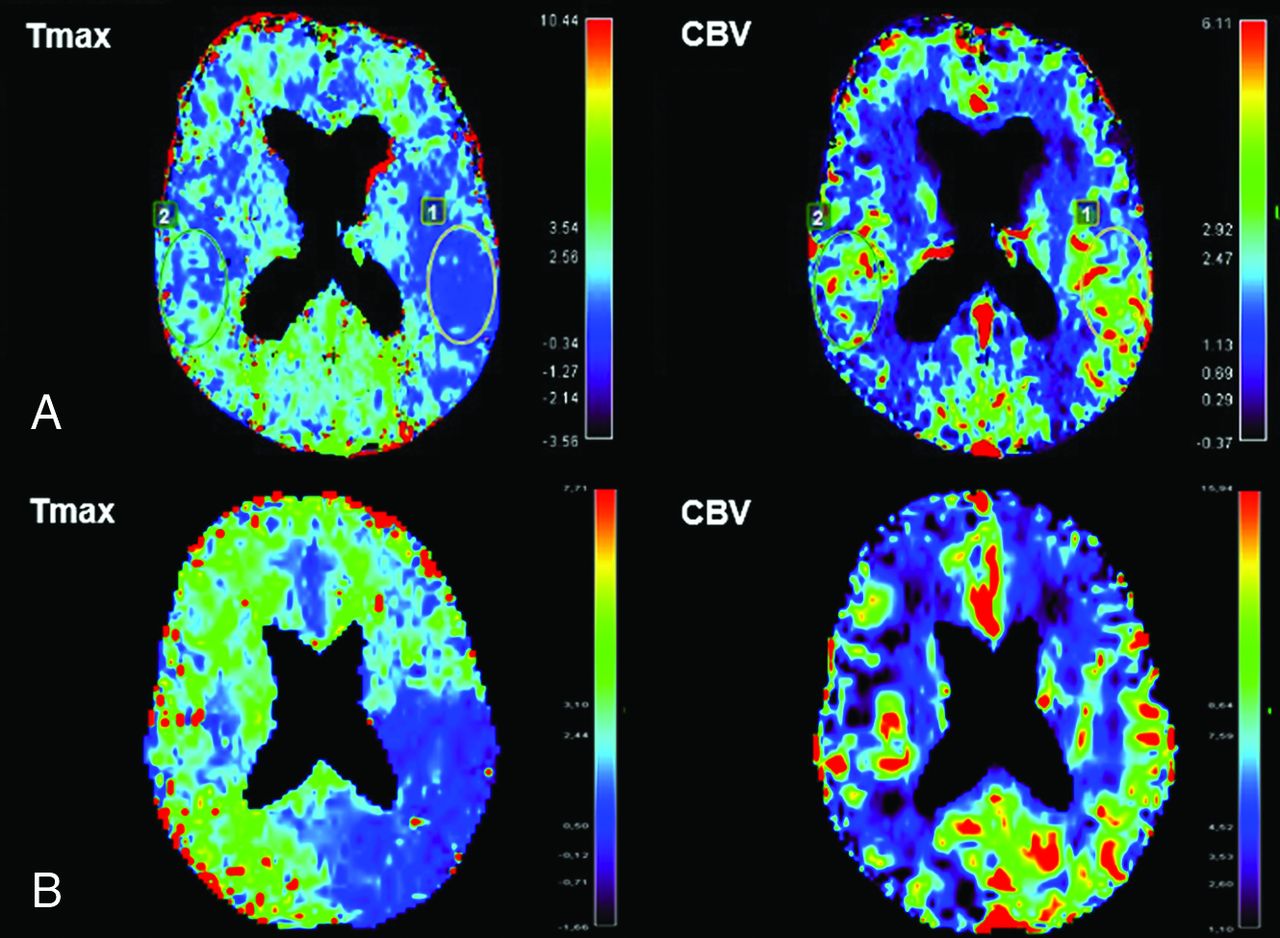
- FIG 4.
Comparison of CTP hypoperfusion patterns between seizure and stroke groups. A, A 61-year-old man presenting with seizure-related aphasia and right face and arm palsy: left holohemispheric hypoperfusion, absence of modification of the CBV parameter. B, A 47-year-old woman presenting with seizure-related aphasia and right-arm palsy: hypoperfusion, which may correspond with a vascular territory, and a relative decrease (33%) of the CBV parameter in the same area. C, A 60-year-old man with stroke-related aphasia and right-arm and facial palsy: hypoperfusion corresponding with posterior territory of the left middle cerebral artery, absence of modification of the CBV parameter, and MCA acute ischemic stroke within the hypoperfusion region on follow-up MR imaging. D, A 77-year-old man with stroke-related dysarthria and left facial palsy: hypoperfusion corresponding to a part of the cortical ribbon of the right medium cerebral artery, no modification of the CBV parameter, and MCA acute ischemic stroke within the hypoperfusion territory on follow-up MR imaging.


Tables
Stroke Group (n = 50) Seizure Group (n = 45) P Age (yr) 63.68 [SD, 14.45] 73.81 [SD, 12.6 <.01 Female sex 20 (40) 22 (49) <.68 Cardiovascular risk factors 37 (74) 35 (78) <.66 Hypertension 22 (44) 33 (73) .01 Current smoking 24 (48) 5 (11) .01 Diabetes 5 (10) 9 (20) <.17 Dyslipidemia 15 (30) 16 (36) .57 History of epileptic seizures 2 (4) 11 (24) .<01 History of stroke 6 (12) 16 (36) <.01 Initial NIHSS score 4.54 [SD, 4.10] 7.44 [SD, 6.34] .01 Symptoms Aphasia 13 (26) 37 (82) <.01 Motor deficit 39 (78) 27 (60) .06 Sensitive deficit 16 (32) 8 (18) .12 Impaired consciousness 0 (0) 4 (9) .025 Hemianopsia 4(8) 8 (18) .15 Dysarthria 15 (30) 1 (2) <.01 Ataxia 16 (32) 1 (2) <.01 CTP delay from symptom onset (min) 153.08 [SD, 73.24] 154.98 [SD, 62.71] .77 Received thrombolysis 26 (52) 8 (18) <.01 Stroke mechanisms (according to ASCOD classification) Cardioembolism 20 (40) – Small-vessel disease 15 (30) – Atherothrombosis 5 (10) – Other causes 4 (8) – Dissection 0 (0) – Undetermined 6 (12) – Note:—ASCOD indicates ?????; –, ???????.
↵a Values are mean [SD] or No. (%).
Stroke Group(n = 50) Seizure Group(n = 45) P Perfusion abnormality 20 (40) 33 (73) .01 Perfusion abnormality not restricted to the vascular territory 1 (2) 20 (44) <.01 Hypoperfusion 19 (38) 17 (38) .1 Restricted to vascular territory 18 (36) 6 (13) .01 Not restricted to vascular territory 1 (2) 11 (24) <.01 Hyperperfusion 1 (2) 16 (36) <.01 Restricted to vascular territory 1 (2) 7 (16) .02 Not restricted to vascular territory 0 (0) 9 (20) <.01 ↵a Values are numbers (%).
Stroke Group (n = 19) Seizure Group (n = 17) P Hypoperfusion pattern Symptomatic ROI Tmax ratio 1.98 [SD, 0.62] 1.50 [SD, 0.36] .01 MTT ratio 1.15 [SD, 0.14] 0.99 [SD, 0.16] .002 CBV ratio 0.88 [SD, 0.19] 0.86 [SD, 0.31] .42 CBF ratio 0.97 [SD, 0.39] 0.84 [SD, 0.19] .38 Frontal ROI Tmax ratio 1.01 [SD, 0.24] 1.18 [SD, 0.27] .05 MTT ratio 1.02 [SD, 0.09] 0.95 [SD, 0.19] .12 CBV ratio 1.03 [SD, 0.18] 0.90 [SD, 0.26] .23 CBF ratio 1.01 [SD, 0.13] 0.93 [SD, 0.15] .36 Temporal ROI Tmax ratio 0.99 [SD, 0.24] 1.35 [SD, 0.45] <.01 MTT ratio 0.92 [SD, 0.21] 0.95 [SD, 0.24] 1 CBV ratio 0.90 [SD, 0.27] 0.92 [SD, 0.32] .67 CBF ratio 0.98 [SD, 0.14] 0.93 [SD, 0.24] .12 Thalamic ROI Tmax ratio 1.05 [SD, 0.41] 1.32 [SD, 0.70] .20 MTT ratio 0.99 [SD, 0.23] 1.03 [SD, 0.21] .53 CBV ratio 0.94 [SD, 0.26] 0.97 [SD, 0.22] .66 CBF ratio 0.99 [SD, 0.21] 0.97 [SD, 0.24] .57 Cerebellar ROI n = 10b n = 8b Tmax ratio 0.95 [SD, 0.32] 0.98 [SD, 0.21] 1 MTT ratio 1.02 [SD, 0.14] 1.09 [SD, 0.3] .69 CBV ratio 0.99 [SD, 0.21]] 1.14 [SD, 0.45] .62 CBF ratio 0.97 [SD, 0.13] 1.03 [SD, 0.21] .46 Normoperfusion pattern n = 30 n = 12 Symptomatic ROI Tmax ratio 1.15 [SD, 0.51] 1.00 [SD, 0.45] .55 MTT ratio 0.99 [SD, 0.12] 1.04 [SD, 0.24] .85 CBV ratio 1.08 [SD, 0.53] 1.18 [SD, 0.41] .67 CBF ratio 1.06 [SD, 0.39] 1.14 [SD, 0.34] .45 Frontal ROI Tmax ratio 1.01 [SD, 0.12] 1.02 [SD, 0.14] .72 MTT ratio 1 [SD, 0.05] 1.01 [SD, 0.06] .46 CBV ratio 1.01 [SD, 0.08] 0.99 [SD, 0.13] .61 CBF ratio 1 [SD, 0.06] 0.98 [SD, 0.08] .60 Temporal ROI Tmax ratio 1.15 [SD, 0.3] 0.96 [SD, 0.20 .07 MTT ratio 1 [SD, 0.19] 1.02 [SD, 0.19] .99 CBV ratio 0.97 [SD, 0.23] 0.99 [SD, 0.19] .38 CBF ratio 0.96 [SD, 0.13] 0.99 [SD, 0.14] .30 Thalamic ROI Tmax ratio 1.11 [SD, 0.36] 1.04 [SD, 0.34] .94 MTT ratio 0.95 [SD, 0.17] 0.95 [SD, 0.23] .99 CBV ratio 0.97 [SD, 0.25] 1.09 [SD, 0.40] .52 CBF ratio 0.98 [SD, 0.14] 1.08 [SD, 0.18] .1 Cerebellar ROI n = 10b n = 8b Tmax ratio 1.01 [SD, 0.16] 0.94 [SD, 0.08] .14 MTT ratio 1.02 [SD, 0.15] 1.10 [SD, 0.30] .93 CBV ratio 1.04 [SD, 0.17] 1.13 [SD, 0.33] .87 CBF ratio 1.02 [SD, 0.09] 1.07 [SD, 0.10] .21 a Values are mean [SD].
↵b For 9 patients in each group, perfusion analysis of the cerebellar ROI could not be performed because of the poor quality of perfusion maps.


Jump to section
Related Articles
Cited By...
- Serum Lactate-Based Stratification for Seizure Diagnosis in Resource-Limited Neurologic Emergency Settings
- Time is Brain: Detection of Nonconvulsive Seizures and Status Epilepticus During Acute Stroke Evaluation Using Point-of-Care Electroencephalography
- Stroke Mimics in the Acute Setting: Role of Multimodal CT Protocol