Advertisement

Main menu

  • Home
  • Content
    • Current Issue
    • Accepted Manuscripts
    • Article Preview
    • Past Issue Archive
    • Video Articles
    • AJNR Case Collection
    • Case of the Week Archive
    • Case of the Month Archive
    • Classic Case Archive
  • Special Collections
    • AJNR Awards
    • Low-Field MRI
    • Alzheimer Disease
    • ASNR Foundation Special Collection
    • Photon-Counting CT
    • View All
  • Multimedia
    • AJNR Podcasts
    • AJNR SCANtastic
    • Trainee Corner
    • MRI Safety Corner
    • Imaging Protocols
  • For Authors
    • Submit a Manuscript
    • Submit a Video Article
    • Submit an eLetter to the Editor/Response
    • Manuscript Submission Guidelines
    • Statistical Tips
    • Fast Publishing of Accepted Manuscripts
    • Graphical Abstract Preparation
    • Imaging Protocol Submission
    • Author Policies
  • About Us
    • About AJNR
    • Editorial Board
    • Editorial Board Alumni
  • More
    • Become a Reviewer/Academy of Reviewers
    • Subscribers
    • Permissions
    • Alerts
    • Feedback
    • Advertisers
    • ASNR Home

User menu

  • Alerts
  • Log in

Search

  • Advanced search
American Journal of Neuroradiology

American Journal of Neuroradiology

ASHNR American Society of Functional Neuroradiology ASHNR American Society of Pediatric Neuroradiology ASSR
  • Alerts
  • Log in

Advanced Search

  • Home
  • Content
    • Current Issue
    • Accepted Manuscripts
    • Article Preview
    • Past Issue Archive
    • Video Articles
    • AJNR Case Collection
    • Case of the Week Archive
    • Case of the Month Archive
    • Classic Case Archive
  • Special Collections
    • AJNR Awards
    • Low-Field MRI
    • Alzheimer Disease
    • ASNR Foundation Special Collection
    • Photon-Counting CT
    • View All
  • Multimedia
    • AJNR Podcasts
    • AJNR SCANtastic
    • Trainee Corner
    • MRI Safety Corner
    • Imaging Protocols
  • For Authors
    • Submit a Manuscript
    • Submit a Video Article
    • Submit an eLetter to the Editor/Response
    • Manuscript Submission Guidelines
    • Statistical Tips
    • Fast Publishing of Accepted Manuscripts
    • Graphical Abstract Preparation
    • Imaging Protocol Submission
    • Author Policies
  • About Us
    • About AJNR
    • Editorial Board
    • Editorial Board Alumni
  • More
    • Become a Reviewer/Academy of Reviewers
    • Subscribers
    • Permissions
    • Alerts
    • Feedback
    • Advertisers
    • ASNR Home
  • Follow AJNR on Twitter
  • Visit AJNR on Facebook
  • Follow AJNR on Instagram
  • Join AJNR on LinkedIn
  • RSS Feeds

AJNR Awards, New Junior Editors, and more. Read the latest AJNR updates

Research ArticleSpine

Respiratory Phase Affects the Conspicuity of CSF–Venous Fistulas in Spontaneous Intracranial Hypotension

T.J. Amrhein, L. Gray, M.D. Malinzak and P.G. Kranz
American Journal of Neuroradiology September 2020, 41 (9) 1754-1756; DOI: https://doi.org/10.3174/ajnr.A6663
T.J. Amrhein
aFrom the Department of Radiology, Duke University Medical Center, Durham, North Carolina.
  • Find this author on Google Scholar
  • Find this author on PubMed
  • Search for this author on this site
  • ORCID record for T.J. Amrhein
L. Gray
aFrom the Department of Radiology, Duke University Medical Center, Durham, North Carolina.
  • Find this author on Google Scholar
  • Find this author on PubMed
  • Search for this author on this site
  • ORCID record for L. Gray
M.D. Malinzak
aFrom the Department of Radiology, Duke University Medical Center, Durham, North Carolina.
  • Find this author on Google Scholar
  • Find this author on PubMed
  • Search for this author on this site
  • ORCID record for M.D. Malinzak
P.G. Kranz
aFrom the Department of Radiology, Duke University Medical Center, Durham, North Carolina.
  • Find this author on Google Scholar
  • Find this author on PubMed
  • Search for this author on this site
  • ORCID record for P.G. Kranz
  • Article
  • Figures & Data
  • Info & Metrics
  • Responses
  • References
  • PDF
Loading

Abstract

SUMMARY: Spinal CSF–venous fistulas are a cause of spontaneous intracranial hypotension that can be difficult to detect on imaging. We describe how the respiratory phase affects the visibility of CSF–venous fistulas during myelography.

ABBREVIATIONS:

CTM
CT myelography
CVF
CSF–venous fistula
SIH
spontaneous intracranial hypotension

Spontaneous intracranial hypotension (SIH) is a debilitating condition caused by spinal CSF leaks and CSF–venous fistulas (CVFs).1 The localization of these spinal causes of SIH is critical for successful treatment.2,3 CVFs are particularly difficult to identify on traditional myelographic imaging.1,4 Technical modifications to the myelography technique such as decubitus imaging during dynamic myelography, digital subtraction myelography, and CT myelography (CTM) have been shown to improve the conspicuity of CVFs, which are often subtle.5 However, additional methods for maximizing CVF identification remain desirable. We have observed that the ability to visualize CVFs varies with different phases of respiration, suggesting that attention to the respiratory phase of imaging may improve their detection in some patients.

MATERIALS AND METHODS

Subjects

This was a retrospective case series of 5 patients with SIH treated at a single major referral institution from January 2018 to February 2020. All patients met the International Classification of Headache Disorders-3 criteria for SIH and had preprocedural contrast-enhanced brain MR imaging that was positive for SIH.6,7 Patients were included when the conspicuity of a spinal CVF was affected by the respiratory phase during image acquisition. In all cases, an initial prone CTM demonstrated findings suggestive of a spinal CVF, necessitating further myelography in the ipsilateral decubitus position. This study was Health Insurance Portability and Accountability Act–compliant and was approved by the institutional internal review board with a waiver of informed consent.

Myelogram Technique

Decubitus myelography was performed in all cases due to subtle findings suggestive of a spinal CVF on a prior prone CTM. Decubitus myelograms were obtained as previously described using 10 mL of iopamidol containing 300 mg/mL of iodine (Isovue-M 300; Bracco).5 Dynamic myelograms were all obtained on the same C-arm fluoroscopy unit equipped with a tilting table (MultiDiagnost Eleva; Philips Healthcare). Digital subtraction was not used. CTMs were all performed on a single 64–detector row CT scanner (Discovery 750HD; GE Healthcare) with the following parameters: helical scan mode, rotation time = 0.8 seconds, pitch = 0.969, tube voltage = 120 kV (peak), automated exposure control, tube current = 300–800 mA, noise index = 19.5, section thickness = 2.5 mm, interval = 2.5 mm, reconstruction thickness = 0.625 mm. Imaging was focused over the nerve root associated with the suspected CVF to limit the radiation dose. In 4 of the 5 patients, both a dynamic myelogram and a CTM in the decubitus position were obtained. In the other patient, only a decubitus CTM was performed. Images were obtained during breath-hold for all cases at various phases of the respiratory cycle (eg, neutral, full inspiration, and full expiration).

RESULTS

Subjects

The mean patient age was 60 years (range, 44–68 years), and 40% were women. All patients in this series coincidentally had undergone 2 CTMs before images were acquired with attention to the phase of the respiratory cycle. In all patients, 1 of the 2 prior CTMs had negative findings, and the other demonstrated a subtle finding suspicious for a CVF, which formed the basis for the decision to acquire the images depicted in this report. CVFs all originated from nerve root sleeves, consistent with previous reports, and were identified on the right at T9 and on the left at C8, T2, T8, and T10.8

Myelogram Findings

Images were acquired with breath-hold during various phases of the respiratory cycle, with the patient in the lateral decubitus position. Inspiratory imaging was compared with additional phases of respiration including expiration (n = 3), neutral breath hold (n = 1), and Valsalva (n = 1). In all 5 patients, the CVF was more conspicuous during the inspiratory phase (Figs 1⇓⇓⇓-5).

FIG 1.
  • Download figure
  • Open in new tab
  • Download powerpoint
FIG 1.

Maximum-intensity-projection CT myelograms of a right T9 nerve root sleeve CSF–venous fistula. A, Image acquisition during inspiration. Marked increased conspicuity of the CSF–venous fistula and hyperdense paraspinal vein (arrows). B, Image during expiration. The CSF–venous fistula is no longer visible.

FIG 2.
  • Download figure
  • Open in new tab
  • Download powerpoint
FIG 2.

Spot-magnified radiographs of a left T2 nerve root sleeve CSF–venous fistula during an ipsilateral decubitus dynamic myelogram. A, Image acquired during inspiration demonstrates well the contrast-opacified CSF–venous fistula (arrows). B, Image during a Valsalva maneuver results in considerably reduced visualization of the CSF–venous fistula.

FIG 3.
  • Download figure
  • Open in new tab
  • Download powerpoint
FIG 3.

Parasagittal maximum-intensity-projection CT myelograms through the neuroforamen of a left T10 CSF–venous fistula. A, Image acquired during inspiration clearly captures contrast within the CSF–venous fistula (arrowhead). B, Image acquired during expiration. Note the markedly reduced conspicuity of the CSF–venous fistula (arrowhead).

FIG 4.
  • Download figure
  • Open in new tab
  • Download powerpoint
FIG 4.

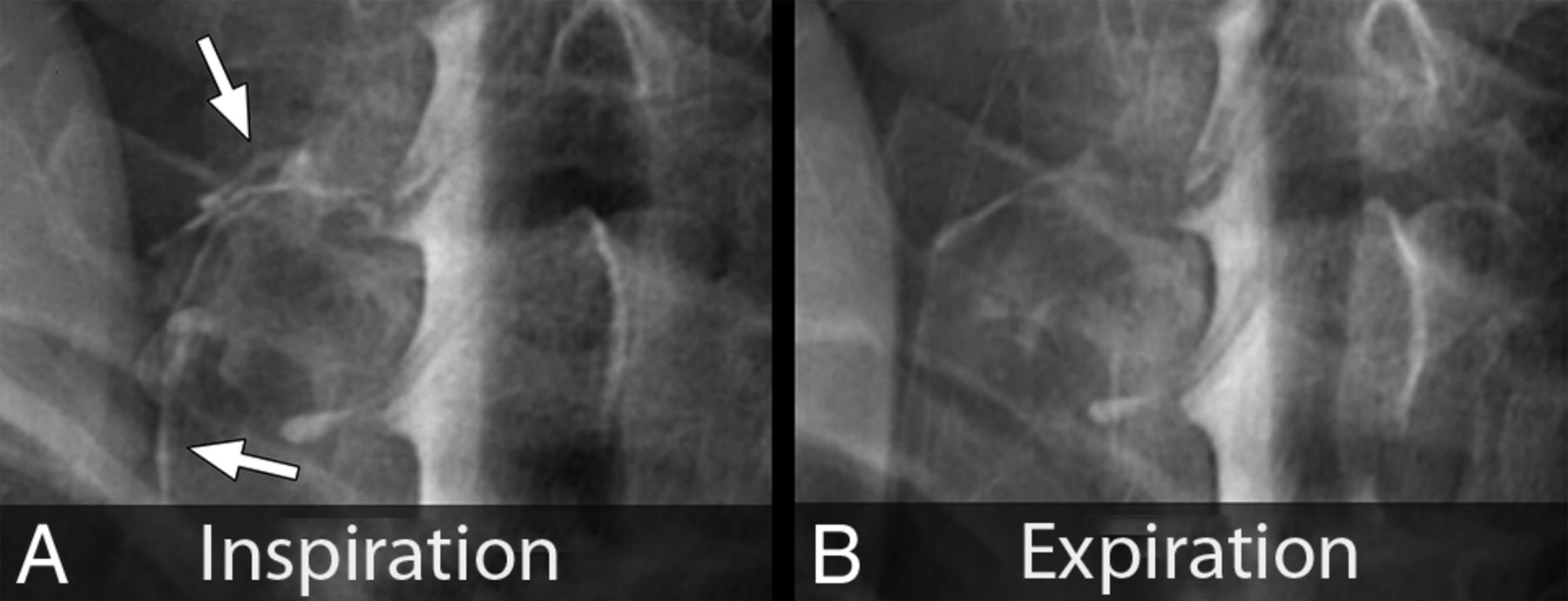
Spot-magnified radiographs of a left C8 CSF–venous fistula during an ipsilateral decubitus dynamic myelogram. A, Image acquired during inspiration demonstrates increased visibility and extent of the CSF–venous fistula (arrows). B, Image acquired during expiration leads to reduced visibility and extent of the CSF–venous fistula.

FIG 5.
  • Download figure
  • Open in new tab
  • Download powerpoint
FIG 5.

Spot-magnified radiographs of a left T8 CSF–venous fistula during an ipsilateral decubitus dynamic myelogram. A, Image acquired during inspiration demonstrates contrast opacification of the CSF–venous fistula extending out over the transverse process (arrows). B, Image acquired during quiet breath-hold during the mid-respiratory cycle. The CSF–venous fistula is no longer visible over the transverse process.

DISCUSSION

We found that the conspicuity of some CVFs varied during different phases of the respiratory cycle and that in all cases, they were most conspicuous during inspiration. This finding suggests that for patients with SIH, myelography performed during maximum inspiration may aid in the detection of CVFs, thereby improving the overall diagnostic yield.

It is well-known that venous return is highly dependent on the respiratory phase.9 Inspiration results in descent of the diaphragm causing negative intrathoracic pressure as well as increased intraabdominal pressure. This result creates a pressure gradient driving blood from the inferior vena cava to the right atrium, increasing venous return to the heart.10 This blood flow also results in decreased intravascular pressure within the inferior vena cava, which would produce a gradient of pressure between the higher pressure CSF and the lower pressure epidural venous plexus and paraspinal veins. In the setting of a CVF, we hypothesize that this gradient would result in rapid unregulated egress of CSF back into the circulatory system, with resultant increased conspicuity on myelography.11 Conversely, during a Valsalva maneuver, the intrathoracic and inferior vena cava pressure considerably increases. This increase markedly reduces venous return and also eliminates the aforementioned pressure gradient between the CSF and the infradiaphragmatic venous system, which we hypothesize would reduce flow through a CVF.12

Our findings suggest that in cases in which a CVF is suspected, the diagnostic yield of spinal imaging may increase if it is performed during inspiration. In our practice, we now routinely perform our initial image acquisitions in this manner. This modification to the myelography technique is broadly applicable across modalities and can be used even in centers without the resources and equipment necessary to perform digital subtraction myelography.

This investigation is limited by the small number of cases reported and by its retrospective nature, which introduces the potential for selection bias. Furthermore, in our practice, some patients with CVF have not demonstrated changes in imaging conspicuity that are dependent on the phase of respiration. The proportion of patients with CVFs that are affected by differences in respiration remains unknown. Effort to answer this question as well as to determine the factors predictive of CVFs that exhibit imaging changes dependent on respiration is a logical direction for future research. Additionally, there may be other unknown factors related to respiration that result in changes to CVF conspicuity. For example, it is possible that there is a threshold at which substantially increased rates of venous return to the heart would result in rapid washout of contrast from a CVF, paradoxically reducing its conspicuity during inspiration.

CONCLUSIONS

The conspicuity of CVFs is improved during inspiration in some cases. Further investigation into the improved diagnostic performance of myelography performed during inspiration is warranted.

Footnotes

  • Disclosures: Linda Gray—UNRELATED: Board Membership: Spinal CSF Leak Foundation.

References

  1. 1.↵
    1. Amrhein TJ,
    2. Kranz PG
    . Spontaneous intracranial hypotension: imaging in diagnosis and treatment. Radiol Clin North Am 2019;57:439–51 doi:10.1016/j.rcl.2018.10.004 pmid:30709479
    CrossRefPubMed
  2. 2.↵
    1. Wang TY,
    2. Karikari IO,
    3. Amrhein TJ, et al
    . Clinical outcomes following surgical ligation of cerebrospinal fluid-venous fistula in patients with spontaneous intracranial hypotension: a prospective case series. Oper Neurosurg (Hagerstown) 2020;18:239–45 doi:10.1093/ons/opz134 pmid:31134267
    CrossRefPubMed
  3. 3.↵
    1. Kranz PG,
    2. Gray L,
    3. Malinzak MD, et al
    . Spontaneous intracranial hypotension: pathogenesis, diagnosis, and treatment. Neuroimaging Clin N Am 2019;29:581–94 doi:10.1016/j.nic.2019.07.006 pmid:31677732
    CrossRefPubMed
  4. 4.↵
    1. Schievink WI,
    2. Maya MM,
    3. Jean-Pierre S, et al
    . A classification system of spontaneous spinal CSF leaks. Neurology 2016;87:673–79 doi:10.1212/WNL.0000000000002986 pmid:27440149
    Abstract/FREE Full Text
  5. 5.↵
    1. Kranz PG,
    2. Gray L,
    3. Amrhein TJ
    . Decubitus CT myelography for detecting subtle CSF leaks in spontaneous intracranial hypotension. AJNR Am J Neuroradiol 2019;40:754–56 doi:10.3174/ajnr.A5995 pmid:30819772
    Abstract/FREE Full Text
  6. 6.↵
    Headache Classification Committee of the International Headache Society (IHS). The International Classification of Headache Disorders, 3rd Edition (Beta Version). Cephalalgia 2013;33:629–808 doi:10.1177/0333102413485658 pmid:23771276
    CrossRefPubMed
  7. 7.↵
    1. Dobrocky T,
    2. Grunder L,
    3. Breiding PS, et al
    . Assessing spinal cerebrospinal fluid leaks in spontaneous intracranial hypotension with a scoring system based on brain magnetic resonance imaging findings. JAMA Neurol 2019;76:580–87 doi:10.1001/jamaneurol.2018.4921 pmid:30776059
    CrossRefPubMed
  8. 8.↵
    1. Kranz PG,
    2. Amrhein TJ,
    3. Gray L
    . CSF venous fistulas in spontaneous intracranial hypotension: imaging characteristics on dynamic and CT myelography. AJR Am J Roentgenol 2017;209:1360–66 doi:10.2214/AJR.17.18351 pmid:29023155
    CrossRefPubMed
  9. 9.↵
    1. Pinsky MR
    . Cardiopulmonary interactions: physiologic basis and clinical applications. Ann Am Thorac Soc 2018;15:S45–48 doi:10.1513/AnnalsATS.201704-339FR pmid:28820609
    CrossRefPubMed
  10. 10.↵
    1. Willeput R,
    2. Rondeux C,
    3. De Troyer A
    . Breathing affects venous return from legs in humans. J Appl Physiol Respir Environ Exerc Physiol 1984;57:971–76 doi:10.1152/jappl.1984.57.4.971 pmid:6238925
    CrossRefPubMed
  11. 11.↵
    1. Kumar N,
    2. Neidert NB,
    3. Diehn FE, et al
    . A novel etiology for craniospinal hypovolemia: a case of inferior vena cava obstruction. J Neurosurg Spine 2018;29:452–55 doi:10.3171/2018.2.SPINE171373 pmid:30004315
    CrossRefPubMed
  12. 12.↵
    1. Laborda A,
    2. Sierre S,
    3. Malve M, et al
    . Influence of breathing movements and Valsalva maneuver on vena caval dynamics. World J Radiol 2014;6:833–39 doi:10.4329/wjr.v6.i10.833 pmid:25349666
    CrossRefPubMed
  • Received March 28, 2020.
  • Accepted after revision May 5, 2020.
  • © 2020 by American Journal of Neuroradiology
View Abstract
PreviousNext
Back to top

In this issue

American Journal of Neuroradiology: 41 (9)
American Journal of Neuroradiology
Vol. 41, Issue 9
1 Sep 2020
  • Table of Contents
  • Index by author
  • Complete Issue (PDF)
Advertisement
Print
Download PDF
Email Article

Thank you for your interest in spreading the word on American Journal of Neuroradiology.

NOTE: We only request your email address so that the person you are recommending the page to knows that you wanted them to see it, and that it is not junk mail. We do not capture any email address.

Enter multiple addresses on separate lines or separate them with commas.
Respiratory Phase Affects the Conspicuity of CSF–Venous Fistulas in Spontaneous Intracranial Hypotension
(Your Name) has sent you a message from American Journal of Neuroradiology
(Your Name) thought you would like to see the American Journal of Neuroradiology web site.
CAPTCHA
This question is for testing whether or not you are a human visitor and to prevent automated spam submissions.
Cite this article
T.J. Amrhein, L. Gray, M.D. Malinzak, P.G. Kranz
Respiratory Phase Affects the Conspicuity of CSF–Venous Fistulas in Spontaneous Intracranial Hypotension
American Journal of Neuroradiology Sep 2020, 41 (9) 1754-1756; DOI: 10.3174/ajnr.A6663

Citation Manager Formats

  • BibTeX
  • Bookends
  • EasyBib
  • EndNote (tagged)
  • EndNote 8 (xml)
  • Medlars
  • Mendeley
  • Papers
  • RefWorks Tagged
  • Ref Manager
  • RIS
  • Zotero
0 Responses
Respond to this article
Share
Bookmark this article
Respiratory Phase Affects the Conspicuity of CSF–Venous Fistulas in Spontaneous Intracranial Hypotension
T.J. Amrhein, L. Gray, M.D. Malinzak, P.G. Kranz
American Journal of Neuroradiology Sep 2020, 41 (9) 1754-1756; DOI: 10.3174/ajnr.A6663
del.icio.us logo Twitter logo Facebook logo Mendeley logo
  • Tweet Widget
  • Facebook Like
  • Google Plus One
Purchase

Jump to section

  • Article
    • Abstract
    • ABBREVIATIONS:
    • MATERIALS AND METHODS
    • RESULTS
    • DISCUSSION
    • CONCLUSIONS
    • Footnotes
    • References
  • Figures & Data
  • Info & Metrics
  • Responses
  • References
  • PDF

Related Articles

  • No related articles found.
  • PubMed
  • Google Scholar

Cited By...

  • Additional Diagnostic Value of Conebeam CT Myelography Performed after Digital Subtraction Myelography for Detecting CSF-Venous Fistulas
  • Density and Time Characteristics of CSF-Venous Fistulas on CT Myelography in Patients with Spontaneous Intracranial Hypotension
  • Azygos Vein Stenosis in Frontotemporal Dementia Sagging Brain Syndrome
  • Spinal CSF Leaks: The Neuroradiologist Transforming Care
  • Direct comparison of digital subtraction myelography versus CT myelography in lateral decubitus position: evaluation of diagnostic yield for cerebrospinal fluid-venous fistulas
  • Diagnostic Yield of Decubitus CT Myelography for Detection of CSF-Venous Fistulas
  • Myelographic Techniques for the Localization of CSF-Venous Fistulas: Updates in 2024
  • Perspectives from the Inaugural "Spinal CSF Leak: Bridging the Gap" Conference: A Convergence of Clinical and Patient Expertise
  • Lateral Decubitus Dynamic CT Myelography with Real-Time Bolus Tracking (dCTM-BT) for Evaluation of CSF-Venous Fistulas: Diagnostic Yield Stratified by Brain Imaging Findings
  • Temporal Characteristics of CSF-Venous Fistulas on Dynamic Decubitus CT Myelography: A Retrospective Multi-Institution Cohort Study
  • Resisted Inspiration Improves Visualization of CSF-Venous Fistulas in Spontaneous Intracranial Hypotension
  • Utility of Photon-Counting Detector CT Myelography for the Detection of CSF-Venous Fistulas
  • Utility of Photon-Counting Detector CT Myelography for the Detection of CSF-Venous Fistulas
  • Temporal Characteristics of CSF-Venous Fistulas on Digital Subtraction Myelography
  • Resisted Inspiration: A New Technique to Aid in the Detection of CSF-Venous Fistulas
  • Spinal Compliance Curves: Preliminary Experience with a New Tool for Evaluating Suspected CSF Venous Fistulas on CT Myelography in Patients with Spontaneous Intracranial Hypotension
  • A Novel Endovascular Therapy for CSF Hypotension Secondary to CSF-Venous Fistulas
  • Decubitus CT Myelography for CSF-Venous Fistulas: A Procedural Approach
  • Crossref (19)
  • Google Scholar

This article has been cited by the following articles in journals that are participating in Crossref Cited-by Linking.

  • A Novel Endovascular Therapy for CSF Hypotension Secondary to CSF-Venous Fistulas
    W. Brinjikji, L.E. Savastano, J.L.D. Atkinson, I. Garza, R. Farb, J.K. Cutsforth-Gregory
    American Journal of Neuroradiology 2021 42 5
  • Decubitus CT Myelography for CSF-Venous Fistulas: A Procedural Approach
    M.D. Mamlouk, R.P. Ochi, P. Jun, P.Y. Shen
    American Journal of Neuroradiology 2021 42 1
  • Utility of Photon-Counting Detector CT Myelography for the Detection of CSF-Venous Fistulas
    A.A. Madhavan, L. Yu, W. Brinjikji, J.K. Cutsforth-Gregory, F.R. Schwartz, I.T. Mark, J.C. Benson, T.J. Amrhein
    American Journal of Neuroradiology 2023 44 6
  • Temporal Characteristics of CSF-Venous Fistulas on Digital Subtraction Myelography
    I. Mark, A. Madhavan, M. Oien, J. Verdoorn, J.C. Benson, J. Cutsforth-Gregory, W. Brinjikji, P. Morris
    American Journal of Neuroradiology 2023 44 4
  • Resisted Inspiration: A New Technique to Aid in the Detection of CSF-Venous Fistulas
    I.T. Mark, M.R. Amans, V.N. Shah, K.H. Narsinh, M.T. Caton, S. Teixeira, W.P. Dillon
    American Journal of Neuroradiology 2022 43 10
  • CSF-Venous Fistula
    Michelle Roytman, Gayle Salama, Matthew S. Robbins, J. Levi Chazen
    Current Pain and Headache Reports 2021 25 1
  • Resisted Inspiration Improves Visualization of CSF-Venous Fistulas in Spontaneous Intracranial Hypotension
    P.G. Kranz, M.D. Malinzak, L. Gray, J. Willhite, T.J. Amrhein
    American Journal of Neuroradiology 2023 44 8
  • Direct comparison of digital subtraction myelography versus CT myelography in lateral decubitus position: evaluation of diagnostic yield for cerebrospinal fluid-venous fistulas
    Niklas Lützen, Theo Demerath, Urs Würtemberger, Nebiyat Filate Belachew, Enrique Barvulsky Aleman, Katharina Wolf, Amir El Rahal, Florian Volz, Christian Fung, Jürgen Beck, Horst Urbach
    Journal of NeuroInterventional Surgery 2024 16 10
  • Spinal Compliance Curves: Preliminary Experience with a New Tool for Evaluating Suspected CSF Venous Fistulas on CT Myelography in Patients with Spontaneous Intracranial Hypotension
    M.T. Caton, B. Laguna, K.A. Soderlund, W.P. Dillon, V.N. Shah
    American Journal of Neuroradiology 2021 42 5
  • Lateral Decubitus Dynamic CT Myelography with Real-Time Bolus Tracking (dCTM-BT) for Evaluation of CSF-Venous Fistulas: Diagnostic Yield Stratified by Brain Imaging Findings
    Thien J. Huynh, Donna Parizadeh, Ahmed K. Ahmed, Christopher T. Gandia, Hal C. Davison, John V. Murray, Ian T. Mark, Ajay A. Madhavan, Darya Shlapak, Todd D. Rozen, Waleed Brinjikji, Prasanna Vibhute, Vivek Gupta, Kacie Brewer, Olga Fermo
    American Journal of Neuroradiology 2024 45 1

More in this TOC Section

  • MP2RAGE 7T in MS Lesions of the Cervical Spine
  • Bern Score Validity for SIH
  • Resisted Inspiration for CSF-Venous Fistula
Show more Spine

Similar Articles

Advertisement

Indexed Content

  • Current Issue
  • Accepted Manuscripts
  • Article Preview
  • Past Issues
  • Editorials
  • Editor's Choice
  • Fellows' Journal Club
  • Letters to the Editor
  • Video Articles

Cases

  • Case Collection
  • Archive - Case of the Week
  • Archive - Case of the Month
  • Archive - Classic Case

More from AJNR

  • Trainee Corner
  • Imaging Protocols
  • MRI Safety Corner
  • Book Reviews

Multimedia

  • AJNR Podcasts
  • AJNR Scantastics

Resources

  • Turnaround Time
  • Submit a Manuscript
  • Submit a Video Article
  • Submit an eLetter to the Editor/Response
  • Manuscript Submission Guidelines
  • Statistical Tips
  • Fast Publishing of Accepted Manuscripts
  • Graphical Abstract Preparation
  • Imaging Protocol Submission
  • Evidence-Based Medicine Level Guide
  • Publishing Checklists
  • Author Policies
  • Become a Reviewer/Academy of Reviewers
  • News and Updates

About Us

  • About AJNR
  • Editorial Board
  • Editorial Board Alumni
  • Alerts
  • Permissions
  • Not an AJNR Subscriber? Join Now
  • Advertise with Us
  • Librarian Resources
  • Feedback
  • Terms and Conditions
  • AJNR Editorial Board Alumni

American Society of Neuroradiology

  • Not an ASNR Member? Join Now

© 2025 by the American Society of Neuroradiology All rights, including for text and data mining, AI training, and similar technologies, are reserved.
Print ISSN: 0195-6108 Online ISSN: 1936-959X

Powered by HighWire