
Article Figures & Data
Figures
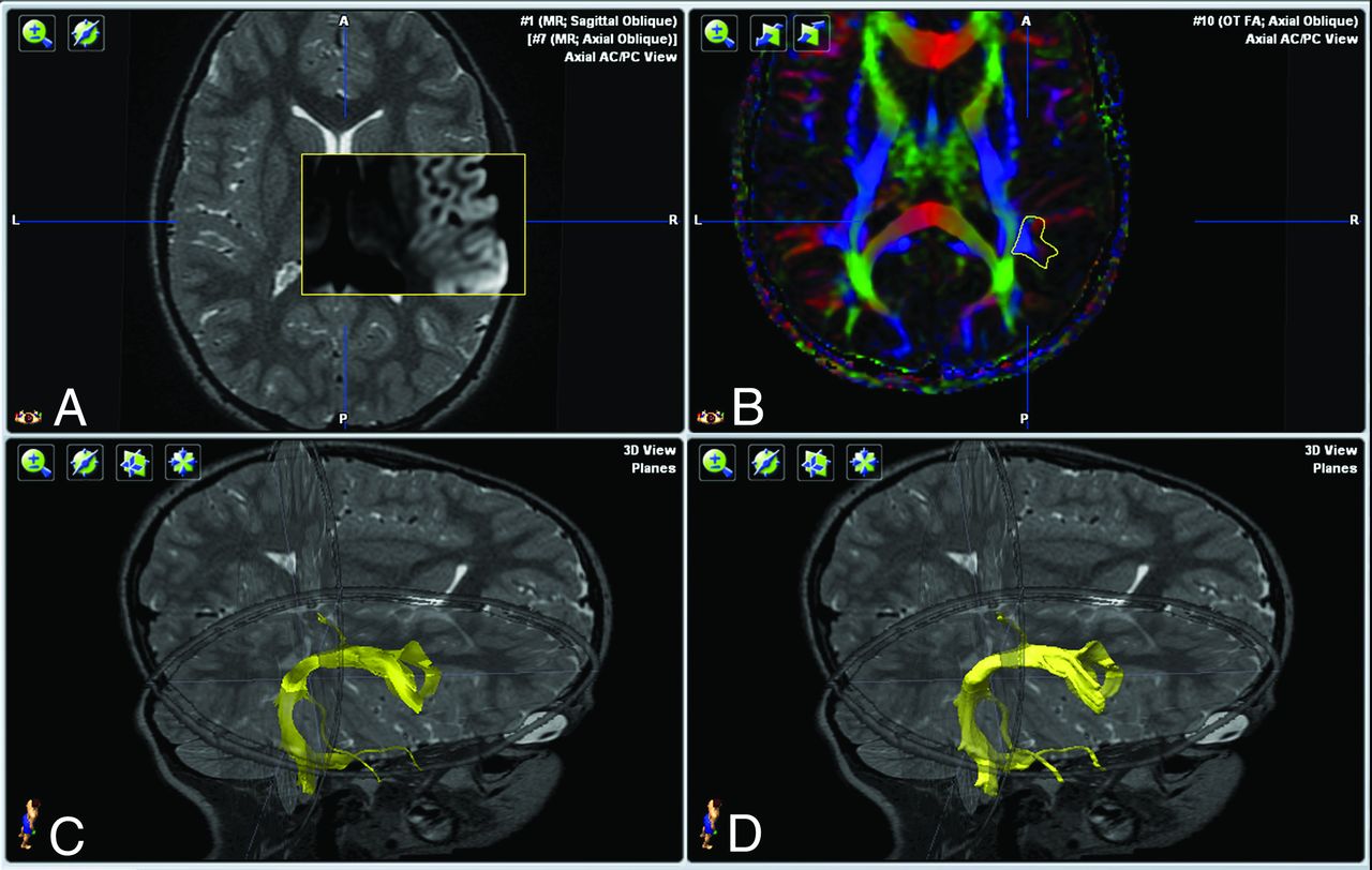
- FIG 1.
Image processing in a 7-year-old girl. Image datasets. A, Coregistration (mutual information algorithm) after anterior/posterior commissure alignment using iPlan Stereotaxy 3.0. T2-3D-weighted sequence. FA color-coded fiber-direction maps. B, Positioning of the ROI on the FA color-coded map to track the right arcuate fasciculus. C, Generating the right arcuate fasciculus. D, Generating the volume corresponding to the tractogram on a T2-weighted 3D dataset.
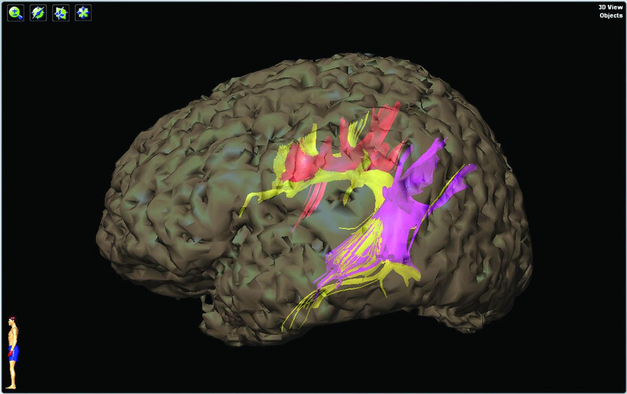
- FIG 2.
3D surface renderings of the telencephalon in a 7-year-old girl from T2-weighted MR images used to map the left superior longitudinal fasciculus (anterior part in red, arcuate part in yellow, and posterior part in pink).
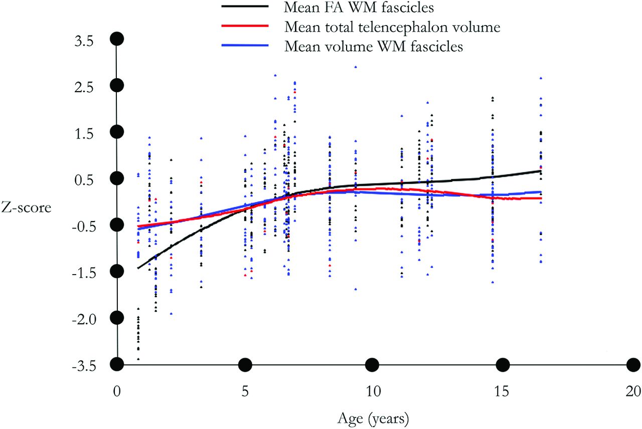
- FIG 3.
Lowess smoothing curve with a bandwidth of 0.8. Standardized. Black indicates mean WM fascicle FA; red, mean total telencephalon volume; blue, mean WM fascicle volume.
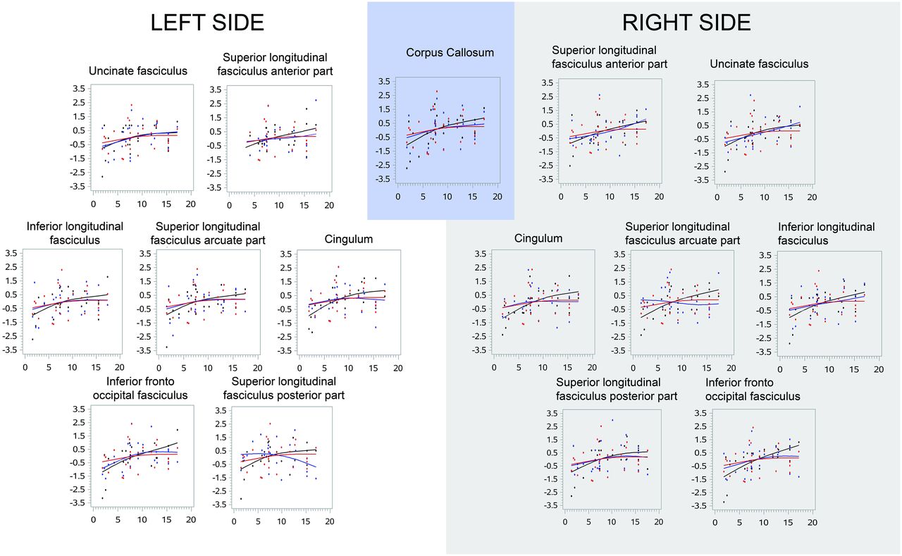
- FIG 4.
Lowess smoothing curve for each WM bundle with a bandwidth of 0.8. Standardized. Black indicates mean WM bundle FA; red, mean telencephalon volume; blue, mean WM bundle volume.


Tables
Effect of age on WM fascicle FA and volume, and test for difference in slope between the effect of age on WM fascicle FA and the effect of age on WM fascicle volume, in the subsamples of participants older than 8 years of age
Effect of Age on WM Fascicle FA Effect of Age on WM Fascicle Volume Interaction between the Effect of Age on WM Fascicle FA and WM Fascicle Volume β SD P Value β SD P Value P Value F1, corpus callosum 0.05 0.06 .447 −0.01 0.09 .901 .770 F2, right cingulum 0.02 0.08 .822 0.02 0.12 .847 .734 F3, left cingulum 0.03 0.09 .736 −0.10 0.11 .396 .459 F4, superior longitudinal fasciculus, right anterior part 0.03 0.08 .718 0.19 0.09 .060 .175 F5, superior longitudinal fasciculus, left anterior part 0.07 0.11 .514 0.01 0.13 .925 .852 F6, superior longitudinal fasciculus, right arcuate part 0.15a 0.06a .042a 0.17 0.10 .111 .506 F7, superior longitudinal fasciculus, left arcuate part 0.10 0.06 .142 0.01 0.07 .860 .308 F8, superior longitudinal fasciculus, right posterior part 0.01 0.07 .868 −0.13 0.14 .360 .434 F9, superior longitudinal fasciculus, left posterior part 0.02a 0.09a .842a −0.22 0.12 .109 .547 F10, right inferior fronto-occipital fasciculus 0.13 0.05 .043 −0.10 0.09 .296 .072 F11, left inferior fronto-occipital fasciculus 0.14 0.07 .083 −0.16 0.07 .062 .017 F12, right inferior longitudinal fasciculus 0.06 0.08 .480 0.09 0.12 .471 .802 F13, left inferior longitudinal fasciculus 0.01 0.10 .951 0.01 0.10 .900 .983 F14, right uncinate fasciculus 0.00 0.07 .963 0.06 0.09 .533 .521 F15, left uncinate fasciculus −0.03 0.09 .731 −0.06 0.07 .431 .641 ↵a P value < .05, without correction for multiple testing.


