
Article Figures & Data
Figures
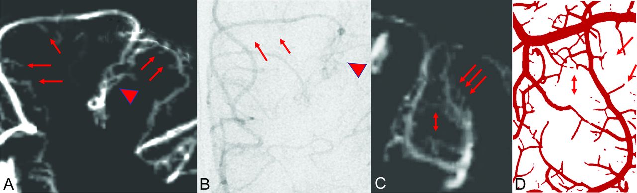
- Fig 1.
Coronal 4D-FPCBCTA reconstruction of the arterial microvasculature at the superior and middle frontal gyri and the superior frontal sulcus (A) and the corresponding DSA image, late arterial phase (B). Main arterial branches (red arrows) and the sulcal pial arterial network (red arrowhead) can be identified. Perpendicular branches to the cortex are difficult to see on DSA. C, A more detailed view shows arteries bending from the cortical surface into the depth the superior frontal sulcus. Perpendicular branches can be followed in the direction of the cortex of the adjacent middle frontal gyrus (red arrows). Note network-like intra-arterial connections (double red arrow). D, Similar findings are detected in injection specimens of cortical arteries (corresponding arrows). Drawing reprinted with permission from Duvernoy et al.1
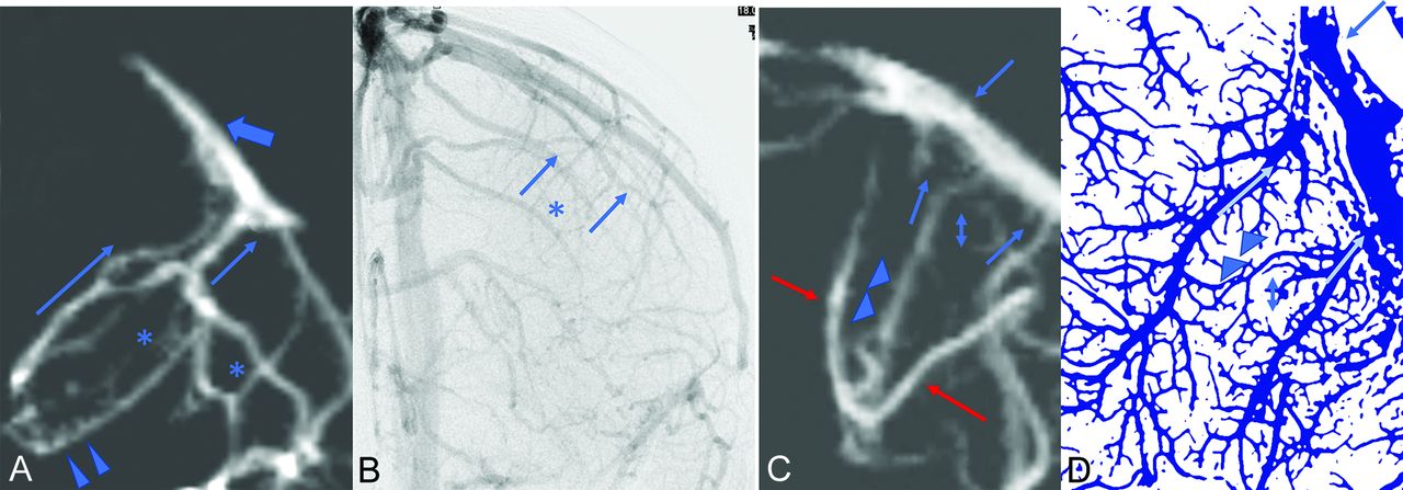
- Fig 2.
Coronal 4D-FPCBCTA reconstruction of veins in the middle frontal sulcus (A) and corresponding DSA image, late venous phase (B). Collecting veins (thin blue arrows) and the sulcal venous network (asterisks) can be identified on DSA. Small veins draining into the collecting veins are only partly visible (blue arrowheads). Further drainage of the collecting veins into a superficial cortical vein (thick blue arrow). Note considerable overlay of venous structures in the DSA projection image. C, Cortical veins are also fed by perpendicular branches from the cortex (blue arrowheads). Collecting veins show a straight course toward a larger cortical vein (blue arrows). Network-like connections are weakly accentuated (blue double arrow). Note the overlay by a sulcal artery (red arrows). D, Corresponding findings on a drawing from an injection specimen with small cortical draining veins (blue arrowheads) perpendicular to straight collecting veins to a cortical vein (thin blue arrows). Drawing reprinted with permission from Duvernoy et al.1


