Advertisement

Main menu

  • Home
  • Content
    • Current Issue
    • Accepted Manuscripts
    • Article Preview
    • Past Issue Archive
    • Video Articles
    • AJNR Case Collection
    • Case of the Week Archive
    • Case of the Month Archive
    • Classic Case Archive
  • Special Collections
    • Advancing NeuroMRI with High-Relaxivity Contrast Agents
    • AJNR Awards
    • Low-Field MRI
    • Alzheimer Disease
    • ASNR Foundation Special Collection
    • Photon-Counting CT
    • View All
  • Multimedia
    • AJNR Podcasts
    • AJNR SCANtastic
    • Trainee Corner
    • MRI Safety Corner
    • Imaging Protocols
  • For Authors
    • Submit a Manuscript
    • Submit a Video Article
    • Submit an eLetter to the Editor/Response
    • Manuscript Submission Guidelines
    • Statistical Tips
    • Fast Publishing of Accepted Manuscripts
    • Graphical Abstract Preparation
    • Imaging Protocol Submission
    • Author Policies
  • About Us
    • About AJNR
    • Editorial Board
    • Editorial Board Alumni
  • More
    • Become a Reviewer/Academy of Reviewers
    • Subscribers
    • Permissions
    • Alerts
    • Feedback
    • Advertisers
    • ASNR Home

User menu

  • Alerts
  • Log in

Search

  • Advanced search
American Journal of Neuroradiology

American Journal of Neuroradiology

ASHNR American Society of Functional Neuroradiology ASHNR American Society of Pediatric Neuroradiology ASSR
  • Alerts
  • Log in

Advanced Search

  • Home
  • Content
    • Current Issue
    • Accepted Manuscripts
    • Article Preview
    • Past Issue Archive
    • Video Articles
    • AJNR Case Collection
    • Case of the Week Archive
    • Case of the Month Archive
    • Classic Case Archive
  • Special Collections
    • Advancing NeuroMRI with High-Relaxivity Contrast Agents
    • AJNR Awards
    • Low-Field MRI
    • Alzheimer Disease
    • ASNR Foundation Special Collection
    • Photon-Counting CT
    • View All
  • Multimedia
    • AJNR Podcasts
    • AJNR SCANtastic
    • Trainee Corner
    • MRI Safety Corner
    • Imaging Protocols
  • For Authors
    • Submit a Manuscript
    • Submit a Video Article
    • Submit an eLetter to the Editor/Response
    • Manuscript Submission Guidelines
    • Statistical Tips
    • Fast Publishing of Accepted Manuscripts
    • Graphical Abstract Preparation
    • Imaging Protocol Submission
    • Author Policies
  • About Us
    • About AJNR
    • Editorial Board
    • Editorial Board Alumni
  • More
    • Become a Reviewer/Academy of Reviewers
    • Subscribers
    • Permissions
    • Alerts
    • Feedback
    • Advertisers
    • ASNR Home
  • Follow AJNR on Twitter
  • Visit AJNR on Facebook
  • Follow AJNR on Instagram
  • Join AJNR on LinkedIn
  • RSS Feeds

AJNR Awards, New Junior Editors, and more. Read the latest AJNR updates


Improved Turnaround Times | Median time to first decision: 12 days

Research ArticleAdult Brain
Open Access

Value of 3T Susceptibility-Weighted Imaging in the Diagnosis of Multiple Sclerosis

M.A. Clarke, D. Pareto, L. Pessini-Ferreira, G. Arrambide, M. Alberich, F. Crescenzo, S. Cappelle, M. Tintoré, J. Sastre-Garriga, C. Auger, X. Montalban, N. Evangelou and À. Rovira
American Journal of Neuroradiology June 2020, 41 (6) 1001-1008; DOI: https://doi.org/10.3174/ajnr.A6547
M.A. Clarke
aFrom the Vall d′Hebron Research Institute (M.A.C., D.P., M.A., M.T., J.S.-G., C.A., X.M., A.R.), Barcelona, Spain
  • Find this author on Google Scholar
  • Find this author on PubMed
  • Search for this author on this site
  • ORCID record for M.A. Clarke
D. Pareto
aFrom the Vall d′Hebron Research Institute (M.A.C., D.P., M.A., M.T., J.S.-G., C.A., X.M., A.R.), Barcelona, Spain
bSection of Neuroradiology, Department of Radiology (D.P., L.P.-F., C.A., A.R.), Hospital Universitari Vall d’Hebron, Universitat Autònoma de Barcelona, Barcelona, Spain
  • Find this author on Google Scholar
  • Find this author on PubMed
  • Search for this author on this site
  • ORCID record for D. Pareto
L. Pessini-Ferreira
bSection of Neuroradiology, Department of Radiology (D.P., L.P.-F., C.A., A.R.), Hospital Universitari Vall d’Hebron, Universitat Autònoma de Barcelona, Barcelona, Spain
  • Find this author on Google Scholar
  • Find this author on PubMed
  • Search for this author on this site
  • ORCID record for L. Pessini-Ferreira
G. Arrambide
cDepartment of Neurology-Neuroimmunology (G.A., M.T., J.S.-G., X.M.), Centre d’Esclerosi Múltiple de Catalunya (Cemcat), Hospital Universitari Vall d’Hebron, Universitat Autònoma de Barcelona, Barcelona, Spain
  • Find this author on Google Scholar
  • Find this author on PubMed
  • Search for this author on this site
  • ORCID record for G. Arrambide
M. Alberich
aFrom the Vall d′Hebron Research Institute (M.A.C., D.P., M.A., M.T., J.S.-G., C.A., X.M., A.R.), Barcelona, Spain
  • Find this author on Google Scholar
  • Find this author on PubMed
  • Search for this author on this site
  • ORCID record for M. Alberich
F. Crescenzo
dDepartment of Neurosciences, Biomedicine and Movement Sciences (F.C.), University of Verona, Verona, Italy
  • Find this author on Google Scholar
  • Find this author on PubMed
  • Search for this author on this site
  • ORCID record for F. Crescenzo
S. Cappelle
eDivision of Radiology (S.C.), University Hospital Leuven, Leuven, Belgium
  • Find this author on Google Scholar
  • Find this author on PubMed
  • Search for this author on this site
  • ORCID record for S. Cappelle
M. Tintoré
aFrom the Vall d′Hebron Research Institute (M.A.C., D.P., M.A., M.T., J.S.-G., C.A., X.M., A.R.), Barcelona, Spain
cDepartment of Neurology-Neuroimmunology (G.A., M.T., J.S.-G., X.M.), Centre d’Esclerosi Múltiple de Catalunya (Cemcat), Hospital Universitari Vall d’Hebron, Universitat Autònoma de Barcelona, Barcelona, Spain
  • Find this author on Google Scholar
  • Find this author on PubMed
  • Search for this author on this site
  • ORCID record for M. Tintoré
J. Sastre-Garriga
aFrom the Vall d′Hebron Research Institute (M.A.C., D.P., M.A., M.T., J.S.-G., C.A., X.M., A.R.), Barcelona, Spain
cDepartment of Neurology-Neuroimmunology (G.A., M.T., J.S.-G., X.M.), Centre d’Esclerosi Múltiple de Catalunya (Cemcat), Hospital Universitari Vall d’Hebron, Universitat Autònoma de Barcelona, Barcelona, Spain
  • Find this author on Google Scholar
  • Find this author on PubMed
  • Search for this author on this site
  • ORCID record for J. Sastre-Garriga
C. Auger
aFrom the Vall d′Hebron Research Institute (M.A.C., D.P., M.A., M.T., J.S.-G., C.A., X.M., A.R.), Barcelona, Spain
bSection of Neuroradiology, Department of Radiology (D.P., L.P.-F., C.A., A.R.), Hospital Universitari Vall d’Hebron, Universitat Autònoma de Barcelona, Barcelona, Spain
  • Find this author on Google Scholar
  • Find this author on PubMed
  • Search for this author on this site
  • ORCID record for C. Auger
X. Montalban
aFrom the Vall d′Hebron Research Institute (M.A.C., D.P., M.A., M.T., J.S.-G., C.A., X.M., A.R.), Barcelona, Spain
cDepartment of Neurology-Neuroimmunology (G.A., M.T., J.S.-G., X.M.), Centre d’Esclerosi Múltiple de Catalunya (Cemcat), Hospital Universitari Vall d’Hebron, Universitat Autònoma de Barcelona, Barcelona, Spain
fDivision of Neurology (X.M.), St. Michael’s Hospital, University of Toronto, Toronto, Ontario, Canada
  • Find this author on Google Scholar
  • Find this author on PubMed
  • Search for this author on this site
  • ORCID record for X. Montalban
N. Evangelou
gDivision of Clinical Neuroscience (N.E.), University of Nottingham, Nottingham, UK.
  • Find this author on Google Scholar
  • Find this author on PubMed
  • Search for this author on this site
  • ORCID record for N. Evangelou
À. Rovira
aFrom the Vall d′Hebron Research Institute (M.A.C., D.P., M.A., M.T., J.S.-G., C.A., X.M., A.R.), Barcelona, Spain
bSection of Neuroradiology, Department of Radiology (D.P., L.P.-F., C.A., A.R.), Hospital Universitari Vall d’Hebron, Universitat Autònoma de Barcelona, Barcelona, Spain
  • Find this author on Google Scholar
  • Find this author on PubMed
  • Search for this author on this site
  • ORCID record for À. Rovira
  • Article
  • Figures & Data
  • Supplemental
  • Info & Metrics
  • Responses
  • References
  • PDF
Loading

Article Figures & Data

Figures

  • Tables
  • Fig 1.
    • Download figure
    • Open in new tab
    • Download powerpoint
    Fig 1.

    Patient flow diagram showing how patients were selected for the study and the reasons for exclusion. We excluded patients for the following reasons: 1) missing either their SWI or T2-FLAIR scan; 2) lesions <3 mm or infratentorial lesions that could not be identified on the SWI due to artifacts; 3) automated lesion masks not passing quality checks; and 4) diagnostic doubt by the end of the study follow-up. RRMS indicates relapsing-remitting multiple sclerosis.

  • Fig 2.
    • Download figure
    • Open in new tab
    • Download powerpoint
    Fig 2.

    Lesion appearance on axial T2-FLAIR (upper row) and the corresponding susceptibility-weighted (lower row) images in patients with Sjögren disease (A and C) and MS (B and D). The patient with Sjögren disease has no visible CVs or IRs on the SWI. The patient with MS has clearly visible IRs, which correspond to the lesion edges visible on the T2-FLAIR. CVs are also visible inside the lesions as hypointense dots or lines.

  • Fig 3.
    • Download figure
    • Open in new tab
    • Download powerpoint
    Fig 3.

    The location of lesions with central veins in the CIS and non-MS groups. Error bars represent standard error.
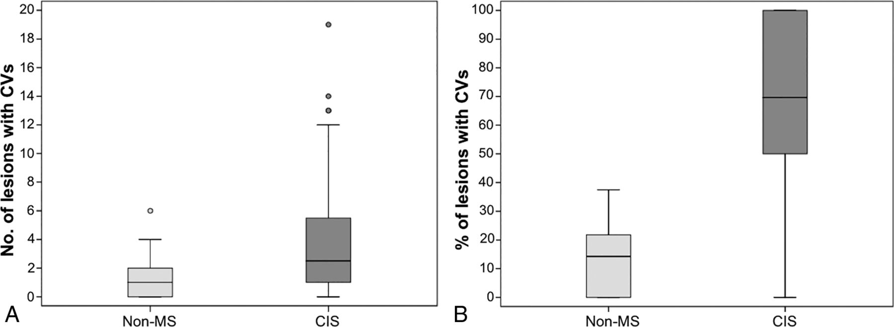
  • Fig 4.
    • Download figure
    • Open in new tab
    • Download powerpoint
    Fig 4.

    Summary of the incidence of lesions with CVs in the CIS and non-MS groups. A, The number of lesions with CVs (per patient) in the 2 groups. B, The percentage of lesions with CVs (per patient) in the 2 groups.

  • Fig 5.
    • Download figure
    • Open in new tab
    • Download powerpoint
    Fig 5.

    Summary of the incidence of lesions with iron rings in the CIS and non-MS groups. A, The number of lesions with iron rings (per patient) in the 2 groups. B, The location of lesions with iron rings in the CIS group.

Tables

  • Figures
  • Demographic and clinical data of the patients included in the study

    CIS Group (n = 112)Non-MS Group (n = 35)
    Mean age (SD, range) (yr)35.4 (7.9, 19–49)41.7 (11.5, 20–67)
    Sex, female No. (%)70 (70.5%)23 (65.7%)
    Clinical diagnosis at the end of the study No. (%)MS = 94 (83.9%) (including CDMS = 42 [37.5%]) CIS = 18 (16.1%)Autoimmune disease = 13 (37.1%)Vascular disease = 8 (22.9%)Incidental findings = 1 (2.9%)Infectious disease = 1 (2.9%)Headache = 3 (8.6%)Other = 9 (25.7%)
    +/− OCBs No. (not performed)80/21 (11)2/15 (18)
    Median EDSS (range)1.5 (0–4.5)NA
    Median WM lesion No. (range)4 (1–31)7 (1–31)
    • Note:—EDSS indicates Expanded Disability Status Scale; NA, not available.

PreviousNext
Back to top

In this issue

American Journal of Neuroradiology: 41 (6)
American Journal of Neuroradiology
Vol. 41, Issue 6
1 Jun 2020
  • Table of Contents
  • Index by author
  • Complete Issue (PDF)
Advertisement
Print
Download PDF
Email Article

Thank you for your interest in spreading the word on American Journal of Neuroradiology.

NOTE: We only request your email address so that the person you are recommending the page to knows that you wanted them to see it, and that it is not junk mail. We do not capture any email address.

Enter multiple addresses on separate lines or separate them with commas.
Value of 3T Susceptibility-Weighted Imaging in the Diagnosis of Multiple Sclerosis
(Your Name) has sent you a message from American Journal of Neuroradiology
(Your Name) thought you would like to see the American Journal of Neuroradiology web site.
CAPTCHA
This question is for testing whether or not you are a human visitor and to prevent automated spam submissions.
Cite this article
M.A. Clarke, D. Pareto, L. Pessini-Ferreira, G. Arrambide, M. Alberich, F. Crescenzo, S. Cappelle, M. Tintoré, J. Sastre-Garriga, C. Auger, X. Montalban, N. Evangelou, À. Rovira
Value of 3T Susceptibility-Weighted Imaging in the Diagnosis of Multiple Sclerosis
American Journal of Neuroradiology Jun 2020, 41 (6) 1001-1008; DOI: 10.3174/ajnr.A6547

Citation Manager Formats

  • BibTeX
  • Bookends
  • EasyBib
  • EndNote (tagged)
  • EndNote 8 (xml)
  • Medlars
  • Mendeley
  • Papers
  • RefWorks Tagged
  • Ref Manager
  • RIS
  • Zotero
0 Responses
Respond to this article
Share
Bookmark this article
Value of 3T Susceptibility-Weighted Imaging in the Diagnosis of Multiple Sclerosis
M.A. Clarke, D. Pareto, L. Pessini-Ferreira, G. Arrambide, M. Alberich, F. Crescenzo, S. Cappelle, M. Tintoré, J. Sastre-Garriga, C. Auger, X. Montalban, N. Evangelou, À. Rovira
American Journal of Neuroradiology Jun 2020, 41 (6) 1001-1008; DOI: 10.3174/ajnr.A6547
del.icio.us logo Twitter logo Facebook logo Mendeley logo
  • Tweet Widget
  • Facebook Like
  • Google Plus One
Purchase

Jump to section

  • Article
    • Abstract
    • ABBREVIATIONS:
    • MATERIALS AND METHODS
    • RESULTS
    • DISCUSSION
    • CONCLUSIONS
    • ACKNOWLEDGMENTS
    • Footnotes
    • References
  • Figures & Data
  • Supplemental
  • Info & Metrics
  • Responses
  • References
  • PDF

Related Articles

  • PubMed
  • Google Scholar

Cited By...

  • The Presence of a Paramagnetic Phase Rim is Linked to Lesion Age in Multiple Sclerosis
  • Metabolic Insights into Iron Deposition in Relapsing-Remitting Multiple Sclerosis via 7T Magnetic Resonance Spectroscopic Imaging
  • Paramagnetic rim lesions are associated with pathogenic CSF profiles and worse clinical outcomes in multiple sclerosis: a retrospective cross-sectional study
  • Evaluation of Ultrafast Wave-Controlled Aliasing in Parallel Imaging 3D-FLAIR in the Visualization and Volumetric Estimation of Cerebral White Matter Lesions
  • Chronic White Matter Inflammation and Serum Neurofilament Levels in Multiple Sclerosis
  • Evaluation of Ultrafast Wave-CAIPI 3D FLAIR in the Visualization and Volumetric Estimation of Cerebral White Matter Lesions
  • Crossref
  • Google Scholar

This article has not yet been cited by articles in journals that are participating in Crossref Cited-by Linking.

More in this TOC Section

  • Diagnostic Neuroradiology of Monoclonal Antibodies
  • Clinical Outcomes After Chiari I Decompression
  • Segmentation of Brain Metastases with BLAST
Show more Adult Brain

Similar Articles

Advertisement

Indexed Content

  • Current Issue
  • Accepted Manuscripts
  • Article Preview
  • Past Issues
  • Editorials
  • Editor's Choice
  • Fellows' Journal Club
  • Letters to the Editor
  • Video Articles

Cases

  • Case Collection
  • Archive - Case of the Week
  • Archive - Case of the Month
  • Archive - Classic Case

More from AJNR

  • Trainee Corner
  • Imaging Protocols
  • MRI Safety Corner
  • Book Reviews

Multimedia

  • AJNR Podcasts
  • AJNR Scantastics

Resources

  • Turnaround Time
  • Submit a Manuscript
  • Submit a Video Article
  • Submit an eLetter to the Editor/Response
  • Manuscript Submission Guidelines
  • Statistical Tips
  • Fast Publishing of Accepted Manuscripts
  • Graphical Abstract Preparation
  • Imaging Protocol Submission
  • Evidence-Based Medicine Level Guide
  • Publishing Checklists
  • Author Policies
  • Become a Reviewer/Academy of Reviewers
  • News and Updates

About Us

  • About AJNR
  • Editorial Board
  • Editorial Board Alumni
  • Alerts
  • Permissions
  • Not an AJNR Subscriber? Join Now
  • Advertise with Us
  • Librarian Resources
  • Feedback
  • Terms and Conditions
  • AJNR Editorial Board Alumni

American Society of Neuroradiology

  • Not an ASNR Member? Join Now

© 2025 by the American Society of Neuroradiology All rights, including for text and data mining, AI training, and similar technologies, are reserved.
Print ISSN: 0195-6108 Online ISSN: 1936-959X

Powered by HighWire