Advertisement

Main menu

  • Home
  • Content
    • Current Issue
    • Accepted Manuscripts
    • Article Preview
    • Past Issue Archive
    • Video Articles
    • AJNR Case Collection
    • Case of the Week Archive
    • Case of the Month Archive
    • Classic Case Archive
  • Special Collections
    • AJNR Awards
    • Low-Field MRI
    • Alzheimer Disease
    • ASNR Foundation Special Collection
    • Photon-Counting CT
    • View All
  • Multimedia
    • AJNR Podcasts
    • AJNR SCANtastic
    • Trainee Corner
    • MRI Safety Corner
    • Imaging Protocols
  • For Authors
    • Submit a Manuscript
    • Submit a Video Article
    • Submit an eLetter to the Editor/Response
    • Manuscript Submission Guidelines
    • Statistical Tips
    • Fast Publishing of Accepted Manuscripts
    • Graphical Abstract Preparation
    • Imaging Protocol Submission
    • Author Policies
  • About Us
    • About AJNR
    • Editorial Board
    • Editorial Board Alumni
  • More
    • Become a Reviewer/Academy of Reviewers
    • Subscribers
    • Permissions
    • Alerts
    • Feedback
    • Advertisers
    • ASNR Home

User menu

  • Alerts
  • Log in
  • Log out

Search

  • Advanced search
American Journal of Neuroradiology

American Journal of Neuroradiology

ASHNR American Society of Functional Neuroradiology ASHNR American Society of Pediatric Neuroradiology ASSR
  • Alerts
  • Log in
  • Log out

Advanced Search

  • Home
  • Content
    • Current Issue
    • Accepted Manuscripts
    • Article Preview
    • Past Issue Archive
    • Video Articles
    • AJNR Case Collection
    • Case of the Week Archive
    • Case of the Month Archive
    • Classic Case Archive
  • Special Collections
    • AJNR Awards
    • Low-Field MRI
    • Alzheimer Disease
    • ASNR Foundation Special Collection
    • Photon-Counting CT
    • View All
  • Multimedia
    • AJNR Podcasts
    • AJNR SCANtastic
    • Trainee Corner
    • MRI Safety Corner
    • Imaging Protocols
  • For Authors
    • Submit a Manuscript
    • Submit a Video Article
    • Submit an eLetter to the Editor/Response
    • Manuscript Submission Guidelines
    • Statistical Tips
    • Fast Publishing of Accepted Manuscripts
    • Graphical Abstract Preparation
    • Imaging Protocol Submission
    • Author Policies
  • About Us
    • About AJNR
    • Editorial Board
    • Editorial Board Alumni
  • More
    • Become a Reviewer/Academy of Reviewers
    • Subscribers
    • Permissions
    • Alerts
    • Feedback
    • Advertisers
    • ASNR Home
  • Follow AJNR on Twitter
  • Visit AJNR on Facebook
  • Follow AJNR on Instagram
  • Join AJNR on LinkedIn
  • RSS Feeds

AJNR Awards, New Junior Editors, and more. Read the latest AJNR updates

Research ArticleAdult Brain

Prognostic Value of Preoperative MRI Metrics for Diffuse Lower-Grade Glioma Molecular Subtypes

P. Darvishi, P.P. Batchala, J.T. Patrie, L.M. Poisson, M.-B. Lopes, R. Jain, C.E. Fadul, D. Schiff and S.H. Patel
American Journal of Neuroradiology May 2020, 41 (5) 815-821; DOI: https://doi.org/10.3174/ajnr.A6511
P. Darvishi
aFrom the Departments of Radiology and Medical Imaging (P.D., P.P.B., S.H.P.)
  • Find this author on Google Scholar
  • Find this author on PubMed
  • Search for this author on this site
  • ORCID record for P. Darvishi
P.P. Batchala
aFrom the Departments of Radiology and Medical Imaging (P.D., P.P.B., S.H.P.)
  • Find this author on Google Scholar
  • Find this author on PubMed
  • Search for this author on this site
  • ORCID record for P.P. Batchala
J.T. Patrie
bPublic Health Sciences (J.T.P.)
  • Find this author on Google Scholar
  • Find this author on PubMed
  • Search for this author on this site
  • ORCID record for J.T. Patrie
L.M. Poisson
eDepartment of Public Health (L.M.P.), Henry Ford Health System, Detroit, Michigan
  • Find this author on Google Scholar
  • Find this author on PubMed
  • Search for this author on this site
  • ORCID record for L.M. Poisson
M.-B. Lopes
cPathology, Divisions of Neuropathology and Molecular Diagnostics (M.-B.L.)
  • Find this author on Google Scholar
  • Find this author on PubMed
  • Search for this author on this site
  • ORCID record for M.-B. Lopes
R. Jain
fDepartments of Radiology (R.J.) and Neurosurgery (R.J.), New York University School of Medicine, New York, New York.
  • Find this author on Google Scholar
  • Find this author on PubMed
  • Search for this author on this site
  • ORCID record for R. Jain
C.E. Fadul
dDivision of Neuro-Oncology (C.E.F., D.S.), University of Virginia Health System, Charlottesville, Virginia
  • Find this author on Google Scholar
  • Find this author on PubMed
  • Search for this author on this site
  • ORCID record for C.E. Fadul
D. Schiff
dDivision of Neuro-Oncology (C.E.F., D.S.), University of Virginia Health System, Charlottesville, Virginia
  • Find this author on Google Scholar
  • Find this author on PubMed
  • Search for this author on this site
  • ORCID record for D. Schiff
S.H. Patel
aFrom the Departments of Radiology and Medical Imaging (P.D., P.P.B., S.H.P.)
  • Find this author on Google Scholar
  • Find this author on PubMed
  • Search for this author on this site
  • ORCID record for S.H. Patel
  • Article
  • Figures & Data
  • Info & Metrics
  • Responses
  • References
  • PDF
Loading

Abstract

BACKGROUND AND PURPOSE: Despite the improved prognostic relevance of the 2016 WHO molecular-based classification of lower-grade gliomas, variability in clinical outcome persists within existing molecular subtypes. Our aim was to determine prognostically significant metrics on preoperative MR imaging for lower-grade gliomas within currently defined molecular categories.

MATERIALS AND METHODS: We undertook a retrospective analysis of 306 patients with lower-grade gliomas accrued from an institutional data base and The Cancer Genome Atlas. Two neuroradiologists in consensus analyzed preoperative MRIs of each lower-grade glioma to determine the following: tumor size, tumor location, number of involved lobes, corpus callosum involvement, hydrocephalus, midline shift, eloquent cortex involvement, ependymal extension, margins, contrast enhancement, and necrosis. Adjusted hazard ratios determined the association between MR imaging metrics and overall survival per molecular subtype, after adjustment for patient age, patient sex, World Health Organization grade, and surgical resection status.

RESULTS: For isocitrate dehydrogenase (IDH) wild-type lower-grade gliomas, tumor size (hazard ratio, 3.82; 95% CI, 1.94–7.75; P < .001), number of involved lobes (hazard ratio, 1.70; 95% CI, 1.28–2.27; P < .001), hydrocephalus (hazard ratio, 4.43; 95% CI, 1.12–17.54; P = .034), midline shift (hazard ratio, 1.16; 95% CI, 1.03–1.30; P = .013), margins (P = .031), and contrast enhancement (hazard ratio, 0.34; 95% CI, 0.13–0.90; P = .030) were associated with overall survival. For IDH-mutant 1p/19q-codeleted lower-grade gliomas, tumor size (hazard ratio, 2.85; 95% CI, 1.06–7.70; P = .039) and ependymal extension (hazard ratio, 6.34; 95% CI, 1.07–37.59; P = .042) were associated with overall survival.

CONCLUSIONS: MR imaging metrics offers prognostic information for patients with lower-grade gliomas within molecularly defined classes, with the greatest prognostic value for IDH wild-type lower-grade gliomas.

ABBREVIATIONS:

LGG
lower-grade glioma
HR
hazard ratio
IDH
isocitrate dehydrogenase
IDHmut-Codel
IDH mutation and a whole-arm deletion of chromosome arms 1p and 19q
IDHmut-Noncodel
IDH-mutant lacking 1p/19q codeletion
IDHwt
IDH wild-type
IQR
interquartile range
OS
overall survival
TCIA
The Cancer Imaging Archive
TERT
telomerase reverse transcriptase
WHO
World Health Organization

Recognition of the biologic and prognostic significance of molecular-based characterization of diffuse lower-grade gliomas (LGGs) heralded major revisions to their classification by the World Health Organization (WHO) in 2016.1⇓-3 LGGs encompass WHO grade II and III astrocytic and oligodendroglial tumors, most of which have a prognostically favorable mutation in the isocitrate dehydrogenase (IDH) gene.2,4 Oligodendrogliomas are defined by the presence of both an IDH mutation and a whole-arm deletion of chromosome arms 1p and 19q (IDHmut-Codel), which confer added prognostic and therapeutic favorability.5,6 IDH-mutant astrocytomas (IDHmut-Noncodel) lack 1p/19q codeletion and typically have tumor protein p53 and alpha-thalassemia/mental retardation syndrome X-linked (ATRX chromatin remodeler, [ATRX]) gene mutations.7,8 IDH wild-type LGGs (IDHwt) are generally associated with far worse clinical outcomes and largely comprise neoplasms that are genotypically similar to primary glioblastoma.1,9

Despite improved clinical applicability of the revised classification system, there remains substantial heterogeneity in clinical outcomes within existing subtypes of LGGs.10⇓-12 While numerous studies have reported neuroimaging features that predict currently defined glioma molecular subtypes,13⇓⇓⇓⇓⇓⇓-20 neuroimaging features might additionally contain information that allows prognostic stratification of gliomas within currently defined molecular categories. The purpose of our investigation was to determine whether neuroimaging features on preoperative anatomic MR imaging have prognostic significance for LGGs within currently defined molecular categories.

MATERIALS AND METHODS

This was a retrospective study with institutional review board approval (University of Virginia Health System, Charlottesville, Virginia) as well as Health Insurance Portability and Accountability Act compliance.

Patient Selection

Cases were accrued from a diffuse LGG data base maintained at our institution (n = 255) as well as from The Cancer Imaging Archive (TCIA) (n = 198).21 Inclusion criteria were the following: 1) known IDH mutation status and 1p/19q-codeletion status; 2) known surgical resection status (gross total resection, subtotal resection, or biopsy); and 3) available preoperative MR imaging including (at a minimum) precontrast T1WI, contrast-enhanced T1WI, and either T2WI or FLAIR.

From the institutional dataset, 178 cases met the inclusion criteria; 43 cases were excluded for lack of IDH and/or 1p/19q-codeletion status, and 34 cases were excluded for lack of requisite preoperative MR imaging. From the TCIA cohort, 128 cases met the inclusion criteria; 1 case was excluded for lack of IDH and 1p/19q-codeletion status and 69 cases lacked requisite preoperative MR imaging. In total, our study cohort included 306 cases. Overall survival times were available for all included patients, defined as the time between the date of pathologic diagnosis to the date of death or last contact.

Neuroimaging and Analysis

Two neuroradiologists with 6 and 14 years of experience, blinded to molecular status, WHO grade, patient demographics, and clinical outcome, analyzed the preoperative MR imaging to determine the following metrics:—1) tumor size: maximum long-axis diameter (centimeters); 2) location: any glioma signal abnormality/enhancement involving the basal ganglia, thalamus, or brain stem (yes = central; no = peripheral); 3) number (n) of involved lobes: each of the following counted as 1 lobe (per hemisphere)—frontal lobe, parietal lobe, temporal lobe, insula, occipital lobe, brain stem/cerebellum; 4) corpus callosum involvement: any glioma signal abnormality/enhancement involving the corpus callosum (yes or no); 5) hydrocephalus: (yes or no); 6) midline shift: greatest degree of contralateral brain displacement (centimeters); 7) eloquent cortex involvement: any glioma signal abnormality/enhancement involving the eloquent cortex, defined per Chang et al22 (yes or no); 8) ependymal extension: any glioma signal abnormality/enhancement involving the ventricular ependyma (yes or no); 9) margins: <33%, 33%–66%, or >66% sharp/circumscribed glioma margins; 10) contrast enhancement: any glioma contrast enhancement (yes or no); and 11) necrosis: any region of glioma necrosis characterized by peripheral contrast enhancement and central nonenhancement (yes or no).

Neuropathology

Pathologic data for patients in the institutional dataset were retrieved from the electronic medical record. Molecular markers were tested in the Clinical Laboratory Improvement Amendments–certified molecular pathology laboratory at our institution. A clinically validated IDH1 antibody was used to determine IDH mutation status.23,24 Clinically validated pyrosequencing assays (PyroMark Q24 system; https://www.qiagen.com/us/shop/automated-solutions/pyro-mark-q24/) were used in immunohistochemistry-negative cases in accordance with the manufacturers to determine IDH1/IDH2 mutation status. The 1p/19q-codeletion status was determined by fluorescence in situ hybridization; dual-color human probes localizing the 1p, 1q, 19p, and 19q loci were used (Vysis LSI 1p36/1q25 and LSI 19q13/19p13 FISH Probe Kit; Abbott Laboratories, Abbott Park, IL). Histopathologic and molecular data for the TCIA cohort were retrieved from supplemental material in Ceccarelli et al.25

Statistical Analysis

Data Summarization.

Categoric variables are summarized by frequencies and percentages, and continuous scaled data are summarized by the median, the interquartile range (IQR), and the range of the empiric distribution.

Survival Analyses.

Cox proportional hazards regression was used to examine the prognostic utility of the preoperative MR imaging metrics to predict survival within the currently defined molecular categories of LGGs. The survival analyses were conducted per molecular category (ie, IDHmut-Codel, IDHmut-NonCodel, and IDHwt) and per MR imaging metrics. For each MR imaging metric, a multivariate Cox proportional hazards regression analysis was conducted in which the MR imaging metric of interest served as the primary predictor variable and patient age, patient sex, WHO grade, and surgical resection status served as concomitant adjustment variables. The follow-up times of survivors were treated as right-censored survival times in the Cox model. With regard to hypothesis testing, the Wald χ2 test was used to test the null hypothesis that the instantaneous risk of death (ie, hazard) is not associated with the values/categories of the MR imaging metric of interest after adjustment for patient age, patient sex, WHO grade, and surgical resection status. A P ≤ .05 decision rule was used as the null hypothesis rejection criterion for the tests of association.

Tumor Grade Analyses.

MR imaging metrics versus WHO grade association were examined via multivariate logistic regression, per LGG molecular subtype. The dependent variable of the multivariate logistic regression model was an indicator variable (Y) that distinguished WHO grade III tumors (Y = 1) from WHO grade II tumors (Y = 0), and the MR imaging metrics served as the predictor variables. Regarding hypothesis testing, the type III Wald χ2 test was used to test the null hypothesis that the adjusted odds for a tumor being WHO grade III are not associated with the MR imaging metric. A P ≤ .05 decision rule was used as the null hypothesis rejection criterion.

RESULTS

The study population included 306 patients, with 154 women (50.3%) and 152 men (49.7%). Patient characteristics per LGG molecular subtype are shown in Table 1, and preoperative MR imaging metric empiric distribution summaries per LGG molecular subtype are shown in Table 2.

View this table:
  • View inline
  • View popup
Table 1:

Patient characteristics for each molecular subtype

View this table:
  • View inline
  • View popup
Table 2:

Empiric distribution summaries for the preoperative MR imaging metrics according to LGG molecular subtype

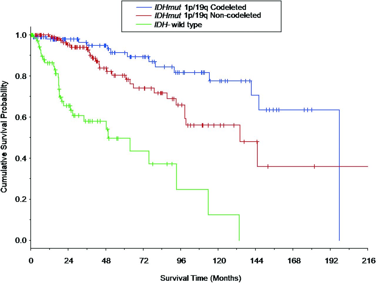
Survival curves for overall survival (OS) are shown in Fig 1 for the LGG molecular subtypes. OS differed among all the LGG molecular subtypes (P < .022 for all comparisons), with patients with IDHwt LGGs having the shortest median OS (49.5 months; 95% CI lower bound, 25.5 months), followed by the patients with IDHmut-Noncodel (134.6 months; 95% CI lower bound, 98.7 months), and patients with IDHmut-Codel (196.6 months, 95% CI lower bound, 141.4 months).

Fig 1.
  • Download figure
  • Open in new tab
  • Download powerpoint
Fig 1.

Kaplan-Meier survival curves based on the LGG molecular subtype. Vertical tick marks identify right-censored survival times. The survival curves differed among all 3 LGG molecular subtypes (IDH-mutant 1p/19q-codeleted molecular subtype versus IDH-mutant 1p/19q-noncodeleted molecular subtype: P = .021; IDH-mutant 1p/19q-codeleted molecular subtype versus IDH wild-type molecular subtype: P < .001; and IDH-mutant 1p/19q-noncodeleted molecular subtype versus IDH-wild type molecular subtype: P < .001). Survival curves for patients who composed a subset of the current patient cohort are shown in Patel et al,46 in 2019.

The associations between preoperative MR imaging metrics and OS per LGG molecular subtype are expressed as hazard ratios (HRs) in Table 3, after adjustment for patient age, patient sex, tumor grade, and surgical resection status. For IDHwt LGGs, greater tumor size (HR, 3.82; 95% CI, 1.94–7.75; P < .001), greater number of involved lobes (HR, 1.70; 95% CI, 1.28–2.27; P < .001), hydrocephalus (HR, 4.43; 95% CI, 1.12–17.54; P = .034), greater degree of midline shift (HR, 1.16; 95% CI, 1.03–1.30; P = .013), and less circumscribed margins (P = .031) were associated with shorter OS, whereas the presence of contrast enhancement (HR, 0.34; 95% CI, 0.13–0.90; P = .030) was associated with longer OS (Fig 2). For IDHmut-Noncodel LGGs, greater tumor size (HR, 2.90; 95% CI, 1.54–5.48; P = .001) was associated with shorter OS. For IDHmut-Codel LGGs, greater tumor size (HR, 2.85; 95% CI, 1.06–7.70; P = .039) and ependymal extension (HR, 6.34; 95% CI, 1.07–37.59; P = .042) were associated with shorter OS.

Fig 2.
  • Download figure
  • Open in new tab
  • Download powerpoint
Fig 2.

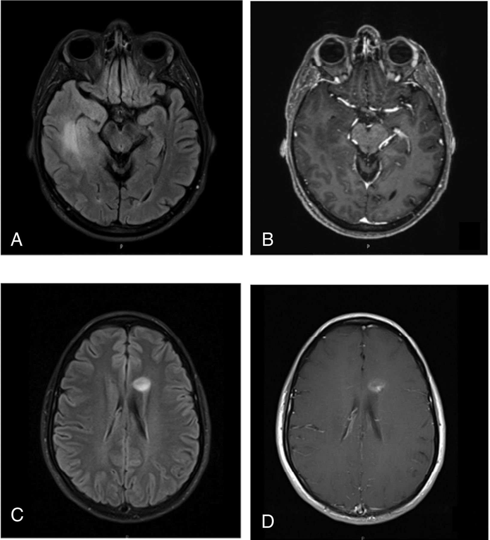
Representative cases of IDHwt LGGs. A 51-year-old man with an IDH wild-type diffuse astrocytoma. FLAIR (A) shows a 6-cm mass in the right temporal lobe with ill-defined margins. Contrast-enhanced T1WI (B) shows no contrast enhancement of the mass. This patient is deceased, with a survival time of 262 days. Further molecular testing in this case was positive for TERT promoter (−124 C > T) mutation. A 26-year-old woman with an IDH wild-type diffuse astrocytoma. FLAIR (C) and contrast-enhanced T1WI (D) show a 1.6-cm left frontal lobe mass with fairly well-circumscribed margins and contrast enhancement. This patient was alive at last follow-up, with a survival time of 2757 days. Further molecular testing in this case was positive for the BRAF V600E mutation.

View this table:
  • View inline
  • View popup
Table 3:

Associations between preoperative MR imaging metrics and overall survival per LGG molecular subtype, after adjustment for patient age, sex, tumor grade, and surgical resection statusa

A secondary analysis determined unique associations between MR imaging metrics and tumor grade (II or III) per LGG molecular subtype (Table 4). Contrast enhancement was associated with grade III for both IDHwt LGGs (OR, 16.75; 95% CI, 3.47–80.86; P < .001) and IDHmut-Noncodel LGGs (OR, 6.08; 95% CI, 2.12–17.41; P = .001), but not for IDHmut-Codel LGGs. For IDHwt LGGs, ependymal extension was associated with grade II (OR, 0.09; 95% CI, 0.01–0.59; P = .012), and for IDHmut-Noncodel LGGs, central location was associated with grade II (OR, 0.21; 95% CI, 0.05–0.80; P = .035).

View this table:
  • View inline
  • View popup
Table 4:

Associations between preoperative MR imaging metrics and WHO grade III per molecular subtypea

DISCUSSION

Despite the markedly improved prognostic relevance of the 2016 WHO classification of LGGs, clinical outcome variability persists for LGGs within existing molecular subtypes.26⇓⇓-29 For each LGG molecular subtype, we separately explored the association between preoperative MR imaging metrics and patient OS after adjustment for WHO grade, surgical resection status, patient age, and patient sex. We further explored the relationship between MR imaging metrics and tumor grade separately for each LGG subtype. Among our results, we found that preoperative MR imaging features have more prognostic value for IDHwt LGGs than for IDH-mutant LGGs. Tumor size is significantly associated with OS in all LGG molecular subtypes, and MR imaging associations with tumor grade vary among LGG molecular subtypes.

Our finding that preoperative MR imaging has the greatest prognostic value for the IDHwt subtype is potentially explained by the increasingly well-recognized biologic and clinical heterogeneity of IDHwt LGGs, in particular based on molecular alterations such as telomerase reverse transcriptase (TERT) promoter mutation (Fig 2A, -B), epidermal growth factor receptor (EGFR) gene amplification, and chromosome 7/10 alterations.12,26,27,30 These molecular alterations have not yet been formally incorporated into the WHO classification scheme, and it is conceivable that some of our neuroimaging metrics correlated with these prognostically significant molecular alterations among our IDHwt cohort. We further speculate that our apparently counterintuitive finding of a positive correlation between contrast enhancement and survival time for the IDHwt cohort could be explained by the potential inclusion of B-raf proto-oncogene (BRAF)-mutant IDHwt gliomas in our cohort (Fig 2C, -D). BRAF-mutant diffuse gliomas commonly demonstrate contrast enhancement and are associated with favorable clinical outcomes.31⇓-33

On the other hand, we found relatively few significant associations with overall survival for the IDH-mutant LGG subtypes. These results might reflect that the IDH-mutant LGG subtypes represent more uniform genomic entities compared with the IDHwt subtype. Nonetheless, for both IDHmut-Noncodel and IDHmut-Codel subtypes, larger tumor size was associated with worse OS. Moreover, we found that ependymal extension is associated with worse OS for the IDHmut-Codel subtype. It is known that subventricular zone extension by glioblastoma portends a worse prognosis, possibly due to recruitment of migratory progenitor cells in this location.34 It is unclear whether a similar process contributes to a more aggressive clinical course among the IDHmut-Codel LGGs, and further investigation into the impact of ependymal extension in the IDHmut-Codel subtype could build on our results.

To our knowledge, the literature investigating prognostically relevant imaging metrics in molecularly defined LGG subtypes is sparse. Perhaps the most applicable study is by Wu et al,35 in 2019, who found that tumor size and elevated relative cerebral blood volume on preoperative imaging correlate with a more aggressive subtype of IDHmut-Noncodel gliomas. While our results support these findings in regard to tumor size, we observed a similar relationship for IDHmut-Codel and IDHwt subgroups as well. Small sample size and lack of adjustment for WHO grade by Wu et al are limitations that may have contributed to differences observed between our studies. Suchorska et al,36 in 2019, found that contrast enhancement is a viable prognostic metric for IDH-mutant subgroups, a finding that was not reproduced in our study. Finally, the apparent diffusion coefficient has been reported as a potential prognostic marker for IDHwt LGGs.37

Multiple prior studies have investigated imaging findings that correlate with WHO grade.38⇓⇓⇓-42 However, to the best of our knowledge, we are the first to report the relationship between neuroimaging metrics and WHO grade separately for each LGG molecular subtype. Our results indicate that contrast enhancement predicts WHO grade III for the IDHmut-Noncodel and IDHwt LGGs, but not for IDHmut-Codel LGGs. A previous study of oligodendrogliomas by White et al,43 in 2005, also reported no association between contrast enhancement and WHO grade; however, 1p/19q testing was not undertaken in their study, and their cohort was small (n = 24). Our results strengthen this conclusion and may serve to modify the well-established dictum that contrast enhancement correlates with tumor grade in adult diffuse gliomas.44,45

While efforts to identify preoperative neuroimaging predictors of LGG molecular status are valuable, the clinical utility is frequently limited because nearly all such cases undergo biopsy or resection for a definitive pathologic diagnosis. Our investigation aimed to uncover simple neuroimaging metrics that could relay prognostic information for LGGs beyond what can be inferred from their molecular and histologic characterization. An added strength of our study design was the adjustment of our analysis for both tumor grade and surgical resection status. The extent of surgical resection in particular is well-recognized for its impact on the overall survival of a patient with LGG.46,47

Nonetheless, our study has limitations. This is a retrospective study and thus inherently limited by design in its ability to infer relationships beyond association. Further prospective multi-institutional investigations would be necessary to confirm our results. Another potential limitation is the binary method of analysis for several metrics (eg, yes or no contrast enhancement). While this method was chosen to reduce ambiguity in the data acquisition and maintain a simple set of imaging metrics, a more graded approach to measurement may allow a more nuanced and potentially revealing analysis. Similarly, we measured “tumor size” as a single long-axis diameter, and volumetric measurements based on 3D MR imaging acquisitions would be preferable. Furthermore, we investigated a limited set of MR imaging pulse sequences (precontrast T1WI, contrast-enhanced T1WI, and either T2WI or FLAIR), and additional study into the prognostic value of more advanced techniques (eg, perfusion-weighted imaging, diffusion tensor imaging, susceptibility-weighted imaging) might be fruitful. We did not adjust our analysis for nonsurgical treatment (eg, chemotherapy and radiation therapy), which presumably varied across our cohort. Finally, future investigations evaluating links between MR imaging metrics and molecular alterations pertinent to IDHwt LGGs, such as BRAF or TERT mutations, would be highly valuable.

CONCLUSIONS

Preoperative MR imaging metrics offer prognostic information for patients with LGG within molecularly defined classes. Multiple neuroimaging features had unique prognostic significance for the IDHwt subtype, including hydrocephalus, midline shift, margin features, and contrast enhancement, likely reflecting the known biologic and clinical heterogeneity of IDHwt LGGs. Contrast enhancement was associated with WHO grade III among IDHwt and IDHmut-Noncodel LGGs, but not IDHmut-Codel LGGs.

Footnotes

  • Disclosures: Laila M. Poisson—UNRELATED: Grants/Grants Pending: National Cancer Institute, Comments: R01CA222146.* Maria-Beatriz Lopes—UNRELATED: Grants/Grants Pending: Focus Ultrasound Foundation*; Royalties: Elsevier, Comments: textbook chapter. Rajan Jain—UNRELATED: Consultancy: Cancer Panels; Royalties: Thieme. Camilo E. Fadul—UNRELATED: Grants/Grants Pending: Novartis.* David Schiff—UNRELATED: Board Membership: Orbus Therapeutics, Comments: Data and Safety Monitoring Committee Chair for clinical trial; Royalties: UpToDate. Sohil H. Patel—RELATED: Grant: Radiological Society of North America Scholar Grant, Comments: RSCH1819.* *Money paid to the institution.

  • S.H. Patel was supported by the Radiological Society of North America Research Scholar Grant (RSCH1819).

References

  1. 1.↵
    1. Brat DJ,
    2. Verhaak RG,
    3. Aldape KD, et al
    ; Cancer Genome Atlas Research Network. Comprehensive, integrative genomic analysis of diffuse lower-grade gliomas. N Engl J Med 2015;372:2481–98 doi:10.1056/NEJMoa1402121 pmid:26061751
    CrossRefPubMed
  2. 2.↵
    1. Eckel-Passow JE,
    2. Lachance DH,
    3. Molinaro AM, et al
    . Glioma groups based on 1p/19q, IDH, and TERT promoter mutations in tumors. N Engl J Med 2015;372:2499–2508 doi:10.1056/NEJMoa1407279 pmid:26061753
    CrossRefPubMed
  3. 3.↵
    1. Louis DN,
    2. Perry A,
    3. Reifenberger G, et al
    . The 2016 World Health Organization classification of tumors of the central nervous system: a summary. Acta Neuropathol 2016;131:803–20 doi:10.1007/s00401-016-1545-1 pmid:27157931
    CrossRefPubMed
  4. 4.↵
    1. Yan H,
    2. Parsons DW,
    3. Jin G, et al
    . IDH1 and IDH2 mutations in gliomas. N Engl J Med 2009;360:765–73 doi:10.1056/NEJMoa0808710 pmid:19228619
    CrossRefPubMed
  5. 5.↵
    1. Chamberlain MC,
    2. Born D
    . Prognostic significance of relative 1p/19q codeletion in oligodendroglial tumors. J Neurooncol 2015;125:249–51 doi:10.1007/s11060-015-1906-y pmid:26341371
    CrossRefPubMed
  6. 6.↵
    1. Aldape K,
    2. Burger PC,
    3. Perry A
    . Clincopathologic aspects of 1p/19q loss and the diagnosis of oligodendroglioma. Arch Pathol Lab Med 2007;131:242–51 pmid:17284109
    PubMed
  7. 7.↵
    1. Liu X,
    2. Gerges N,
    3. Korshunov A, et al
    . Frequent ATRX mutations and loss of expression in adult diffuse astrocytic tumors carrying IDH1/IDH2 and TP53 mutations. Acta Neuropathol 2012;124:615–25 doi:10.1007/s00401-012-1031-3 pmid:22886134
    CrossRefPubMed
  8. 8.↵
    1. Kannan K,
    2. Inagaki A,
    3. Silber J, et al
    . Whole-exome sequencing identifies ATRX mutation as a key molecular determinant in lower-grade glioma. Oncotarget 2012;3:1194–1203 doi:10.18632/oncotarget.689 pmid:23104868
    CrossRefPubMed
  9. 9.↵
    1. Reuss DE,
    2. Kratz A,
    3. Sahm F, et al
    . Adult IDH wild type astrocytomas biologically and clinically resolve into other tumor entities. Acta Neuropathol 2015;130:407–17 doi:10.1007/s00401-015-1454–58 pmid:26087904
    CrossRefPubMed
  10. 10.↵
    1. Gleize V,
    2. Alentorn A,
    3. Connen de Kérillis L, et al
    ; POLA network. CIC inactivating mutations identify aggressive subset of 1p19q codeleted gliomas. Ann Neurol 2015;78:355–74 doi:10.1002/ana.24443 pmid:26017892
    CrossRefPubMed
  11. 11.↵
    1. Reis GF,
    2. Pekmezci M,
    3. Hansen HM, et al
    . CDKN2A loss is associated with shortened overall survival in lower-grade (World Health Organization grades II-III) astrocytomas. J Neuropathol Exp Neurol 2015;74:442–42 doi:10.1097/NEN.0000000000000188 pmid:25853694
    CrossRefPubMed
  12. 12.↵
    1. Aibaidula A,
    2. Chan AK,
    3. Shi Z, et al
    . Adult IDH wild-type lower-grade gliomas should be further stratified. Neuro Oncol 2017;19:1327–37 doi:10.1093/neuonc/nox078 pmid:28575485
    CrossRefPubMed
  13. 13.↵
    1. Park YW,
    2. Han K,
    3. Ahn SS, et al
    . Prediction of IDH1-mutation and 1p/19q-codeletion status using preoperative MR imaging phenotypes in lower grade gliomas. AJNR Am J Neuroradiol 2018;39:37–42 doi:10.3174/ajnr.A5421 pmid:29122763
    Abstract/FREE Full Text
  14. 14.↵
    1. Lasocki A,
    2. Gaillard F,
    3. Gorelik A, et al
    . MRI features can predict 1p/19q status in intracranial gliomas. AJNR Am J Neuroradiol 2018;39:687–92 doi:10.3174/ajnr.A5572 pmid:29519793
    Abstract/FREE Full Text
  15. 15.↵
    1. Smits M,
    2. van den Bent MJ
    . Imaging correlates of adult glioma genotypes. Radiology 2017;284:316–31 doi:10.1148/radiol.2017151930 pmid:28723281
    CrossRefPubMed
  16. 16.↵
    1. Jenkinson MD,
    2. Du Plessis DG,
    3. Smith TS, et al
    . Histological growth patterns and genotype in oligodendroglial tumours: correlation with MRI features. Brain 2006;129:1884–91 doi:10.1093/brain/awl108 pmid:16670176
    CrossRefPubMed
  17. 17.↵
    1. Megyesi JF,
    2. Kachur E,
    3. Lee DH, et al
    . Imaging correlates of molecular signatures in oligodendrogliomas. Clin Cancer Res 2004;10:4303–06 doi:10.1158/1078-0432.CCR-04-0209 pmid:15240515
    Abstract/FREE Full Text
  18. 18.↵
    1. Patel SH,
    2. Poisson LM,
    3. Brat DJ, et al
    . T2–FLAIR mismatch, an imaging biomarker for IDH and 1p/19q status in lower-grade gliomas: a TCGA/TCIA project. Clin Cancer Res 2017;23:6078–85 doi:10.1158/1078-0432.CCR-17-0560 pmid:28751449
    Abstract/FREE Full Text
  19. 19.↵
    1. Batchala PP,
    2. Muttikkal TJ,
    3. Donahue JH, et al
    . Neuroimaging-based classification algorithm for predicting 1p/19q-codeletion status in IDH-mutant lower grade gliomas. AJNR Am J Neuroradiol 2019;40:426–32 doi:10.3174/ajnr.A5957 pmid:30705071
    Abstract/FREE Full Text
  20. 20.↵
    1. Aliotta E,
    2. Nourzadeh H,
    3. Batchala P, et al
    . Molecular subtype classification in lower-grade glioma with accelerated DTI. AJNR Am J Neuroradiol 2019;40:1458–63 doi:10.3174/ajnr.A6162 pmid:31413006
    Abstract/FREE Full Text
  21. 21.↵
    Radiology Data from The Cancer Genome Atlas Low Grade Glioma [TCGA-LGG] collection. The Cancer Imaging Archive. https://www.cancerimagingarchive.net. Accessed April 11, 2019
  22. 22.↵
    1. Chang EF,
    2. Smith JS,
    3. Chang SM, et al
    . Preoperative prognostic classification system for hemispheric low-grade gliomas in adults. J Neurosurg 2008;109:817–24 doi:10.3171/JNS/2008/109/11/0817 pmid:18976070
    CrossRefPubMed
  23. 23.↵
    1. Capper D,
    2. Weissert S,
    3. Balss J, et al
    . Characterization of R132H mutation-specific IDH1 antibody binding in brain tumors. Brain Pathol 2010;20:245–54 doi:10.1111/j.1750-3639.2009.00352.x pmid:19903171
    CrossRefPubMed
  24. 24.↵
    1. Capper D,
    2. Zentgraf H,
    3. Balss J, et al
    . Monoclonal antibody specific for IDH1 R132H mutation. Acta Neuropathol 2009;118:599–601 doi:10.1007/s00401-009-0595-z pmid:19798509
    CrossRefPubMed
  25. 25.↵
    1. Ceccarelli M,
    2. Barthel FP,
    3. Malta TM, et al
    ; TCGA Research Network. Molecular profiling reveals biologically discrete subsets and pathways of progression in diffuse glioma. Cell 2016;164:550–63 doi:10.1016/j.cell.2015.12.028 pmid:26824661
    CrossRefPubMed
  26. 26.↵
    1. Di Carlo DT,
    2. Duffau H,
    3. Cagnazzo F, et al
    . IDH wild-type WHO grade II diffuse low-grade gliomas: a heterogeneous family with different outcomes—systematic review and meta-analysis. Neurosurg Rev 2018 Jun 26. [Epub ahead of print] doi:10.1007/s10143-018-0996-3 pmid:29943141
    CrossRefPubMed
  27. 27.↵
    1. Poulen G,
    2. Goze C,
    3. Rigau V, et al
    . Huge heterogeneity in survival in a subset of adult patients with resected, wild-type isocitrate dehydrogenase status, WHO grade II astrocytomas. J Neurosurg 2018 Apr 20. [Epub ahead of print] doi:10.3171/2017.10.JNS171825 pmid:29676695
    CrossRefPubMed
  28. 28.↵
    1. Wijnenga M,
    2. French PJ,
    3. Dubbink HJ, et al
    . Prognostic relevance of mutations and copy number alterations assessed with targeted next generation sequencing in IDH mutant grade II glioma. J Neurooncol 2018;139:349–57 doi:10.1007/s11060-018-2867-8 pmid:29663171
    CrossRefPubMed
  29. 29.↵
    1. Wijnenga MM,
    2. Dubbink HJ,
    3. French PJ, et al
    . Molecular and clinical heterogeneity of adult diffuse low-grade IDH wild-type gliomas: assessment of TERT promoter mutation and chromosome 7 and 10 copy number status allows superior prognostic stratification. Acta Neuropathol 2017;134:957–59 doi:10.1007/s00401-017-1781-z pmid:29052002
    CrossRefPubMed
  30. 30.↵
    1. Brat DJ,
    2. Aldape K,
    3. Colman H, et al
    . cIMPACT-NOW update 3: recommended diagnostic criteria for “Diffuse astrocytic glioma, IDH-wildtype, with molecular features of glioblastoma, WHO grade IV.” Acta Neuropathol 2018;136:805–10 doi:10.1007/s00401-018-1913-0 pmid:30259105
    CrossRefPubMed
  31. 31.↵
    1. Chi AS,
    2. Batchelor T,
    3. Yang D, et al
    . BRAF V600E mutation identifies a subset of low-grade diffusely infiltrating gliomas in adults. J Clin Oncol 2013;31:e233–36 doi:10.1200/JCO.2012.46.0220 pmid:23547069
    FREE Full Text
  32. 32.↵
    1. Chan AY,
    2. Zhang RQ,
    3. Aibaidula A, et al
    . BRAF mutation marks out specific subgroups of glioma. Glioma 2018;1:168 doi:10.4103/glioma.glioma_33_18
    CrossRef
  33. 33.↵
    1. Aibaidula A,
    2. Chan AK,
    3. Shi Z, et al
    . Adult IDH wild-type lower-grade gliomas should be further stratified. Neuro-Oncology 2017;19:1327–37 doi:10.1093/neuonc/nox078 pmid:28575485
    CrossRefPubMed
  34. 34.↵
    1. Jafri NF,
    2. Clarke JL,
    3. Weinberg V, et al
    . Relationship of glioblastoma multiforme to the subventricular zone is associated with survival. Neuro-Oncology 2013;15:91–96 doi:10.1093/neuonc/nos268 pmid:23095230
    CrossRefPubMed
  35. 35.↵
    1. Wu CC,
    2. Jain R,
    3. Neto L, et al
    . MR imaging phenotype correlates with extent of genome-wide copy number abundance in IDH mutant gliomas. Neuroradiology 2019;61:1023–31 doi:10.1007/s00234-019-02219-8 pmid:31134296
    CrossRefPubMed
  36. 36.↵
    1. Suchorska B,
    2. Schüller U,
    3. Biczok A, et al
    . Contrast enhancement is a prognostic factor in IDH1/2 mutant, but not in wild-type WHO grade II/III glioma as confirmed by machine learning. Eur J Cancer 2019;107:15–27 doi:10.1016/j.ejca.2018.10.019 pmid:30529899
    CrossRefPubMed
  37. 37.↵
    1. Wu C,
    2. Jain R,
    3. Radmanesh A, et al
    . Predicting genotype and survival in glioma using standard clinical MR imaging apparent diffusion coefficient images: a pilot study from the Cancer Genome Atlas. AJNR Am J Neuroradiol 2018;39:1814–20 doi:10.3174/ajnr.A5794 pmid:30190259
    Abstract/FREE Full Text
  38. 38.↵
    1. Hakyemez B,
    2. Erdogan C,
    3. Ercan I, et al
    . High-grade and low-grade gliomas: differentiation by using perfusion MR imaging. Clin Radiol 2005;60:493–502 doi:10.1016/j.crad.2004.09.009 pmid:15767107
    CrossRefPubMed
  39. 39.↵
    1. Komatsu K,
    2. Wanibuchi M,
    3. Mikami T, et al
    . Arterial spin labeling method as a supplemental predictor to distinguish between high- and low-grade gliomas. World Neurosurg 2018;114:e495–500 doi:10.1016/j.wneu.2018.03.015 pmid:29530684
    CrossRefPubMed
  40. 40.↵
    1. Togao O,
    2. Hiwatashi A,
    3. Yamashita K, et al
    . Differentiation of high-grade and low-grade diffuse gliomas by intravoxel incoherent motion MR imaging. Neuro-Oncology 2016;18:132–41 doi:10.1093/neuonc/nov147 pmid:26243792
    CrossRefPubMed
  41. 41.↵
    1. Leu K,
    2. Ott GA,
    3. Lai A, et al
    . Perfusion and diffusion MRI signatures in histologic and genetic subtypes of WHO grade II–III diffuse gliomas. J Neurooncol 2017;134:177–88 doi:10.1007/s11060-017-2506-9 pmid:28547590
    CrossRefPubMed
  42. 42.↵
    1. Delgado AF,
    2. Delgado AF
    . Discrimination between glioma grades II and III using dynamic susceptibility perfusion MRI: a meta-analysis. AJNR Am J Neuroradiol 2017;38:1348–55 doi:10.3174/ajnr.A5218 pmid:28522666
    Abstract/FREE Full Text
  43. 43.↵
    1. White M,
    2. Zhang Y,
    3. Kirby P, et al
    . Can tumor contrast enhancement be used as a criterion for differentiating tumor grades of oligodendrogliomas? AJNR Am J Neuroradiol 2005;26:784–90
    Abstract/FREE Full Text
  44. 44.↵
    1. Smirniotopoulos JG,
    2. Murphy FM,
    3. Rushing EJ, et al
    . Patterns of contrast enhancement in the brain and meninges. Radiographics 2007;27:525–51 doi:10.1148/rg.272065155 pmid:28522666
    CrossRefPubMed
  45. 45.↵
    1. Young GS
    . Advanced MRI of adult brain tumors. Neurol Clin 2007;25:947–73 doi:10.1016/j.ncl.2007.07.010 pmid:17964022
    CrossRefPubMed
  46. 46.↵
    1. Patel SH,
    2. Bansal AG,
    3. Young EB, et al
    . Extent of surgical resection in lower-grade glioma: differential impact based on molecular subtype. AJNR Am J Neuroradiol 2019;40:1149–55 doi:10.3174/ajnr.A6102 pmid:31248860
    Abstract/FREE Full Text
  47. 47.↵
    1. Wijnenga MM,
    2. French PJ,
    3. Dubbink HJ, et al
    . The impact of surgery in molecularly defined low-grade glioma: an integrated clinical, radiological, and molecular analysis. Neuro Oncol 2018;20:103–12 doi:10.1093/neuonc/nox176 pmid:29016833
    CrossRefPubMed
  • Received November 27, 2019.
  • Accepted after revision February 29, 2020.
  • © 2020 by American Journal of Neuroradiology
View Abstract
PreviousNext
Back to top

In this issue

American Journal of Neuroradiology: 41 (5)
American Journal of Neuroradiology
Vol. 41, Issue 5
1 May 2020
  • Table of Contents
  • Index by author
  • Complete Issue (PDF)
Advertisement
Print
Download PDF
Email Article

Thank you for your interest in spreading the word on American Journal of Neuroradiology.

NOTE: We only request your email address so that the person you are recommending the page to knows that you wanted them to see it, and that it is not junk mail. We do not capture any email address.

Enter multiple addresses on separate lines or separate them with commas.
Prognostic Value of Preoperative MRI Metrics for Diffuse Lower-Grade Glioma Molecular Subtypes
(Your Name) has sent you a message from American Journal of Neuroradiology
(Your Name) thought you would like to see the American Journal of Neuroradiology web site.
CAPTCHA
This question is for testing whether or not you are a human visitor and to prevent automated spam submissions.
Cite this article
P. Darvishi, P.P. Batchala, J.T. Patrie, L.M. Poisson, M.-B. Lopes, R. Jain, C.E. Fadul, D. Schiff, S.H. Patel
Prognostic Value of Preoperative MRI Metrics for Diffuse Lower-Grade Glioma Molecular Subtypes
American Journal of Neuroradiology May 2020, 41 (5) 815-821; DOI: 10.3174/ajnr.A6511

Citation Manager Formats

  • BibTeX
  • Bookends
  • EasyBib
  • EndNote (tagged)
  • EndNote 8 (xml)
  • Medlars
  • Mendeley
  • Papers
  • RefWorks Tagged
  • Ref Manager
  • RIS
  • Zotero
0 Responses
Respond to this article
Share
Bookmark this article
Prognostic Value of Preoperative MRI Metrics for Diffuse Lower-Grade Glioma Molecular Subtypes
P. Darvishi, P.P. Batchala, J.T. Patrie, L.M. Poisson, M.-B. Lopes, R. Jain, C.E. Fadul, D. Schiff, S.H. Patel
American Journal of Neuroradiology May 2020, 41 (5) 815-821; DOI: 10.3174/ajnr.A6511
del.icio.us logo Twitter logo Facebook logo Mendeley logo
  • Tweet Widget
  • Facebook Like
  • Google Plus One
Purchase

Jump to section

  • Article
    • Abstract
    • ABBREVIATIONS:
    • MATERIALS AND METHODS
    • RESULTS
    • DISCUSSION
    • CONCLUSIONS
    • Footnotes
    • References
  • Figures & Data
  • Info & Metrics
  • Responses
  • References
  • PDF

Related Articles

  • No related articles found.
  • PubMed
  • Google Scholar

Cited By...

  • Probing individual-level structural atrophy in frontal glioma patients
  • Crossref (14)
  • Google Scholar

This article has been cited by the following articles in journals that are participating in Crossref Cited-by Linking.

  • The biological significance of tumor grade, age, enhancement, and extent of resection in IDH-mutant gliomas: How should they inform treatment decisions in the era of IDH inhibitors?
    Martin J van den Bent, Pim J French, Daniel Brat, Joerg C Tonn, Mehdi Touat, Benjamin M Ellingson, Robert J Young, Johan Pallud, Andreas von Deimling, Felix Sahm, Dominique Figarella Branger, Raymond Y Huang, Michael Weller, Ingo K Mellinghoff, Tim F Cloughsey, Jason T Huse, Kenneth Aldape, Guido Reifenberger, Gilbert Youssef, Philipp Karschnia, Houtan Noushmehr, Katherine B Peters, Francois Ducray, Matthias Preusser, Patrick Y Wen
    Neuro-Oncology 2024 26 10
  • MRI features predict tumor grade in isocitrate dehydrogenase (IDH)–mutant astrocytoma and oligodendroglioma
    David A. Joyner, John Garrett, Prem P. Batchala, Bharath Rama, Joshua R. Ravicz, James T. Patrie, Maria-B. Lopes, Camilo E. Fadul, David Schiff, Rajan Jain, Sohil H. Patel
    Neuroradiology 2023 65 1
  • WHO Grade Loses Its Prognostic Value in Molecularly Defined Diffuse Lower-Grade Gliomas
    Louise Carstam, Alba Corell, Anja Smits, Anna Dénes, Hanna Barchéus, Klara Modin, Helene Sjögren, Sandra Ferreyra Vega, Thomas Olsson Bontell, Helena Carén, Asgeir Store Jakola
    Frontiers in Oncology 2022 11
  • High-throughput analysis of tissue microarrays using automated desorption electrospray ionization mass spectrometry
    Nicolás M. Morato, Hannah Marie Brown, Diogo Garcia, Erik H. Middlebrooks, Mark Jentoft, Kaisorn Chaichana, Alfredo Quiñones-Hinojosa, R. Graham Cooks
    Scientific Reports 2022 12 1
  • PCV chemotherapy alone for WHO grade 2 oligodendroglioma: prolonged disease control with low risk of malignant progression
    Jonathan Weller, Sophie Katzendobler, Philipp Karschnia, Stefanie Lietke, Rupert Egensperger, Niklas Thon, Michael Weller, Bogdana Suchorska, Joerg-Christian Tonn
    Journal of Neuro-Oncology 2021 153 2
  • Long-term follow up of patients with WHO grade 2 oligodendroglioma
    Louise Carstam, Francesco Latini, Ole Solheim, Jiri Bartek, Lars K. Pedersen, Maria Zetterling, Stanislav Beniaminov, Kristin Sjåvik, Mats Ryttlefors, Margret Jensdottir, Bertil Rydenhag, Anja Smits, Asgeir S. Jakola
    Journal of Neuro-Oncology 2023 164 1
  • Preoperative assessment of eloquence in neurosurgery: a systematic review
    Emma Rammeloo, Joost Willem Schouten, Keghart Krikour, Eelke Marijn Bos, Mitchel Stuart Berger, Brian Vala Nahed, Arnaud Jean Pierre Edouard Vincent, Jasper Kees Wim Gerritsen
    Journal of Neuro-Oncology 2023 165 3
  • Diffuse glioma, not otherwise specified: imaging-based risk stratification achieves histomolecular-level prognostication
    Eun Bee Jang, Ho Sung Kim, Ji Eun Park, Seo Young Park, Yeo Kyung Nam, Soo Jung Nam, Young-Hoon Kim, Jeong Hoon Kim
    European Radiology 2022 32 11
  • Prognostic Factors of Low-Grade Gliomas in Adults
    Mariana Deacu, Steliana Popescu, Any Docu Axelerad, Theodor Sebastian Topliceanu, Mariana Aschie, Madalina Bosoteanu, Georgeta Camelia Cozaru, Ana Maria Cretu, Raluca Ioana Voda, Cristian Ionut Orasanu
    Current Oncology 2022 29 10
  • Probing individual-level structural atrophy in frontal glioma patients
    Guobin Zhang, Xiaokang Zhang, Huawei Huang, Yonggang Wang, Haoyi Li, Yunyun Duan, Hongyan Chen, Yaou Liu, Bin Jing, Yanmei Tie, Song Lin
    Neurosurgical Review 2022 45 4

More in this TOC Section

  • Diagnostic Neuroradiology of Monoclonal Antibodies
  • Cerebral ADC Changes in Fabry Disease
  • ML for Glioma Molecular Subtype Prediction
Show more ADULT BRAIN

Similar Articles

Advertisement

Indexed Content

  • Current Issue
  • Accepted Manuscripts
  • Article Preview
  • Past Issues
  • Editorials
  • Editor's Choice
  • Fellows' Journal Club
  • Letters to the Editor
  • Video Articles

Cases

  • Case Collection
  • Archive - Case of the Week
  • Archive - Case of the Month
  • Archive - Classic Case

More from AJNR

  • Trainee Corner
  • Imaging Protocols
  • MRI Safety Corner
  • Book Reviews

Multimedia

  • AJNR Podcasts
  • AJNR Scantastics

Resources

  • Turnaround Time
  • Submit a Manuscript
  • Submit a Video Article
  • Submit an eLetter to the Editor/Response
  • Manuscript Submission Guidelines
  • Statistical Tips
  • Fast Publishing of Accepted Manuscripts
  • Graphical Abstract Preparation
  • Imaging Protocol Submission
  • Evidence-Based Medicine Level Guide
  • Publishing Checklists
  • Author Policies
  • Become a Reviewer/Academy of Reviewers
  • News and Updates

About Us

  • About AJNR
  • Editorial Board
  • Editorial Board Alumni
  • Alerts
  • Permissions
  • Not an AJNR Subscriber? Join Now
  • Advertise with Us
  • Librarian Resources
  • Feedback
  • Terms and Conditions
  • AJNR Editorial Board Alumni

American Society of Neuroradiology

  • Not an ASNR Member? Join Now

© 2025 by the American Society of Neuroradiology All rights, including for text and data mining, AI training, and similar technologies, are reserved.
Print ISSN: 0195-6108 Online ISSN: 1936-959X

Powered by HighWire