Advertisement

Main menu

  • Home
  • Content
    • Current Issue
    • Accepted Manuscripts
    • Article Preview
    • Past Issue Archive
    • Video Articles
    • AJNR Case Collection
    • Case of the Week Archive
    • Case of the Month Archive
    • Classic Case Archive
  • Special Collections
    • AJNR Awards
    • Low-Field MRI
    • Alzheimer Disease
    • ASNR Foundation Special Collection
    • Photon-Counting CT
    • View All
  • Multimedia
    • AJNR Podcasts
    • AJNR SCANtastic
    • Trainee Corner
    • MRI Safety Corner
    • Imaging Protocols
  • For Authors
    • Submit a Manuscript
    • Submit a Video Article
    • Submit an eLetter to the Editor/Response
    • Manuscript Submission Guidelines
    • Statistical Tips
    • Fast Publishing of Accepted Manuscripts
    • Graphical Abstract Preparation
    • Imaging Protocol Submission
    • Author Policies
  • About Us
    • About AJNR
    • Editorial Board
    • Editorial Board Alumni
  • More
    • Become a Reviewer/Academy of Reviewers
    • Subscribers
    • Permissions
    • Alerts
    • Feedback
    • Advertisers
    • ASNR Home

User menu

  • Alerts
  • Log in

Search

  • Advanced search
American Journal of Neuroradiology

American Journal of Neuroradiology

ASHNR American Society of Functional Neuroradiology ASHNR American Society of Pediatric Neuroradiology ASSR
  • Alerts
  • Log in

Advanced Search

  • Home
  • Content
    • Current Issue
    • Accepted Manuscripts
    • Article Preview
    • Past Issue Archive
    • Video Articles
    • AJNR Case Collection
    • Case of the Week Archive
    • Case of the Month Archive
    • Classic Case Archive
  • Special Collections
    • AJNR Awards
    • Low-Field MRI
    • Alzheimer Disease
    • ASNR Foundation Special Collection
    • Photon-Counting CT
    • View All
  • Multimedia
    • AJNR Podcasts
    • AJNR SCANtastic
    • Trainee Corner
    • MRI Safety Corner
    • Imaging Protocols
  • For Authors
    • Submit a Manuscript
    • Submit a Video Article
    • Submit an eLetter to the Editor/Response
    • Manuscript Submission Guidelines
    • Statistical Tips
    • Fast Publishing of Accepted Manuscripts
    • Graphical Abstract Preparation
    • Imaging Protocol Submission
    • Author Policies
  • About Us
    • About AJNR
    • Editorial Board
    • Editorial Board Alumni
  • More
    • Become a Reviewer/Academy of Reviewers
    • Subscribers
    • Permissions
    • Alerts
    • Feedback
    • Advertisers
    • ASNR Home
  • Follow AJNR on Twitter
  • Visit AJNR on Facebook
  • Follow AJNR on Instagram
  • Join AJNR on LinkedIn
  • RSS Feeds

AJNR Awards, New Junior Editors, and more. Read the latest AJNR updates

Research ArticleAdult Brain
Open Access

MRI Vessel Wall Imaging after Intra-Arterial Treatment for Acute Ischemic Stroke

A. Lindenholz, I.C. van der Schaaf, A.G. van der Kolk, H.B. van der Worp, A.A. Harteveld, L.J. Kappelle and J. Hendrikse
American Journal of Neuroradiology April 2020, 41 (4) 624-631; DOI: https://doi.org/10.3174/ajnr.A6460
A. Lindenholz
aFrom the Departments of Radiology (A.L., I.C.v.d.S., A.G.v.d.K., A.A.H., J.H.)
  • Find this author on Google Scholar
  • Find this author on PubMed
  • Search for this author on this site
  • ORCID record for A. Lindenholz
I.C. van der Schaaf
aFrom the Departments of Radiology (A.L., I.C.v.d.S., A.G.v.d.K., A.A.H., J.H.)
  • Find this author on Google Scholar
  • Find this author on PubMed
  • Search for this author on this site
  • ORCID record for I.C. van der Schaaf
A.G. van der Kolk
aFrom the Departments of Radiology (A.L., I.C.v.d.S., A.G.v.d.K., A.A.H., J.H.)
  • Find this author on Google Scholar
  • Find this author on PubMed
  • Search for this author on this site
  • ORCID record for A.G. van der Kolk
H.B. van der Worp
bNeurology and Neurosurgery (H.B.v.d.W., L.J.K.), University Medical Center Utrecht, Utrecht, the Netherlands
  • Find this author on Google Scholar
  • Find this author on PubMed
  • Search for this author on this site
  • ORCID record for H.B. van der Worp
A.A. Harteveld
aFrom the Departments of Radiology (A.L., I.C.v.d.S., A.G.v.d.K., A.A.H., J.H.)
  • Find this author on Google Scholar
  • Find this author on PubMed
  • Search for this author on this site
  • ORCID record for A.A. Harteveld
L.J. Kappelle
bNeurology and Neurosurgery (H.B.v.d.W., L.J.K.), University Medical Center Utrecht, Utrecht, the Netherlands
  • Find this author on Google Scholar
  • Find this author on PubMed
  • Search for this author on this site
  • ORCID record for L.J. Kappelle
J. Hendrikse
aFrom the Departments of Radiology (A.L., I.C.v.d.S., A.G.v.d.K., A.A.H., J.H.)
  • Find this author on Google Scholar
  • Find this author on PubMed
  • Search for this author on this site
  • ORCID record for J. Hendrikse
  • Article
  • Figures & Data
  • Supplemental
  • Info & Metrics
  • Responses
  • References
  • PDF
Loading

Article Figures & Data

Figures

  • Tables
  • Fig 1.
    • Download figure
    • Open in new tab
    • Download powerpoint
    Fig 1.

    Flowchart of study inclusion.

  • Fig 2.
    • Download figure
    • Open in new tab
    • Download powerpoint
    Fig 2.

    An 85-year-old man with an occlusion of the left M1–M2 segment (arrow), successfully treated with a thrombosuction device (patient 9 in Table 2). A, Digital subtraction angiography directly before and after the thrombectomy procedure. B, Subtraction image from coregistered pre- and postcontrast vessel wall images confirms a hyperintense configuration at the thrombectomy site. The transverse pre- (C) and postcontrast (D) MPIR-TSE vessel wall images at 7T (voxel size = 0.8 × 0.8 × 0.8 mm3) were obtained 22 days after thrombectomy procedure. The eccentric vessel wall enhancement present after contrast administration is at the same location as the thrombectomy site (arrow, D).
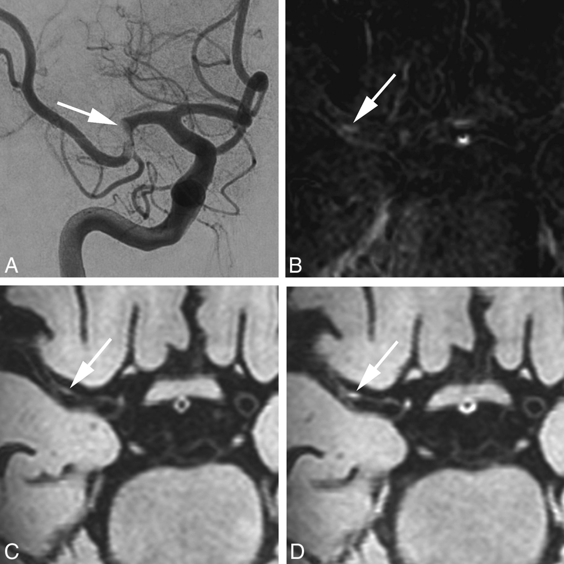
  • Fig 3.
    • Download figure
    • Open in new tab
    • Download powerpoint
    Fig 3.

    A 72-year-old woman with an occlusion of the right M1 segment, successfully treated with a thrombosuction device (patient 11 in Table 2). Digital subtraction angiography directly before the thrombectomy procedure shows an acute occlusion in the right middle cerebral artery (A, arrow). B, Subtraction image from coregistered pre- and postcontrast (C and D) vessel wall images confirms a hyperintense rim (arrow) at the vessel wall. Transverse pre- (C) and postcontrast (D) MPIR-TSE vessel wall images at 7T were obtained 25 days after the thrombectomy procedure. The area of eccentric vessel wall enhancement (D) is seen in the right M1 segment, at the same location as the thrombectomy site, confirmed on the subtraction image in B (arrows).

  • Fig 4.
    • Download figure
    • Open in new tab
    • Download powerpoint
    Fig 4.

    A 67-year-old man with an occlusion of the left M1–M2 segment, successfully treated with intra-arterial thrombectomy (patient 14 in Table 2). The patient was treated with both a stent-retriever device and a thrombosuction device. Axial pre- (A) and postcontrast (B) MPIR-TSE vessel wall images at 7T, 87 days after the thrombectomy procedure. Clear contrast enhancement is present (white arrows) at the same location where the thrombectomy was performed. The carotid and basilar arteries appear normal (white arrowheads, A and B). C, Subtraction image of the pre- and postcontrast vessel wall images confirms the enhancement at the same location. D and E, Coronal views of the postcontrast MPIR-TSE vessel wall images show enhancement over a long trajectory of the left M1 and M2 segments. F, Sagittal view of the postcontrast MPIR-TSE vessel wall image shows that the enhancement has a concentric configuration.
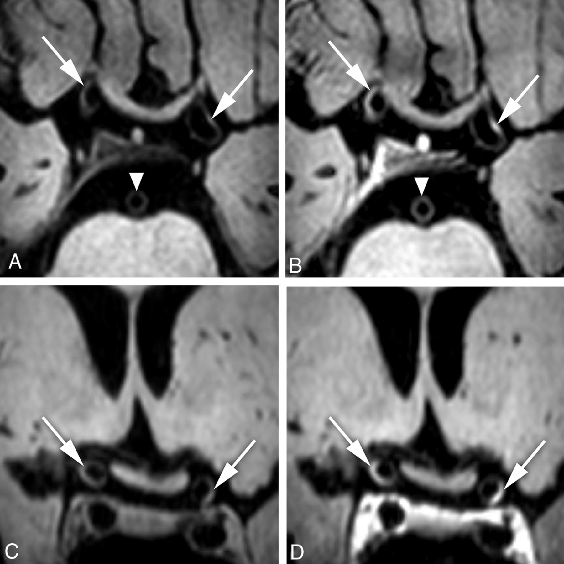
  • Fig 5.
    • Download figure
    • Open in new tab
    • Download powerpoint
    Fig 5.

    A 75-year-old woman with an anterior circulation ischemic infarction of the left MCA territory (non-IAT group, patient 30 in On-line Table). Transverse and coronal precontrast (A and C) and postcontrast (B and D) MPIR-TSE vessel wall images at 7T were obtained 10 days after symptom onset of the ischemic infarction. Note eccentric vessel wall enhancement of the supraclinoid portion of both intracranial carotid arteries (B and D, arrows). The basilar artery appears normal (white arrowheads, A and B).

Tables

  • Figures
    • View popup
    Table 1:

    Baseline characteristics, specified by patients with stroke with and without IAT

    IAT Total (%)Non-IAT Total (%)P Value
    Age (mean) (range) (yr)65 (42–85)60 (35–81).23a
    Sex (male)10 (71%)18 (51%).34b
    Hypertension4 (29%)18 (51%).21b
    Hyperlipidemia7 (50%)18 (51%).99b
    Diabetes mellitus0 (0%)4 (11%).31b
    Current smoking3 (21%)12 (34%).46c
    Former smoking5 (36%)7 (20%).46c
    Atrial fibrillation3 (21%)4 (11%).39b
    NIHSS score (mean) (range)10.5 (3–20)6.3 (0–21).02a
    TOAST criteria30.62c
     Large-artery atherosclerosis7 (50%)21 (60%)
     Cardioembolism4 (29%)5 (14%)
     Small-vessel occlusion0 (0%)0 (0%)
     Other determined etiology1 (7%)5 (14%)
     Undetermined2 (14%)4 (12%)
    Time to 7T MR imaging (mean) (SD) (day)55 (±22)16 (±23)<.001a
    Time to 7T MR imaging (median) (range) (day)58 (22–87)6 (1–84)
    • ↵a Mann-Whitney U test.

    • ↵b Fisher exact test.

    • ↵c Pearson χ2 test.

    • View popup
    Table 2:

    Location of occlusion and treatment details, including detected enhancing vessel wall foci, in the IAT-groupa

    Patient No.Occlusion SiteNIHSS ScoreNOPTime Symptom Onset to IAT (Min)Procedural Time (Min)TICI ScoreAlteplaseTime IAT to MRI (Day)Ipsilateral Enhancing FociContralateral Enhancing Foci
    1M1 right10119035TICI 3Yes33––
    2M1–M2 right11120834TICI 3Yes67–M1-E
    3Distal carotid left8331850TICI 2bNo84M1-C–
    4M2 left5517083TICI 2bYes49ICA-E, M2-EICA-E
    5M2 left5112742TICI 3Yes65––
    6(Large) M3 right3113560TICI 0Yes67ICA-C–
    7Distal carotid left12220160TICI 2bYes80ICA-C–
    8M2 left4327570TICI 2bNo59ICA-C, M1-EICA-E
    9M1–M2 left15115035TICI 3Yes22M1-E–
    10M1 left14115039TICI 3Yes24M1-E–
    11M1 right12114529TICI 3Yes25ICA-E 2x, M1-E–
    12M1–M2 right2017640TICI 3Yes51––
    13M1 right1504442TICI 3Yes57––
    14M1–M2 left14218050TICI 2bYes87M1-C, M2-C–
    • Note:—C indicates concentric; E, eccentric; M, segment of the middle cerebral artery (M1 and M2); NOP, number of passes; –, no enhancing foci detected.

    • ↵a Treatment details of the 14 patients including the number and location of the detected foci of contrast enhancement (by A.G.v.d.K.). In patient 3, the enhancing focus detected in the M1 segment was located distal to the occlusion but directly adjacent to the occlusion site and therefore identified as the same location as the thrombosuction site.

    • View popup
    Table 3:

    Comparison of enhancing foci between the IAT-group and the non-IAT group and between the ipsilateral and contralateral sides

    IAT-GroupNon-IAT GroupP Value
    Total No. of enhancing foci21.530.04a
    Total No. of ipsilateral enhancing foci18.518.0.003a
    Total No. of contralateral enhancing foci3.012.0.74a
    Total No. of concentric ipsilateral foci9.08.5.02a
    Total No. of eccentric ipsilateral foci9.59.5.07a
    Total No. of enhancing foci ipsilateral vs contralateral (proportion) P value(18.5 vs 3.0) .005b(18.0 vs 12.0) .47b
    Total No. of concentric enhancing foci ipsilateral vs. contralateral (proportion) P value(9.0 vs 0.0) .011b(8.5 vs 3.0) .14b
    Total No. of eccentric enhancing foci ipsilateral vs. contralateral (proportion) P value(9.5 vs 3.0) 06b(9.5 vs 9.0) .97b
    • ↵a Mann-Whitney U test.

    • ↵b Wilcoxon signed rank test.

PreviousNext
Back to top

In this issue

American Journal of Neuroradiology: 41 (4)
American Journal of Neuroradiology
Vol. 41, Issue 4
1 Apr 2020
  • Table of Contents
  • Index by author
  • Complete Issue (PDF)
Advertisement
Print
Download PDF
Email Article

Thank you for your interest in spreading the word on American Journal of Neuroradiology.

NOTE: We only request your email address so that the person you are recommending the page to knows that you wanted them to see it, and that it is not junk mail. We do not capture any email address.

Enter multiple addresses on separate lines or separate them with commas.
MRI Vessel Wall Imaging after Intra-Arterial Treatment for Acute Ischemic Stroke
(Your Name) has sent you a message from American Journal of Neuroradiology
(Your Name) thought you would like to see the American Journal of Neuroradiology web site.
CAPTCHA
This question is for testing whether or not you are a human visitor and to prevent automated spam submissions.
Cite this article
A. Lindenholz, I.C. van der Schaaf, A.G. van der Kolk, H.B. van der Worp, A.A. Harteveld, L.J. Kappelle, J. Hendrikse
MRI Vessel Wall Imaging after Intra-Arterial Treatment for Acute Ischemic Stroke
American Journal of Neuroradiology Apr 2020, 41 (4) 624-631; DOI: 10.3174/ajnr.A6460

Citation Manager Formats

  • BibTeX
  • Bookends
  • EasyBib
  • EndNote (tagged)
  • EndNote 8 (xml)
  • Medlars
  • Mendeley
  • Papers
  • RefWorks Tagged
  • Ref Manager
  • RIS
  • Zotero
0 Responses
Respond to this article
Share
Bookmark this article
MRI Vessel Wall Imaging after Intra-Arterial Treatment for Acute Ischemic Stroke
A. Lindenholz, I.C. van der Schaaf, A.G. van der Kolk, H.B. van der Worp, A.A. Harteveld, L.J. Kappelle, J. Hendrikse
American Journal of Neuroradiology Apr 2020, 41 (4) 624-631; DOI: 10.3174/ajnr.A6460
del.icio.us logo Twitter logo Facebook logo Mendeley logo
  • Tweet Widget
  • Facebook Like
  • Google Plus One
Purchase

Jump to section

  • Article
    • Abstract
    • ABBREVIATIONS:
    • MATERIALS AND METHODS
    • RESULTS
    • DISCUSSION
    • CONCLUSIONS
    • Footnotes
    • References
  • Figures & Data
  • Supplemental
  • Info & Metrics
  • Responses
  • References
  • PDF

Related Articles

  • PubMed
  • Google Scholar

Cited By...

  • Report from the society of magnetic resonance angiography: clinical applications of 7T neurovascular MR in the assessment of intracranial vascular disease
  • Unexplained early neurological deterioration after endovascular treatment for acute large vessel occlusion: incidence, predictors, and clinical impact: Data from ANGEL-ACT registry
  • Characterization of Subarachnoid Hyperdensities After Thrombectomy for Acute Stroke Using Dual-Energy CT
  • Crossref (13)
  • Google Scholar

This article has been cited by the following articles in journals that are participating in Crossref Cited-by Linking.

  • MRI with ultrahigh field strength and high-performance gradients: challenges and opportunities for clinical neuroimaging at 7 T and beyond
    Behroze Vachha, Susie Y. Huang
    European Radiology Experimental 2021 5 1
  • European Stroke Organisation (ESO) guidelines on Primary Angiitis of the Central Nervous System (PACNS)
    Rosario Pascarella, Katherina Antonenko, Grégoire Boulouis, Hubert De Boysson, Caterina Giannini, Mirjam R Heldner, Odysseas Kargiotis, Thanh N Nguyen, Claire M Rice, Carlo Salvarani, Antje Schmidt-Pogoda, Daniel Strbian, Salman Hussain, Marialuisa Zedde
    European Stroke Journal 2023 8 4
  • A high resolution scanning electron microscopy analysis of intracranial thrombi embedded along the stent retrievers
    Daniela Dumitriu LaGrange, Gianmarco Bernava, Philippe Reymond, Isabel Wanke, Maria Isabel Vargas, Paolo Machi, Karl-Olof Lövblad
    Scientific Reports 2022 12 1
  • Characterization of Subarachnoid Hyperdensities After Thrombectomy for Acute Stroke Using Dual-Energy CT
    Arturo Renú, Carlos Laredo, Alejandro Rodríguez-Vázquez, Daniel Santana, Mariano Werner, Laura Llull, Antonio Lopez-Rueda, Xabier Urra, Salvatore Rudilosso, Víctor Obach, Sergi Amaro, Ángel Chamorro
    Neurology 2022 98 6
  • Unexplained early neurological deterioration after endovascular treatment for acute large vessel occlusion: incidence, predictors, and clinical impact: Data from ANGEL-ACT registry
    Dapeng Sun, Xu Tong, Xiaochuan Huo, Baixue Jia, Raynald, Anxin Wang, Gaoting Ma, Ning Ma, Feng Gao, Dapeng Mo, Ligang Song, Xuan Sun, Lian Liu, Yiming Deng, Xiaoqing Li, Bo Wang, Gang Luo, Yongjun Wang, Zhongrong Miao
    Journal of NeuroInterventional Surgery 2022 14 9
  • 7T MRI for Intracranial Vessel Wall Lesions and Its Associated Neurological Disorders: A Systematic Review
    Chen Zhang, Jiong Shi
    Brain Sciences 2022 12 5
  • Report from the society of magnetic resonance angiography: clinical applications of 7T neurovascular MR in the assessment of intracranial vascular disease
    Binbin Sui, Bhagya Sannananja, Chengcheng Zhu, Niranjan Balu, Laura Eisenmenger, Hediyeh Baradaran, Myriam Edjlali, Javier M Romero, Prabakhar Shantha Rajiah, Rui Li, Mahmud Mossa-Basha
    Journal of NeuroInterventional Surgery 2024 16 8
  • Predictors of symptomatic intracranial hemorrhage after endovascular treatment for acute large vessel occlusion: data from ANGEL-ACT registry
    Dapeng Sun, Xiaochuan Huo, Raynald, Baixue Jia, Xu Tong, Gaoting Ma, Anxin Wang, Ning Ma, Feng Gao, Dapeng Mo, Zhongrong Miao
    Journal of Thrombosis and Thrombolysis 2022 54 3
  • Practical Aspects of novel MRI Techniques in Neuroradiology: Part 2 – Acceleration Methods and Implications for Individual Regions
    Benedikt Sundermann, Benoit Billebaut, Jochen Bauer, Catalin George Iacoban, Olga Alykova, Christoph Schülke, Maike Gerdes, Harald Kugel, Sojan Neduvakkattu, Holger Bösenberg, Christian Mathys
    RöFo - Fortschritte auf dem Gebiet der Röntgenstrahlen und der bildgebenden Verfahren 2022 194 11
  • Imagerie des Urgences Neuroradiologiques
    Myriam Edjlali-Goujon
    2024

More in this TOC Section

Adult Brain

  • Diagnostic Neuroradiology of Monoclonal Antibodies
  • Clinical Outcomes After Chiari I Decompression
  • Segmentation of Brain Metastases with BLAST
Show more Adult Brain

Neurointervention

  • Contour Neurovascular System: Five Year Follow Up
  • Effect of SARS-CoV2 on Endovascular Thrombectomy
  • A Key Factor Shapes LS-DAVFs EVT Outcome
Show more Neurointervention

Similar Articles

Advertisement

Indexed Content

  • Current Issue
  • Accepted Manuscripts
  • Article Preview
  • Past Issues
  • Editorials
  • Editor's Choice
  • Fellows' Journal Club
  • Letters to the Editor
  • Video Articles

Cases

  • Case Collection
  • Archive - Case of the Week
  • Archive - Case of the Month
  • Archive - Classic Case

More from AJNR

  • Trainee Corner
  • Imaging Protocols
  • MRI Safety Corner
  • Book Reviews

Multimedia

  • AJNR Podcasts
  • AJNR Scantastics

Resources

  • Turnaround Time
  • Submit a Manuscript
  • Submit a Video Article
  • Submit an eLetter to the Editor/Response
  • Manuscript Submission Guidelines
  • Statistical Tips
  • Fast Publishing of Accepted Manuscripts
  • Graphical Abstract Preparation
  • Imaging Protocol Submission
  • Evidence-Based Medicine Level Guide
  • Publishing Checklists
  • Author Policies
  • Become a Reviewer/Academy of Reviewers
  • News and Updates

About Us

  • About AJNR
  • Editorial Board
  • Editorial Board Alumni
  • Alerts
  • Permissions
  • Not an AJNR Subscriber? Join Now
  • Advertise with Us
  • Librarian Resources
  • Feedback
  • Terms and Conditions
  • AJNR Editorial Board Alumni

American Society of Neuroradiology

  • Not an ASNR Member? Join Now

© 2025 by the American Society of Neuroradiology All rights, including for text and data mining, AI training, and similar technologies, are reserved.
Print ISSN: 0195-6108 Online ISSN: 1936-959X

Powered by HighWire