Advertisement

Main menu

  • Home
  • Content
    • Current Issue
    • Accepted Manuscripts
    • Article Preview
    • Past Issue Archive
    • Video Articles
    • AJNR Case Collection
    • Case of the Week Archive
    • Case of the Month Archive
    • Classic Case Archive
  • Special Collections
    • AJNR Awards
    • Low-Field MRI
    • Alzheimer Disease
    • ASNR Foundation Special Collection
    • Photon-Counting CT
    • View All
  • Multimedia
    • AJNR Podcasts
    • AJNR SCANtastic
    • Trainee Corner
    • MRI Safety Corner
    • Imaging Protocols
  • For Authors
    • Submit a Manuscript
    • Submit a Video Article
    • Submit an eLetter to the Editor/Response
    • Manuscript Submission Guidelines
    • Statistical Tips
    • Fast Publishing of Accepted Manuscripts
    • Graphical Abstract Preparation
    • Imaging Protocol Submission
    • Author Policies
  • About Us
    • About AJNR
    • Editorial Board
    • Editorial Board Alumni
  • More
    • Become a Reviewer/Academy of Reviewers
    • Subscribers
    • Permissions
    • Alerts
    • Feedback
    • Advertisers
    • ASNR Home

User menu

  • Alerts
  • Log in

Search

  • Advanced search
American Journal of Neuroradiology

American Journal of Neuroradiology

ASHNR American Society of Functional Neuroradiology ASHNR American Society of Pediatric Neuroradiology ASSR
  • Alerts
  • Log in

Advanced Search

  • Home
  • Content
    • Current Issue
    • Accepted Manuscripts
    • Article Preview
    • Past Issue Archive
    • Video Articles
    • AJNR Case Collection
    • Case of the Week Archive
    • Case of the Month Archive
    • Classic Case Archive
  • Special Collections
    • AJNR Awards
    • Low-Field MRI
    • Alzheimer Disease
    • ASNR Foundation Special Collection
    • Photon-Counting CT
    • View All
  • Multimedia
    • AJNR Podcasts
    • AJNR SCANtastic
    • Trainee Corner
    • MRI Safety Corner
    • Imaging Protocols
  • For Authors
    • Submit a Manuscript
    • Submit a Video Article
    • Submit an eLetter to the Editor/Response
    • Manuscript Submission Guidelines
    • Statistical Tips
    • Fast Publishing of Accepted Manuscripts
    • Graphical Abstract Preparation
    • Imaging Protocol Submission
    • Author Policies
  • About Us
    • About AJNR
    • Editorial Board
    • Editorial Board Alumni
  • More
    • Become a Reviewer/Academy of Reviewers
    • Subscribers
    • Permissions
    • Alerts
    • Feedback
    • Advertisers
    • ASNR Home
  • Follow AJNR on Twitter
  • Visit AJNR on Facebook
  • Follow AJNR on Instagram
  • Join AJNR on LinkedIn
  • RSS Feeds

AJNR Awards, New Junior Editors, and more. Read the latest AJNR updates

Research ArticlePatient Safety
Open Access

Nephrogenic Systemic Fibrosis Risk Assessment and Skin Biopsy Quantification in Patients with Renal Disease following Gadobenate Contrast Administration

E. Kanal, T.J. Patton, I. Krefting and C. Wang
American Journal of Neuroradiology March 2020, 41 (3) 393-399; DOI: https://doi.org/10.3174/ajnr.A6448
E. Kanal
aDepartments of Radiology (E.K.)
  • Find this author on Google Scholar
  • Find this author on PubMed
  • Search for this author on this site
  • ORCID record for E. Kanal
T.J. Patton
bDermatology (T.J.P.), University of Pittsburgh Medical Center and University of Pittsburgh, Pittsburgh, Pennsylvania
  • Find this author on Google Scholar
  • Find this author on PubMed
  • Search for this author on this site
  • ORCID record for T.J. Patton
I. Krefting
cDivision of Medical Imaging and Radiation Medicine (I.K.)
  • Find this author on Google Scholar
  • Find this author on PubMed
  • Search for this author on this site
  • ORCID record for I. Krefting
C. Wang
dOffice of Pharmacovigilance and Epidemiology (C.W.), US Food and Drug Administration, Silver Spring, Maryland.
  • Find this author on Google Scholar
  • Find this author on PubMed
  • Search for this author on this site
  • ORCID record for C. Wang
  • Article
  • Figures & Data
  • Info & Metrics
  • Responses
  • References
  • PDF
Loading

Abstract

BACKGROUND AND PURPOSE: Nephrogenic systemic fibrosis following administration of intravenous gadobenate during MR imaging is rare. This study aimed to analyze any nephrogenic systemic fibrosis–related risks and quantify skin gadolinium levels in patients with impaired renal function but without nephrogenic systemic fibrosis who had received gadobenate.

MATERIALS AND METHODS: In this retrospective study with a prospective skin biopsy phase, patients with estimated glomerular filtration rates of <60 mL/min/1.73 m2 undergoing contrast-enhanced MR imaging from July 2007 through June 2014 were screened for nephrogenic systemic fibrosis using a questionnaire. This was highly sensitive but not specific and reliably excluded nephrogenic systemic fibrosis if responses to at least 6 of the 8 questions were negative. If no nephrogenic systemic fibrosis was detected, a skin biopsy was requested.

RESULTS: Of 2914 patients who met these criteria, 1988 were excluded for various reasons. Of the remaining 926 patients, 860 were screened negative for nephrogenic systemic fibrosis. Of these, 17 (2%) had estimated glomerular filtration rates of <15 mL/min/1.73 m2, 51 (6%) had levels of 15 < 30 mL/min/1.73 m2, 234 (27%) had levels of 30 < 45 mL/min/1.73 m2, and 534 (62%) had levels of 45 < 60 mL/min/1.73 m2. Of the 66 who were not cleared of nephrogenic systemic fibrosis by the questionnaire, 6 patients were evaluated by a dermatologist and confirmed not to have nephrogenic systemic fibrosis (no biopsy required).

CONCLUSIONS: A diagnosis of nephrogenic systemic fibrosis was excluded in 860 patients with impaired renal function who were followed up and received gadobenate during MR imaging. In 14 such patients who underwent at least 1 gadobenate-enhanced MR imaging examination and did not have nephrogenic systemic fibrosis, gadolinium levels in the skin were exceedingly low.

ABBREVIATIONS:

eGFR
estimated glomerular filtration rate
GBCA
gadolinium-based contrast agents
ICP-MS
inductively coupled plasma mass spectrometry
NSF
nephrogenic systemic fibrosis

While the relative nephrogenic systemic fibrosis (NSF)–related safety of at least some of the macrocyclic agents has been established, there do not seem to be any confirmed cases of NSF following the prior unconfounded administration of the linear gadolinium-based contrast agent gadobenate.1 Since gadolinium-based contrast agents (GBCA) received regulatory approval in 1988, >450 million doses have been administered worldwide.1 While GBCA have a favorable pharmacologic safety profile, evidence of long-term retention in human tissues and occurrence of NSF in some patients with severely compromised renal function have raised concerns.2,3

The connection between gadolinium exposure and NSF was recognized by Grobner,4 in 2006. Gadolinium levels are elevated in the biopsies of skin lesions in patients with NSF.3,5⇓⇓-8 In 2014, Kanda et al9 noted retention of gadolinium in the brain parenchyma of patients with normal renal function following repeat gadolinium-enhanced MRIs. Gadolinium has also been detected in the skin, bones, and livers of patients with normal renal function following repeat MRIs with commercially available GBCA.

The theory of transmetallation has been quoted as a rationale for patients receiving GBCA and developing NSF. Also known as the dechelation or dissociation theory, this theory recognizes the notable differences in the comparative bond strength and kinetics of dissociation of the bonds between the gadolinium ion and ligand molecules used in GBCA. The kinetics of the dissociation strength of these bonds among the linear GBCA in general is faster than that for the macrocyclic GBCA, suggesting that dissociation of the gadolinium ion from its ligand molecule would be greater for linear and especially nonionic linear agents than for macrocyclic agents. Precisely such results have been shown in vivo.10-22

Only low single-digit numbers of cases of NSF have been seen following unconfounded administration of macrocyclic GBCA. As discussed at the December 8, 2009, FDA Advisory Committee,1 there are several hundred confirmed cases of NSF following unconfounded administration of the linear nonionic gadolinium-based contrast agent, gadodiamide, with several dozen following unconfounded administration of the other linear nonionic gadolinium-based contrast agent, gadoversetamide.

However, the ability of this theory alone to explain clinical observations is called into question with roughly 100 cases of NSF following unconfounded administration of the linear ionic agent gadopentetate, yet zero or 1 possibly case following unconfounded administration of the linear ionic agent gadobenate.23,24 These differences cannot be ascribed to the relative market share differences alone.25

The lack of clarity as to causation is compounded by the concern that perhaps NSF might actually represent one extreme on a spectrum of gadolinium-related issues. In other words, there is a concern that a so-called “subclinical NSF” might exist in which some changes may be histologically present but not clinically manifest at lower total exposure levels to various GBCA.

To that end, Phase I of our study was designed to evaluate the incidence of NSF following administration of gadobenate in patients with chronic renal disease who would be expected to be at the highest risk for NSF. Phase II involved measuring gadolinium levels in the skin of patients with impaired renal function who did not have NSF but had undergone at least 1 contrast-enhanced MR imaging study with gadobenate.

This study was performed to attempt to further analyze and quantify any NSF-related risks associated with this linear GBCA.

MATERIALS AND METHODS

Phase I

As part of a clinical quality-assurance project initiated in 2007 at the University of Pittsburgh Medical Center, we recorded data for all clinical patients with impaired renal function (defined as estimated glomerular filtration rate [eGFR] values of <60 mL/min/1.73 m2) who underwent clinically requested contrast-enhanced MR imaging examinations following a clinical benefit-risk assessment in which the outcome was to proceed with the examination.

For this project, all patients who were identified as having at least moderate chronic kidney disease who underwent clinically indicated contrast-enhanced MR imaging with gadobenate were identified from our clinical quality-assurance data base noted above from July 2007 through June 2014 (inclusive). Institutional review board approval for this study was granted in addition to the ongoing clinical quality-assurance project approval and oversight noted above. For each patient with impaired renal function, contemporaneous recording was made of the patient’s name and identification number, weight, the administered MR imaging contrast agent name and dose, and the most recently available eGFR value. A neutral third party reviewed this list of clinical patients and compared it against the medical record data base of deceased patients in the health care system. After we excluded patients identified as deceased, the contact information for the remaining patients was procured from medical records, and attempts were made by the neutral third party to contact each of these patients for NSF-screening purposes. The order in which the patients were contacted was such that the first patients to be contacted were those with the lowest eGFR level at time of gadobenate administration. Because mortality was expected to be highest among those with the greatest levels of impairment of renal function, we opted to begin with those patients to successfully screen as many as possible in the earliest stages of this study.

Using the screening criteria established by Lima et al,26 we verbally screened all contacted patients with an 8-question questionnaire that focused on cutaneous and musculoskeletal manifestations of NSF. Lima et al had demonstrated that affirmative responses to ≥3 of the 8 closed-ended (yes/no) questions yielded a 90% sensitivity and 70% specificity for NSF. On the basis of differences in proposed prevalence values, this outcome yielded a positive predictive value for this questionnaire that ranged from a somewhat disappointing 0.3% to 39.7%, but, at the same time, a negative predictive value, which ranged from 97% to >99%. Therefore, a negative response to this questionnaire was thought to reliably exclude a diagnosis of NSF, whereas a positive response to at least 3 of the questions posed in this questionnaire was deemed worthy of further investigation because specificity and positive predictive values were insufficient for conclusive diagnoses based on the questionnaire alone.

During the execution of Phase I of this study, patients who were screened as negative for NSF were asked if they would permit us to contact them at a later date for possible inclusion in a research study. Only those who responded in the affirmative were advanced to Phase II of this study.

Phase II

After the conclusion of Phase I of this study, all patients who screened negative for NSF and who gave permission for future contact were again contacted and recruited to be possible participants in Phase II of this study. These patients had impaired renal function and had undergone at least 1 MR imaging examination enhanced with intravenously administered gadobenate. Study participation entailed undergoing a purely research-generated punch deep skin biopsy of the lateral thigh. These biopsies were then analyzed using inductively coupled plasma mass spectrometry (ICP-MS) for gadolinium-level quantification in Dr Whitney High’s laboratory (Department of Dermatology & Dermatopathology, University of Colorado School of Medicine). The ICP-MS methodology is outlined in a previous publication by High et al,7 from 2007, which reported gadolinium levels in the skin of patients with NSF; this methodology has not changed.

RESULTS

Phase I

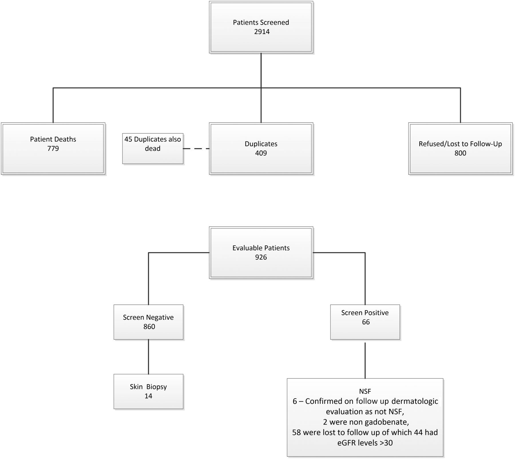
A total of 2914 patients were identified who had eGFR values of <60 mL/min/1.73 m2 and who underwent at least 1 contrast-enhanced MR imaging examination with gadobenate (Fig 1). Of these, 1988 were excluded due to death (779), duplicates (409, deceased patient duplicates numbered 45 of these 409), or patients who refused contact/cooperation or were lost to follow-up (800). Of the remaining 926 patients (Table 1), all except 66 screened negative for NSF. Of the 66 who had responded yes to ≥3 questions in the NSF screening questionnaire, 6 patients were seen and evaluated by a dermatologist and were confirmed to have another diagnosis and not NSF (no biopsy required), 19 lived too far away/could not return for follow-up at our institution, 10 refused follow-up care, 24 did not return multiple (at least 3) separate calls/messages, 3 had dementia/were in nursing home facilities, 2 had died, and 2 had received nongadobenate gadolinium-based contrast agents (1 gadoxetate, 1 gadobutrol).

Fig 1.
  • Download figure
  • Open in new tab
  • Download powerpoint
Fig 1.

Patient selection.

View this table:
  • View inline
  • View popup
Table 1:

Dose distribution of 926 patients

Of the 860 patients screened as not having NSF, 17 (2%) had eGFR levels of <15 mL/min/1.73 m2, 51 (6%) had eGFR levels of 15 < 30 mL/min/1.73 m2, 234 (27%) had eGFR levels of 30 < 45 mL/min/1.73 m2, 534 (62%) had eGFR levels of 45 < 60 mL/min/1.73 m2, and 24 (3%) had no concurrent eGFR data available or had eGFR levels of >60 but did have a history of known renal disease. There were no cases of NSF identified either in the screening process using the questionnaire or among those examined directly by the dermatologist and who had responded yes to ≥3 questions on the screening questionnaire. In 4 patients, there were nonspecific findings that did not require biopsy (1 of these 4 had undergone an unrelated clinical biopsy of her lower leg with nonspecific findings that were not consistent with NSF); in another, there was dyspigmentation of the skin consistent with postinflammatory pigment changes secondary to trauma; and one had induration of the distal lower extremities consistent with lipodermatosclerosis.

Of the 66 patients who had responded positively to ≥3 questions, 2 had received other GBCA (1 gadoxetate, 1 gadobutrol) and 2 patients were found to have had eGFR levels exceeding 59 at time of administration and were thus excluded from further analysis. Of the remaining 62 patients, 6 (10%) had eGFR levels of <15 mL/min/1.73 m2, 11 (18%) had eGFR levels of 15 < 30 mL/min/1.73 m2, 19 (31%) had eGFR levels of 30 < 45 mL/min/1.73 m2, and 25 (40%) had eGFR levels of 45 < 60 mL/min/1.73 m2. One patient with known renal disease did not have an eGFR before GBCA administration and was excluded from the tally.

Of the 6 patients who had responded positively to ≥3 questions and who were examined by a dermatologist, one (17%) had eGFR levels of <15 mL/min/1.73 m2, none (0%) had eGFR levels of 15 < 30 mL/min/1.73 m2, one (17%) had eGFR levels of 30 < 45 mL/min/1.73 m2, and 4 patients (67%) had eGFR levels of 45 < 60 mL/min/1.73 m2.

Phase II

A total of 14 patients with renal impairment who screened negative for NSF on the screening questionnaire consented to participate in this phase of the study and successfully underwent an elective research-motivated deep skin punch biopsy and gadolinium-level quantification using ICP-MS (Fig 2). The measured eGFR values for these 14 research subjects ranged from 26 to 59 mL/min/1.73 m2. The measured gadolinium values in the skin biopsies of these patients ranged from <0.05 to 1.1 µg/g (Table 2). These values were all judged to be consistent with background values for this laboratory and were distinctly lower than gadolinium levels typically measured in this lab for skin biopsies of patients with NSF in whom skin-sample gadolinium levels ranged from 4.8 to 106 μg/g.

Fig 2.
  • Download figure
  • Open in new tab
  • Download powerpoint
Fig 2.

Location marked is representative of the biopsy location on each of the 14 research subjects studied.

View this table:
  • View inline
  • View popup
Table 2:

Subject biopsy details, including gadolinium level and number of prior MRIs

DISCUSSION

The transmetallation theory has been broadly cited to explain how NSF develops and the mechanism for potential safety issues associated with retained/residual gadolinium following clinical gadolinium administration. However, the transmetallation theory itself does not sufficiently explain the relative incidence of NSF. Certain inconsistencies seem to be present if we seek to explain the NSF incidence on the basis of the transmetallation theory alone. For example, gadopentetate and gadobenate are both linear ionic gadolinium-based contrast agents, yet there is a large difference in the number of cases of NSF associated with their prior unconfounded administration that cannot be explained by relative market share differences.25

The lack of clarity as to causation is compounded by the concern that perhaps NSF might actually represent one extreme on a spectrum of gadolinium-related issues. In other words, there is a concern that a so-called subclinical NSF might exist in which some changes may be histologically present but not clinically manifest at lower total exposure levels to various GBCA.

This study was undertaken in an effort to further analyze and quantify the NSF risk associated with gadobenate administration, specifically to the population deemed to be at highest risk for this disease, namely, those with significant renal disease. The findings of this study, revealing no cases of NSF associated with the administration of gadobenate to patients with significant, serious, or end-stage renal disease support those of other publications describing the observed NSF incidence in this specific patient population.27,-,30

With gadolinium having been reported in the skin of patients with NSF (Table 3),5,31 it is interesting to note the lack of elevation of gadolinium levels in the 14 subjects with significant renal disease who had previously undergone at least 1 contrast-enhanced MR imaging examination with gadobenate.

View this table:
  • View inline
  • View popup
Table 3:

Gadolinium deposition in skin samples from patients with NSF (adapted from Roberts et al34)

It remains unclear whether the presence of gadolinium in NSF skin lesions is etiologically related, secondary, or incidental in nature, and this lack of clarity provides further evidence of differences between patients with NSF with renal disease relative to those with significant renal disease who had undergone gadobenate-enhanced MR imaging but did not have clinical NSF.

Limitations

The study is limited in that there are several patients who had responded “yes” to ≥3 screening questions about whom there is no follow-up information available due to patient refusals, death, lack of access to follow-up, and so forth. However, the data remain quite reassuring insofar as most patients in this study with significant renal disease who had undergone a gadobenate-enhanced MR imaging examination were able to be successfully screened, and no cases of NSF were found. Another limitation of the examination is that the vast majority of those with significant renal disease in this study who did receive gadobenate received a half-dose and not a full dose. However, this specific gadolinium-based contrast agent has a high relaxivity, and several publications have already documented the favorable diagnostic sensitivities of partial-dose gadobenate relative to studies performed following full doses of several of the other lower relaxivity GBCA used for neuroradiologic imaging.32,33 Thus, from a clinical point of view, partial-dose administration of this agent may well be an appropriate diagnostic comparator against a full dose of lower relaxivity agents, with the added benefit of having administered to the patient proportionally fewer gadolinium ions by so doing.

The gadolinium levels were obtained after the patient volunteered for the study and therefore at various times following the last gadolinium contrast MR imaging, the date of which was not available to the study team. We, therefore, view these levels as a “snapshot” at an indeterminate time from the last administration.

Gadolinium levels should be considered either negligible or large; exact quantitation, given the small mass of the skin biopsy, may not be achievable. The small mass of the skin biopsies introduces the possibility of contamination during multiple processing procedures to determine gadolinium levels such as the following:

  • Metal in the punch biopsy device

  • Metal in the forceps

  • Non-reagent grade chemicals used in the processing procedures

  • Contamination from the microtomes

  • Contamination from the water baths.

Consistent with other similar reports, in our study of 860 patients with varying levels of impaired renal function (defined as <60 mL./min/1.73 m2) who underwent clinically indicated MR imaging examinations with gadobenate, subsequent screening failed to reveal any cases of nephrogenic systemic fibrosis. In comparison with previous publications, we screened patients with a uniform, previously validated questionnaire and performed skin biopsies. Previous studies also faced the methodologic difficulty of accounting for drop-out and deceased patients. Gadolinium-level quantification by ICP-MS of deep skin punch biopsies of 14 research subjects with impaired renal function who did not have clinical signs or symptoms of NSF and who had previously undergone at least 1 contrast-enhanced MR imaging examination with gadobenate failed to reveal any measurable elevation of skin gadolinium levels beyond that found in healthy controls.

CONCLUSIONS

No evidence of NSF was found in 860 patients with moderate or worse chronic kidney disease for whom follow-up was available and who had received (predominantly half dose) gadobenate for clinical MR imaging examinations. Additionally, in 14 patients with impaired renal function who underwent at least 1 gadobenate-enhanced MR imaging examination and who did not have clinical NSF, ICP-MS determined that gadolinium levels in the skin were exceedingly low and were considered essentially at background levels without evidence of abnormal gadolinium accumulation. Implications for patient care are that NSF was not identified in patients with renal disease undergoing predominantly half-dose gadobenate contrast MR imaging and that gadolinium skin retention is minimal in these patients.

Acknowledgment

The authors wish to thank Dr Whitney High for his assistance in testing skin samplings for gadolinium.

Footnotes

  • Dr Wang is currently with Ascentage Pharma Group, Rockville, Maryland.

  • This work was funded, in part, by FDA Cooperative Agreement U01 FD004713.

  • Disclosures: Emanuel Kanal—UNRELATED: Consultancy: Bracco Diagnostics, GE Healthcare, Guerbet, Comments: MR imaging safety consulting.

Indicates open access to non-subscribers at www.ajnr.org

References

  1. 1.↵
    1. Balzer T
    . Presence of Gadolinium (Gd) in the Brain and Body: Presentation to the Medical Imaging Drugs Advisory Committee, September 8, 2017. FDA. Silver Spring, Md: U.S. Food and Drug Administration. https://www.fda.gov/media/107670/download. Accessed February 5, 2020
  2. 2.↵
    1. McDonald RJ,
    2. Levine D,
    3. Weinreb J, et al
    . Gadolinium retention: a research roadmap from the 2018 NIH/ACR/RSNA Workshop on Gadolinium Chelates. Radiology 2018;289:517–34 doi:10.1148/radiol.2018181151 pmid:30204075
    CrossRefPubMed
  3. 3.↵
    1. Boyd AS,
    2. Zic JA,
    3. Abraham JL
    . Gadolinium deposition in nephrogenic fibrosing dermopathy. J Am Acad Dermatol 2007;56:27–30 doi:10.1016/j.jaad.2006.10.048 pmid:17109993
    CrossRefPubMed
  4. 4.↵
    1. Grobner T
    . Gadolinium: a specific trigger for the development of nephrogenic fibrosing dermopathy and nephrogenic systemic fibrosis? Nephrol Dial Transplant 2006;21:1104–08 doi:10.1093/ndt/gfk062 pmid:16431890
    CrossRefPubMed
  5. 5.↵
    1. Birka M,
    2. Wentker KS,
    3. Lusmoller E, et al
    . Diagnosis of nephrogenic systemic fibrosis by means of elemental bioimaging and speciation analysis. Anal Chem 2015;87:3321–28 doi:10.1021/ac504488k pmid:25708271
    CrossRefPubMed
  6. 6.↵
    1. High WA,
    2. Ayers RA,
    3. Chandler J, et al
    . Gadolinium is detectable within the tissue of patients with nephrogenic systemic fibrosis. J Am Acad Dermatol 2007;56:21–26 doi:10.1016/j.jaad.2006.10.047 pmid:17097388
    CrossRefPubMed
  7. 7.↵
    1. High WA,
    2. Ayers RA,
    3. Cowper SE
    . Gadolinium is quantifiable within the tissue of patients with nephrogenic systemic fibrosis. J Am Acad Dermatol 2007;56:710–12 doi:10.1016/j.jaad.2007.01.022 pmid:17289213
    CrossRefPubMed
  8. 8.↵
    1. Grobner T,
    2. Prischl FC
    . Gadolinium and nephrogenic systemic fibrosis. Kidney Int 2007;72:260–64 doi:10.1038/sj.ki.5002338 pmid:17507905
    CrossRefPubMed
  9. 9.↵
    1. Kanda T,
    2. Ishii K,
    3. Kawaguchi H, et al
    . High signal intensity in the dentate nucleus and globus pallidus on unenhanced T1-weighted MR images: relationship with increasing cumulative dose of a gadolinium-based contrast material. Radiology 2014;270:834–41 doi:10.1148/radiol.13131669 pmid:24475844
    CrossRefPubMed
  10. 10.↵
    1. Kanda T,
    2. Osawa M,
    3. Oba H, et al
    . High signal intensity in dentate nucleus on unenhanced T1-weighted MR images: association with linear versus macrocyclic gadolinium chelate administration. Radiology 2015;275:803–09 doi:10.1148/radiol.14140364 pmid:25633504
    CrossRefPubMed
  11. 11.
    1. Boyken J,
    2. Frenzel T,
    3. Lohrke J, et al
    . Gadolinium accumulation in the deep cerebellar nuclei and globus pallidus after exposure to linear but not macrocyclic gadolinium-based contrast agents in a retrospective pig study with high similarity to clinical conditions. Invest Radiol 2018;53:278–85 doi:10.1097/RLI.0000000000000440 pmid:29319556
    CrossRefPubMed
  12. 12.
    1. Jost G,
    2. Lenhard DC,
    3. Sieber MA, et al
    . Signal increase on unenhanced T1-weighted images in the rat brain after repeated, extended doses of gadolinium-based contrast agents: comparison of linear and macrocyclic agents. Invest Radiol 2016;51:83–89 doi:10.1097/RLI.0000000000000242 pmid:26606548
    CrossRefPubMed
  13. 13.
    1. Lohrke J,
    2. Frisk AL,
    3. Frenzel T, et al
    . Histology and gadolinium distribution in the rodent brain after the administration of cumulative high doses of linear and macrocyclic gadolinium-based contrast agents. Invest Radiol 2017;52:324–33 doi:10.1097/RLI.0000000000000344 pmid:28323657
    CrossRefPubMed
  14. 14.
    1. Kartamihardja AA,
    2. Nakajima T,
    3. Kameo S, et al
    . Distribution and clearance of retained gadolinium in the brain: differences between linear and macrocyclic gadolinium-based contrast agents in a mouse model. Br J Radiol 2016;89:20160509 doi:10.1259/bjr.20160509 pmid:27459250
    CrossRefPubMed
  15. 15.
    1. McDonald RJ,
    2. McDonald JS,
    3. Dai D, et al
    . Comparison of gadolinium concentrations within multiple rat organs after intravenous administration of linear versus macrocyclic gadolinium chelates. Radiology 2017;285:536–45 doi:10.1148/radiol.2017161594 pmid:28640692
    CrossRefPubMed
  16. 16.
    1. Moser FG,
    2. Watterson CT,
    3. Weiss S, et al
    . High signal intensity in the dentate nucleus and globus pallidus on unenhanced T1-weighted MR images: comparison between gadobutrol and linear gadolinium-based contrast agents. AJNR Am J Neuroradiol 2018;39:421–26 doi:10.3174/ajnr.A5538 pmid:29419400
    Abstract/FREE Full Text
  17. 17.
    1. Radbruch A,
    2. Weberling LD,
    3. Kieslich PJ, et al
    . Intraindividual analysis of signal intensity changes in the dentate nucleus after consecutive serial applications of linear and macrocyclic gadolinium-based contrast agents. Invest Radiol 2016;51:683–90 doi:10.1097/RLI.0000000000000308 pmid:27495187
    CrossRefPubMed
  18. 18.
    1. Rasschaert M,
    2. Emerit A,
    3. Fretellier N, et al
    . Gadolinium retention, brain T1 hyperintensity, and endogenous metals: a comparative study of macrocyclic versus linear gadolinium chelates in renally sensitized rats. Invest Radiol 2018;53:328–37 doi:10.1097/RLI.0000000000000447 pmid:29329151
    CrossRefPubMed
  19. 19.
    1. Robert P,
    2. Lehericy S,
    3. Grand S, et al
    . T1-weighted hypersignal in the deep cerebellar nuclei after repeated administrations of gadolinium-based contrast agents in healthy rats: difference between linear and macrocyclic agents. Invest Radiol 2015;50:473–80 doi:10.1097/RLI.0000000000000181 pmid:26107651
    CrossRefPubMed
  20. 20.
    1. Runge VM
    . Macrocyclic versus linear gadolinium chelates. Invest Radiol 2015;50:811 doi:10.1097/RLI.0000000000000229 pmid:26523911
    CrossRefPubMed
  21. 21.
    1. Runge VM
    . Commentary on T1-weighted hypersignal in the deep cerebellar nuclei after repeated administrations of gadolinium-based contrast agents in healthy rats: difference between linear and macrocyclic agents. Invest Radiol 2015;50:481–82 doi:10.1097/RLI.0000000000000182 pmid:26107652
    CrossRefPubMed
  22. 22.↵
    1. Schmitt-Willich H
    . Stability of linear and macrocyclic gadolinium-based contrast agents. Br J Radiol 2007;80:581–82 doi:10.1259/bjr/17326033 pmid:17704318
    FREE Full Text
  23. 23.↵
    1. Edwards BJ,
    2. Laumann AE,
    3. Nardone B, et al
    . Advancing pharmacovigilance through academic-legal collaboration: the case of gadolinium-based contrast agents and nephrogenic systemic fibrosis: a Research on Adverse Drug Events and Reports (RADAR) report. Br J Radiol 2014;87:20140307 doi:10.1259/bjr.20140307 pmid:25230161
    CrossRefPubMed
  24. 24.↵
    1. Lohani S,
    2. Golenbiewski J,
    3. Swami A, et al
    . A unique case of nephrogenic systemic fibrosis from gadolinium exposure in a patient with normal eGFR. BMJ Case Rep 2017;2017 doi:10.1136/bcr-2017-221016 pmid:29025775
    Abstract/FREE Full Text
  25. 25.↵
    FDA. Meeting Materials Cardiovascular and Renal Drugs Advisory Committee. March 19, 2009. Adelphi, Maryland. https://www.federalregister.gov/documents/2009/02/13/E9-3089/cardiovascular-and-renal-drugs-advisory-committee-notice-of-meeting. Accessed February 5, 2020
  26. 26.↵
    1. Lima XT,
    2. Alora-Palli MB,
    3. Kimball AB, et al
    . Validation of a screening instrument for nephrogenic systemic fibrosis. Arthritis Care Res (Hoboken) 2013;65:637–42 doi:10.1002/acr.21877 pmid:23097320
    CrossRefPubMed
  27. 27.↵
    1. Bryant BJ,
    2. Im K,
    3. Broome DR
    . Evaluation of the incidence of nephrogenic systemic fibrosis in patients with moderate renal insufficiency administered gadobenate dimeglumine for MRI. Clin Radiol 2009;64:706–13 doi:10.1016/j.crad.2009.04.004 pmid:19520215
    CrossRefPubMed
  28. 28.↵
    1. Nandwana SB,
    2. Moreno CC,
    3. Osipow MT, et al
    . Gadobenate dimeglumine administration and nephrogenic systemic fibrosis: is there a real risk in patients with impaired renal function? Radiology 2015;276:741–47 doi:10.1148/radiol.2015142423 pmid:25875973
    CrossRefPubMed
  29. 29.↵
    1. Soulez G,
    2. Bloomgarden DC,
    3. Rofsky NM, et al
    . Prospective cohort study of nephrogenic systemic fibrosis in patients with stage 3–5 chronic kidney disease undergoing MRI with injected gadobenate dimeglumine or gadoteridol. AJR Am J Roentgenol 2015;205:469–78 doi:10.2214/AJR.14.14268 pmid:26295633
    CrossRefPubMed
  30. 30.↵
    1. Bruce R,
    2. Wentland AL,
    3. Haemel AK, et al
    . Incidence of nephrogenic systemic fibrosis using gadobenate dimeglumine in 1423 patients with renal insufficiency compared with gadodiamide. Invest Radiol 2016;51:701–05 doi:10.1097/RLI.0000000000000259 pmid:26885631
    CrossRefPubMed
  31. 31.↵
    1. Roberts DR,
    2. Chatterjee AR
    . The critical need for pediatric and juvenile animal research addressing gadolinium retention in the developing body. Invest Radiol 2019;54:72–75 doi:10.1097/RLI.0000000000000516 pmid:30273280
    CrossRefPubMed
  32. 32.↵
    1. Vaneckova M,
    2. Herman M,
    3. Smith MP, et al
    . The benefits of high relaxivity for brain tumor imaging: results of a multicenter intraindividual crossover comparison of gadobenate dimeglumine with gadoterate meglumine (the BENEFIT Study). AJNR Am J Neuroradiol 2015;36:1589–98 doi:10.3174/ajnr.A4468 pmid:26185325
    Abstract/FREE Full Text
  33. 33.↵
    1. Khouri Chalouhi K,
    2. Papini GD,
    3. Bandirali M, et al
    . Less is better? Intraindividual and interindividual comparison between 0.075 mmol/kg of gadobenate dimeglumine and 0.1 mmol/kg of gadoterate meglumine for cranial MRI. Eur J Radiol 2014;83:1245–49 doi:10.1016/j.ejrad.2014.03.030 pmid:24816087
    CrossRefPubMed
  34. 34.↵
    1. Roberts DR,
    2. Lindhorst SM,
    3. Welsh CT, et al
    . High levels of gadolinium deposition in the skin of a patient with normal renal function. Invest Radiol 2016;51:280–89 doi:10.1097/RLI.0000000000000266 pmid:26953564
    CrossRefPubMed
  35. 35.
    1. Khurana A,
    2. Greene JF Jr.,
    3. High WA
    . Quantification of gadolinium in nephrogenic systemic fibrosis: re-examination of a reported cohort with analysis of clinical factors. J Am Acad Dermatol 2008;59:218–24 doi:10.1016/j.jaad.2008.04.010 pmid:18538448
    CrossRefPubMed
  36. 36.
    1. Christensen KN,
    2. Lee CU,
    3. Hanley MM, et al
    . Quantification of gadolinium in fresh skin and serum samples from patients with nephrogenic systemic fibrosis. J Am Acad Dermatol 2011;64:91–96 doi:10.1016/j.jaad.2009.12.044 pmid:21036418
    CrossRefPubMed
  • Received October 24, 2019.
  • Accepted after revision December 18, 2019.
  • © 2020 by American Journal of Neuroradiology
View Abstract
PreviousNext
Back to top

In this issue

American Journal of Neuroradiology: 41 (3)
American Journal of Neuroradiology
Vol. 41, Issue 3
1 Mar 2020
  • Table of Contents
  • Index by author
  • Complete Issue (PDF)
Advertisement
Print
Download PDF
Email Article

Thank you for your interest in spreading the word on American Journal of Neuroradiology.

NOTE: We only request your email address so that the person you are recommending the page to knows that you wanted them to see it, and that it is not junk mail. We do not capture any email address.

Enter multiple addresses on separate lines or separate them with commas.
Nephrogenic Systemic Fibrosis Risk Assessment and Skin Biopsy Quantification in Patients with Renal Disease following Gadobenate Contrast Administration
(Your Name) has sent you a message from American Journal of Neuroradiology
(Your Name) thought you would like to see the American Journal of Neuroradiology web site.
CAPTCHA
This question is for testing whether or not you are a human visitor and to prevent automated spam submissions.
Cite this article
E. Kanal, T.J. Patton, I. Krefting, C. Wang
Nephrogenic Systemic Fibrosis Risk Assessment and Skin Biopsy Quantification in Patients with Renal Disease following Gadobenate Contrast Administration
American Journal of Neuroradiology Mar 2020, 41 (3) 393-399; DOI: 10.3174/ajnr.A6448

Citation Manager Formats

  • BibTeX
  • Bookends
  • EasyBib
  • EndNote (tagged)
  • EndNote 8 (xml)
  • Medlars
  • Mendeley
  • Papers
  • RefWorks Tagged
  • Ref Manager
  • RIS
  • Zotero
0 Responses
Respond to this article
Share
Bookmark this article
Nephrogenic Systemic Fibrosis Risk Assessment and Skin Biopsy Quantification in Patients with Renal Disease following Gadobenate Contrast Administration
E. Kanal, T.J. Patton, I. Krefting, C. Wang
American Journal of Neuroradiology Mar 2020, 41 (3) 393-399; DOI: 10.3174/ajnr.A6448
del.icio.us logo Twitter logo Facebook logo Mendeley logo
  • Tweet Widget
  • Facebook Like
  • Google Plus One
Purchase

Jump to section

  • Article
    • Abstract
    • ABBREVIATIONS:
    • MATERIALS AND METHODS
    • RESULTS
    • DISCUSSION
    • CONCLUSIONS
    • Acknowledgment
    • Footnotes
    • References
  • Figures & Data
  • Info & Metrics
  • Responses
  • References
  • PDF

Related Articles

  • No related articles found.
  • PubMed
  • Google Scholar

Cited By...

  • Group II GBCM Can Be Used Safely for Imaging in Stage 4/5 CKD Patients: COMMENTARY
  • Crossref (5)
  • Google Scholar

This article has been cited by the following articles in journals that are participating in Crossref Cited-by Linking.

  • Risk for Nephrogenic Systemic Fibrosis After Exposure to Newer Gadolinium Agents
    Joseph Lunyera, Dinushika Mohottige, Anastasia-Stefania Alexopoulos, Hilary Campbell, C. Blake Cameron, Nicole Sagalla, Timothy J. Amrhein, Matthew J. Crowley, Jessica R. Dietch, Adelaide M. Gordon, Andrzej S. Kosinski, Sarah Cantrell, John W. Williams, Jennifer M. Gierisch, Belinda Ear, Karen M. Goldstein
    Annals of Internal Medicine 2020 173 2
  • Dose‐Lowering in Contrast‐Enhanced MRI of the Central Nervous System: A Retrospective, Parallel‐Group Comparison Using Gadobenate Dimeglumine
    Mark C. DeLano, Maria Vittoria Spampinato, Eric Y. Chang, Richard G. Barr, Richard J. Lichtenstein, Cesare Colosimo, Josef Vymazal, Zhibo Wen, Doris D. M. Lin, Miles A. Kirchin, Gianpaolo Pirovano
    Journal of Magnetic Resonance Imaging 2021 54 5
  • The magnetic resonance imaging characteristics of radicular cysts and granulomas
    Natnicha Wamasing, Supasith Yomtako, Hiroshi Watanabe, Junichiro Sakamoto, Kou Kayamori, Tohru Kurabayashi
    Dentomaxillofacial Radiology 2023 52 3
  • Can Hybrid Arterial Spin Labeling‐Tagged Zero‐Echo‐Time Magnetic Resonance Angiography Be an Effective Candidate in the Evaluation of Intracranial Artery Diseases? A Clinical Feasibility Study
    Song'an Shang, Lijuan Wang, Jing Ye, Xianfu Luo, Hongying Zhang, Weiqiang Dou, Jingtao Wu, Daixin Li
    Journal of Magnetic Resonance Imaging 2021 54 3
  • Group II GBCM Can Be Used Safely for Imaging in Stage 4/5 CKD Patients: COMMENTARY
    Ali K. Abu-Alfa
    Kidney360 2021 2 1

More in this TOC Section

  • Safety of Intrathecal Gadobutrol in Various Doses
  • Impact of Kidney Function on CNS Gadolinium Deposition in Patients Receiving Repeated Doses of Gadobutrol
  • Contrast-Induced Acute Kidney Injury in Radiologic Management of Acute Ischemic Stroke in the Emergency Setting
Show more PATIENT SAFETY

Similar Articles

Advertisement

Indexed Content

  • Current Issue
  • Accepted Manuscripts
  • Article Preview
  • Past Issues
  • Editorials
  • Editor's Choice
  • Fellows' Journal Club
  • Letters to the Editor
  • Video Articles

Cases

  • Case Collection
  • Archive - Case of the Week
  • Archive - Case of the Month
  • Archive - Classic Case

More from AJNR

  • Trainee Corner
  • Imaging Protocols
  • MRI Safety Corner
  • Book Reviews

Multimedia

  • AJNR Podcasts
  • AJNR Scantastics

Resources

  • Turnaround Time
  • Submit a Manuscript
  • Submit a Video Article
  • Submit an eLetter to the Editor/Response
  • Manuscript Submission Guidelines
  • Statistical Tips
  • Fast Publishing of Accepted Manuscripts
  • Graphical Abstract Preparation
  • Imaging Protocol Submission
  • Evidence-Based Medicine Level Guide
  • Publishing Checklists
  • Author Policies
  • Become a Reviewer/Academy of Reviewers
  • News and Updates

About Us

  • About AJNR
  • Editorial Board
  • Editorial Board Alumni
  • Alerts
  • Permissions
  • Not an AJNR Subscriber? Join Now
  • Advertise with Us
  • Librarian Resources
  • Feedback
  • Terms and Conditions
  • AJNR Editorial Board Alumni

American Society of Neuroradiology

  • Not an ASNR Member? Join Now

© 2025 by the American Society of Neuroradiology All rights, including for text and data mining, AI training, and similar technologies, are reserved.
Print ISSN: 0195-6108 Online ISSN: 1936-959X

Powered by HighWire