
Article Figures & Data
Figures
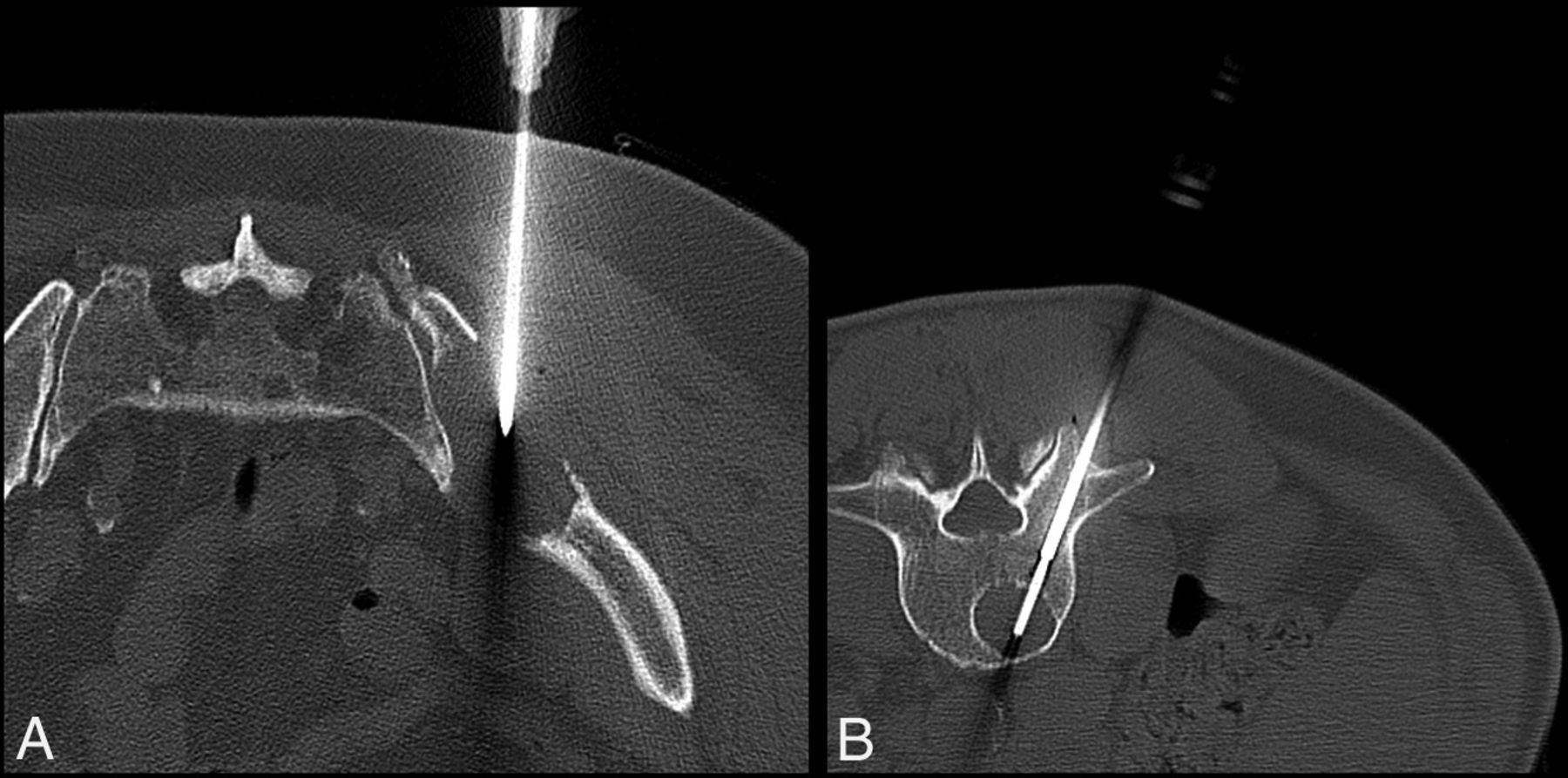
- Fig 1.
Bone biopsy of lytic bone lesions with manual and battery-powered drills. A, A 74-year-old man with multiple myeloma undergoing CT-guided biopsy of a right iliac lytic lesion with the Osteo-site manual system. CT image shows placement of the needle into the right iliac lytic lesion. B, A 58-year-old man with multiple myeloma undergoing CT-guided biopsy of a lytic L3 lesion with the OnControl battery-powered system. CT image shows placement of needle into the L3 vertebral body.
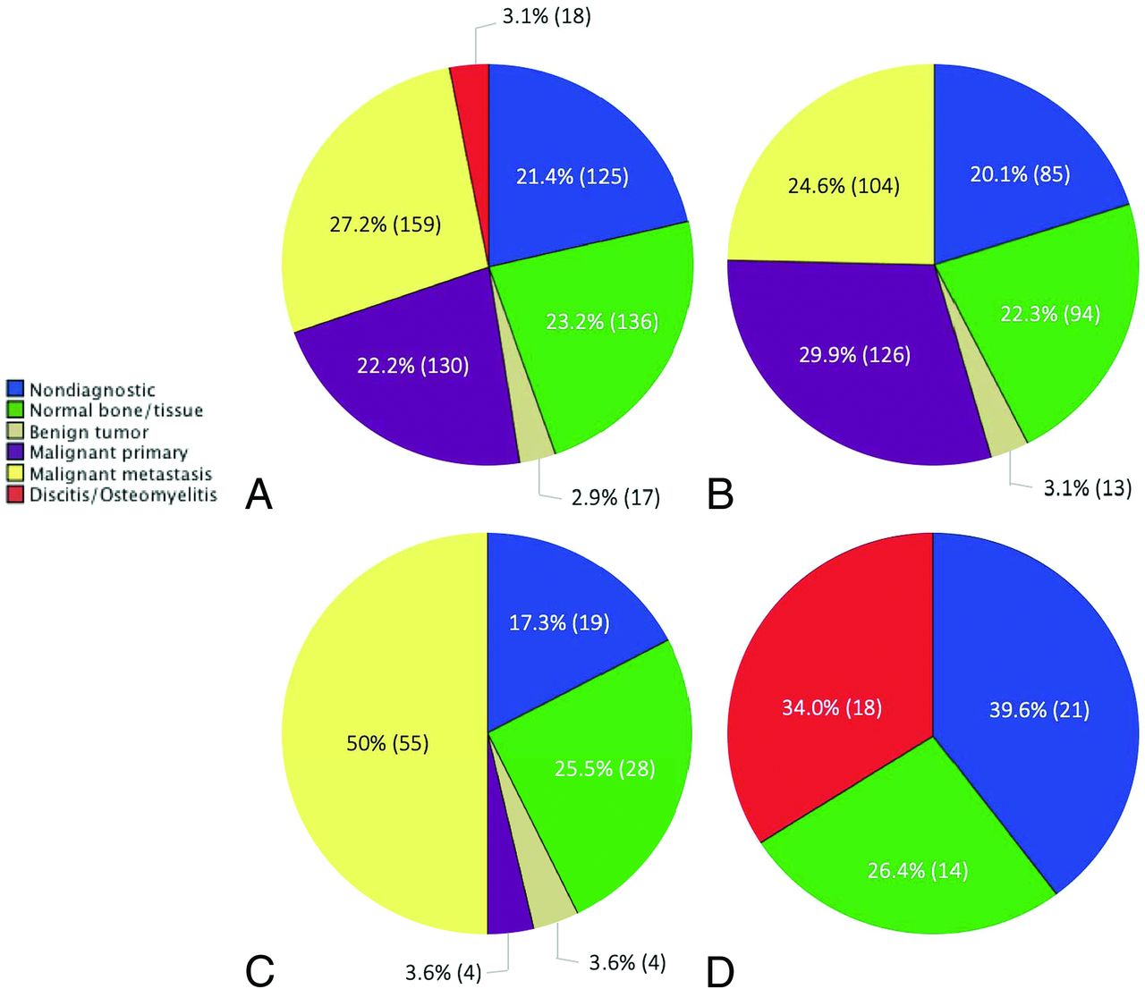
- Fig 2.
Breakdown of the final pathology diagnosis in various bone lesion classes: all bone lesions (A), lytic lesions (B), sclerotic lesions (C), and suspected infectious lesions (D).


Tables
Total Manual Battery-Powered P Value No. 585 314 271 Age (mean ± SD) 62 ± 13 62 ± 13 61 ± 12 .541 Sex (M/F) 305:280 173:141 132:139 .135 Suspected origin of primary disease .103 Boneb 41.7 (244) 38.9 (122) 45 (122) Prostate 3.8 (22) 4.5 (14) 3.0 (8) Breast 10.4 (61) 11.5 (36) 9.2 (25) Lung 2.9 (17) 4.8 (15) 0.7 (2) Kidney 0.7 (4) 1.0 (3) 0.4 (1) Systemicc 0.7 (4) 1.0 (3) 0.4 (1) Liver 0.3 (2) 0.6 (2) 0 (0) Bladder 1.7 (10) 1.6 (5) 1.8 (5) Miscellaneousd 8.5 (50) 9.2 (29) 7.7 (21) Unestablished/unknowne 27 (158) 24.8 (78) 29.5 (80) Blood 2.2 (13) 2.2 (7) 2.2 (6) Location of bone lesion .16 Vertebral column 47.0 (275) 45.2 (142) 49.1 (133) Cervical 0.6 (4) 0.1 (4) 0 (0) Thoracic 17.9 (105) 16.2 (51) 19.9 (54) Lumbar 17.3 (101) 17.5 (55) 17.0 (46) Sacral 11.1 (65) 10.2 (32) 12.2 (33) Pelvis 36.9 (216) 39.5 (124) 33.9 (92) Extremity 8.4 (49) 10.5 (33) 5.9 (16) Miscellaneousf 0.5 (3) 0.6 (2) 0.4 (1) Disc 7.2 (42) 4.1 (13) 10.7 (29) ↵a Values for frequency of suspected origin of primary disease and lesion location represent percentages followed by total number of cases in parentheses.
↵b Primary bone lesions include suspected multiple myeloma from laboratory testing.
↵c Predominantly sarcoidosis.
↵d Includes thyroid, salivary gland, vulvar, tongue, melanoma, and anal cancers.
↵e Includes all cases without known malignancy or suspected primary bone lesion.
↵f Includes sternum, ribs, clavicle, scapula, calvaria, and tarsal bones.
Diagnostic Yield Crush Artifacts Radiation Dose (mGy × cm2) Scanning Time (min) Lytic lesions Manual 83.4 (201/241) 5 (12/241) 845 42 Battery-powered 89.0 (161/181) 7.7 (14/181) 657 35 P value .069 .168 .001b .001b Sclerotic lesions Manual 76.4 (42/55) 7.3 (4/55) 1061 44 Battery-powered 89.1 (49/55) 7.3 (4/55) 804 37 P value .064 1 .028b .012b Infectious lesions Manual 50.0 (9/18) 11.1 (2/18) 1113 46 Battery-powered 51.4 (18/35) 0 (0/35) 811 39 P value .576 .111 .155 .128 All lesions Manual 80.3 (252/314) 5.7 (18/314) 900 43 Battery-powered 84.1 (228/271) 6.6 (18/271) 704 36 P value .538 .387 .001b .001b


Jump to section
Related Articles
Cited By...
- No citing articles found.