Advertisement

Main menu

  • Home
  • Content
    • Current Issue
    • Accepted Manuscripts
    • Article Preview
    • Past Issue Archive
    • Video Articles
    • AJNR Case Collection
    • Case of the Week Archive
    • Case of the Month Archive
    • Classic Case Archive
  • Special Collections
    • AJNR Awards
    • Low-Field MRI
    • Alzheimer Disease
    • ASNR Foundation Special Collection
    • Photon-Counting CT
    • View All
  • Multimedia
    • AJNR Podcasts
    • AJNR SCANtastic
    • Trainee Corner
    • MRI Safety Corner
    • Imaging Protocols
  • For Authors
    • Submit a Manuscript
    • Submit a Video Article
    • Submit an eLetter to the Editor/Response
    • Manuscript Submission Guidelines
    • Statistical Tips
    • Fast Publishing of Accepted Manuscripts
    • Graphical Abstract Preparation
    • Imaging Protocol Submission
    • Author Policies
  • About Us
    • About AJNR
    • Editorial Board
    • Editorial Board Alumni
  • More
    • Become a Reviewer/Academy of Reviewers
    • Subscribers
    • Permissions
    • Alerts
    • Feedback
    • Advertisers
    • ASNR Home

User menu

  • Alerts
  • Log in

Search

  • Advanced search
American Journal of Neuroradiology

American Journal of Neuroradiology

ASHNR American Society of Functional Neuroradiology ASHNR American Society of Pediatric Neuroradiology ASSR
  • Alerts
  • Log in

Advanced Search

  • Home
  • Content
    • Current Issue
    • Accepted Manuscripts
    • Article Preview
    • Past Issue Archive
    • Video Articles
    • AJNR Case Collection
    • Case of the Week Archive
    • Case of the Month Archive
    • Classic Case Archive
  • Special Collections
    • AJNR Awards
    • Low-Field MRI
    • Alzheimer Disease
    • ASNR Foundation Special Collection
    • Photon-Counting CT
    • View All
  • Multimedia
    • AJNR Podcasts
    • AJNR SCANtastic
    • Trainee Corner
    • MRI Safety Corner
    • Imaging Protocols
  • For Authors
    • Submit a Manuscript
    • Submit a Video Article
    • Submit an eLetter to the Editor/Response
    • Manuscript Submission Guidelines
    • Statistical Tips
    • Fast Publishing of Accepted Manuscripts
    • Graphical Abstract Preparation
    • Imaging Protocol Submission
    • Author Policies
  • About Us
    • About AJNR
    • Editorial Board
    • Editorial Board Alumni
  • More
    • Become a Reviewer/Academy of Reviewers
    • Subscribers
    • Permissions
    • Alerts
    • Feedback
    • Advertisers
    • ASNR Home
  • Follow AJNR on Twitter
  • Visit AJNR on Facebook
  • Follow AJNR on Instagram
  • Join AJNR on LinkedIn
  • RSS Feeds

AJNR Awards, New Junior Editors, and more. Read the latest AJNR updates

Research ArticleAdult Brain
Open Access

Brain Imaging of Patients with COVID-19: Findings at an Academic Institution during the Height of the Outbreak in New York City

E. Lin, J.E. Lantos, S.B. Strauss, C.D. Phillips, T.R. Campion, B.B. Navi, N.S. Parikh, A.E. Merkler, S. Mir, C. Zhang, H. Kamel, M. Cusick, P. Goyal and A. Gupta
American Journal of Neuroradiology November 2020, 41 (11) 2001-2008; DOI: https://doi.org/10.3174/ajnr.A6793
E. Lin
aFrom the Department of Radiology (E.L., J.E.L., S.B.S., C.D.P., A.G.)
  • Find this author on Google Scholar
  • Find this author on PubMed
  • Search for this author on this site
  • ORCID record for E. Lin
J.E. Lantos
aFrom the Department of Radiology (E.L., J.E.L., S.B.S., C.D.P., A.G.)
  • Find this author on Google Scholar
  • Find this author on PubMed
  • Search for this author on this site
  • ORCID record for J.E. Lantos
S.B. Strauss
aFrom the Department of Radiology (E.L., J.E.L., S.B.S., C.D.P., A.G.)
  • Find this author on Google Scholar
  • Find this author on PubMed
  • Search for this author on this site
  • ORCID record for S.B. Strauss
C.D. Phillips
aFrom the Department of Radiology (E.L., J.E.L., S.B.S., C.D.P., A.G.)
  • Find this author on Google Scholar
  • Find this author on PubMed
  • Search for this author on this site
  • ORCID record for C.D. Phillips
T.R. Campion Jr.
bDepartment of Population Health Sciences (T.R.C., M.C.)
  • Find this author on Google Scholar
  • Find this author on PubMed
  • Search for this author on this site
  • ORCID record for T.R. Campion Jr.
B.B. Navi
cClinical and Translational Neuroscience Unit (B.B.N., N.S.P., A.E.M., S.M., C.Z., A.G.)
  • Find this author on Google Scholar
  • Find this author on PubMed
  • Search for this author on this site
  • ORCID record for B.B. Navi
N.S. Parikh
cClinical and Translational Neuroscience Unit (B.B.N., N.S.P., A.E.M., S.M., C.Z., A.G.)
  • Find this author on Google Scholar
  • Find this author on PubMed
  • Search for this author on this site
  • ORCID record for N.S. Parikh
A.E. Merkler
cClinical and Translational Neuroscience Unit (B.B.N., N.S.P., A.E.M., S.M., C.Z., A.G.)
  • Find this author on Google Scholar
  • Find this author on PubMed
  • Search for this author on this site
  • ORCID record for A.E. Merkler
S. Mir
cClinical and Translational Neuroscience Unit (B.B.N., N.S.P., A.E.M., S.M., C.Z., A.G.)
  • Find this author on Google Scholar
  • Find this author on PubMed
  • Search for this author on this site
  • ORCID record for S. Mir
C. Zhang
cClinical and Translational Neuroscience Unit (B.B.N., N.S.P., A.E.M., S.M., C.Z., A.G.)
  • Find this author on Google Scholar
  • Find this author on PubMed
  • Search for this author on this site
  • ORCID record for C. Zhang
H. Kamel
  • Find this author on Google Scholar
  • Find this author on PubMed
  • Search for this author on this site
  • ORCID record for H. Kamel
M. Cusick
bDepartment of Population Health Sciences (T.R.C., M.C.)
  • Find this author on Google Scholar
  • Find this author on PubMed
  • Search for this author on this site
  • ORCID record for M. Cusick
P. Goyal
dFeil Family Brain and Mind Research Institute and Department of Neurology, and Department of Medicine (P.G.), Weill Cornell Medicine, New York, New York
  • Find this author on Google Scholar
  • Find this author on PubMed
  • Search for this author on this site
  • ORCID record for P. Goyal
A. Gupta
aFrom the Department of Radiology (E.L., J.E.L., S.B.S., C.D.P., A.G.)
cClinical and Translational Neuroscience Unit (B.B.N., N.S.P., A.E.M., S.M., C.Z., A.G.)
  • Find this author on Google Scholar
  • Find this author on PubMed
  • Search for this author on this site
  • ORCID record for A. Gupta
  • Article
  • Figures & Data
  • Info & Metrics
  • Responses
  • References
  • PDF
Loading

Article Figures & Data

Figures

  • Tables
  • FIG 1.
    • Download figure
    • Open in new tab
    • Download powerpoint
    FIG 1.

    Stroke. A 53-year-old man with COVID-19 with an axial CTA image (A) demonstrating an abrupt cutoff of the proximal M1 segment of the left MCA (arrow), consistent with thrombosis. The patient later underwent MR imaging, with a DWI sequence (B) demonstrating acute infarctions in the left MCA territory (arrows). An 85-year-old woman with COVID-19 and MR imaging with a DWI sequence (C and D) demonstrating acute infarctions in both the anterior (arrow, C) and posterior (arrows, D) circulations, consistent with central embolic etiology. Most patients in our cohort had stroke of either embolic or cryptogenic etiology.

  • FIG 2.
    • Download figure
    • Open in new tab
    • Download powerpoint
    FIG 2.

    Corpus callosum microhemorrhages. A 65-year-old woman (A), a 44-year-old woman (B), and a 69-year-old man (C) all demonstrate microhemorrhages on SWI, with a similar distribution, preferentially involving the corpus callosum (arrows in A–C), particularly the splenium. All patients had undergone mechanical ventilation before imaging. The distribution is similar to that previously described in critically ill, ventilated patients as well as in those with high-altitude cerebral edema.

  • FIG 3.
    • Download figure
    • Open in new tab
    • Download powerpoint
    FIG 3.

    PRES. A 65-year-old woman (A, same patient as in Fig 2A) and 63-year-old man (B and C) demonstrate a typical imaging appearance of PRES on T2-FLAIR images (arrows in A and B), with bilateral subcortical occipital white matter hyperintense signal, as well as more pronounced involvement of the patient in B with thalamic and internal and external capsule involvement. This patient also has evidence of associated right occipital microhemorrhage (arrow in C). Both patients had the typical risk factors for PRES of acute kidney injury and hypertension.
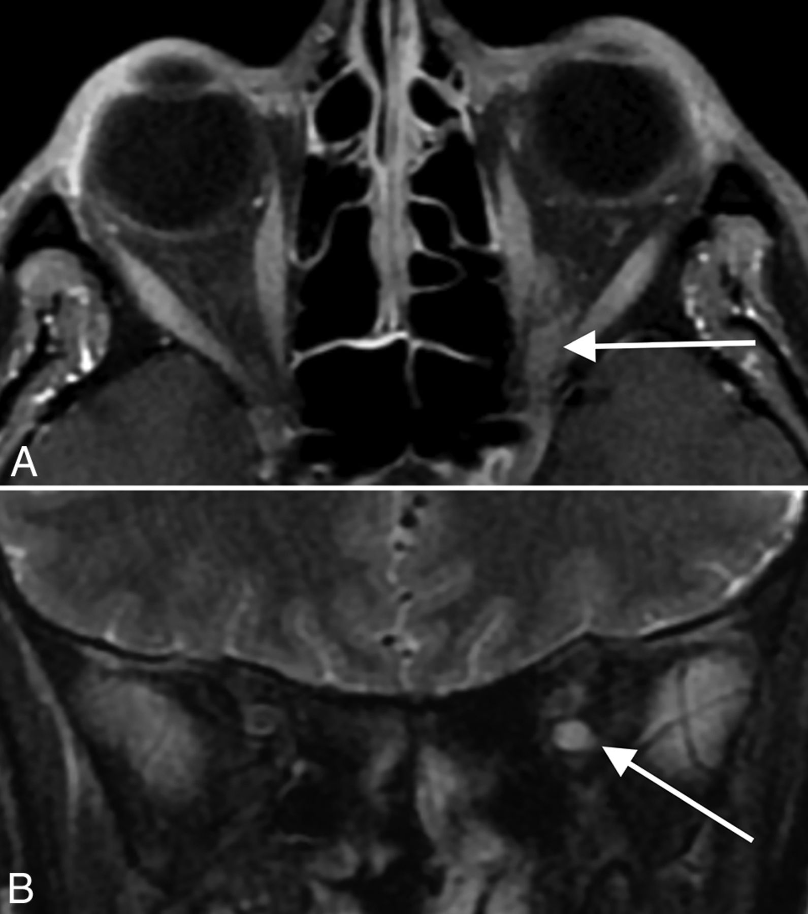
  • FIG 4.
    • Download figure
    • Open in new tab
    • Download powerpoint
    FIG 4.

    Miller Fisher syndrome. A 36-year-old male patient with a history of COVID-19 and diplopia, ataxia, and areflexia. Axial T1 postcontrast (A) and coronal T2 fat-suppressed (B) MR images through the orbits demonstrate striking enlargement, enhancement, and T2 hyperintense signal of cranial nerve III (arrows in A and B). The patient was clinically diagnosed with Miller Fisher syndrome and improved with intravenous immunoglobulin treatment.

  • FIG 5.
    • Download figure
    • Open in new tab
    • Download powerpoint
    FIG 5.

    Olfactory neuritis. Six patients with COVID-19 with coronal T2-FLAIR postcontrast images. A 48-year-old woman (A) demonstrates hyperintense signal in the right (arrow) greater than left olfactory bulbs. A 52-year-old woman (B) and a 62-year-old man (C) demonstrate symmetric hyperintense signal in the olfactory bulbs (arrows). For comparison, 3 different patients (D–F) in our cohort with normal olfactory bulbs (arrows) are included.

Tables

  • Figures
  • Characteristics of patients with COVID-19a

    CharacteristicPatients with COVID-19 Undergoing CT or MR Imaging of the Brain (n = 278)Patients with COVID-19 without Brain Imaging(n = 1776)
    Age (yr)b71.8 (15.4)60.6 (18.1)
    Male165 (59%)1009 (57%)
    Race
     Asian42 (15%)222 (13%)
     Black37 (13%)231 (13%)
     White84 (30%)524 (30%)
     Other58 (21%)400 (23%)
     Unknown57 (21%)399 (22%)
    Medical comorbidities
     Atrial fibrillation79 (28%)244 (14%)
     Hypertension214 (77%)1030 (58%)
     Hyperlipidemia115 (41%)515 (29%)
     Diabetes144 (52%)709 (40%)
     Coronary artery disease93 (33%)421 (24%)
     Chronic kidney disease58 (21%)267 (15%)
     COPD40 (14%)150 (8%)
    Mechanical ventilation53 (19%)292 (16%)
    ICU admission92 (33%)399 (22%)
    Death61 (22%)213 (12%)
    • Note:—COPD indicates chronic obstructive pulmonary disease; ICU, intensive care unit.

    • ↵a Data are reported as number (%) except where otherwise noted.

    • ↵b Age is presented as mean age, with SD in parentheses.

PreviousNext
Back to top

In this issue

American Journal of Neuroradiology: 41 (11)
American Journal of Neuroradiology
Vol. 41, Issue 11
1 Nov 2020
  • Table of Contents
  • Index by author
  • Complete Issue (PDF)
Advertisement
Print
Download PDF
Email Article

Thank you for your interest in spreading the word on American Journal of Neuroradiology.

NOTE: We only request your email address so that the person you are recommending the page to knows that you wanted them to see it, and that it is not junk mail. We do not capture any email address.

Enter multiple addresses on separate lines or separate them with commas.
Brain Imaging of Patients with COVID-19: Findings at an Academic Institution during the Height of the Outbreak in New York City
(Your Name) has sent you a message from American Journal of Neuroradiology
(Your Name) thought you would like to see the American Journal of Neuroradiology web site.
CAPTCHA
This question is for testing whether or not you are a human visitor and to prevent automated spam submissions.
Cite this article
E. Lin, J.E. Lantos, S.B. Strauss, C.D. Phillips, T.R. Campion, B.B. Navi, N.S. Parikh, A.E. Merkler, S. Mir, C. Zhang, H. Kamel, M. Cusick, P. Goyal, A. Gupta
Brain Imaging of Patients with COVID-19: Findings at an Academic Institution during the Height of the Outbreak in New York City
American Journal of Neuroradiology Nov 2020, 41 (11) 2001-2008; DOI: 10.3174/ajnr.A6793

Citation Manager Formats

  • BibTeX
  • Bookends
  • EasyBib
  • EndNote (tagged)
  • EndNote 8 (xml)
  • Medlars
  • Mendeley
  • Papers
  • RefWorks Tagged
  • Ref Manager
  • RIS
  • Zotero
0 Responses
Respond to this article
Share
Bookmark this article
Brain Imaging of Patients with COVID-19: Findings at an Academic Institution during the Height of the Outbreak in New York City
E. Lin, J.E. Lantos, S.B. Strauss, C.D. Phillips, T.R. Campion, B.B. Navi, N.S. Parikh, A.E. Merkler, S. Mir, C. Zhang, H. Kamel, M. Cusick, P. Goyal, A. Gupta
American Journal of Neuroradiology Nov 2020, 41 (11) 2001-2008; DOI: 10.3174/ajnr.A6793
del.icio.us logo Twitter logo Facebook logo Mendeley logo
  • Tweet Widget
  • Facebook Like
  • Google Plus One
Purchase

Jump to section

  • Article
    • Abstract
    • ABBREVIATIONS:
    • MATERIALS AND METHODS
    • RESULTS
    • DISCUSSION
    • CONCLUSIONS
    • Footnotes
    • References
  • Figures & Data
  • Info & Metrics
  • Responses
  • References
  • PDF

Related Articles

  • No related articles found.
  • PubMed
  • Google Scholar

Cited By...

  • Adapting the UK Biobank brain imaging protocol and analysis pipeline for the C-MORE multi-organ study of COVID-19 survivors
  • MR Imaging of the Olfactory Bulbs in Patients with COVID-19 and Anosmia: How to Avoid Misinterpretation
  • Reply:
  • Crossref (83)
  • Google Scholar

This article has been cited by the following articles in journals that are participating in Crossref Cited-by Linking.

  • Neuropathology and virus in brain of SARS-CoV-2 infected non-human primates
    Ibolya Rutkai, Meredith G. Mayer, Linh M. Hellmers, Bo Ning, Zhen Huang, Christopher J. Monjure, Carol Coyne, Rachel Silvestri, Nadia Golden, Krystle Hensley, Kristin Chandler, Gabrielle Lehmicke, Gregory J. Bix, Nicholas J. Maness, Kasi Russell-Lodrigue, Tony Y. Hu, Chad J. Roy, Robert V. Blair, Rudolf Bohm, Lara A. Doyle-Meyers, Jay Rappaport, Tracy Fischer
    Nature Communications 2022 13 1
  • Posterior Reversible Encephalopathy Syndrome
    Alexandra N. Gewirtz, Virginia Gao, Sarah C. Parauda, Matthew S. Robbins
    Current Pain and Headache Reports 2021 25 3
  • Long COVID and neuropsychiatric manifestations (Review)
    Vasiliki Efstathiou, Maria-Ioanna Stefanou, Marina Demetriou, Nikolaos Siafakas, Michael Makris, Georgios Tsivgoulis, Vassilios Zoumpourlis, Stylianos Kympouropoulos, James Tsoporis, Demetrios Spandidos, Nikolaos Smyrnis, Emmanouil Rizos
    Experimental and Therapeutic Medicine 2022 23 5
  • Heterogeneity in Regional Damage Detected by Neuroimaging and Neuropathological Studies in Older Adults With COVID-19: A Cognitive-Neuroscience Systematic Review to Inform the Long-Term Impact of the Virus on Neurocognitive Trajectories
    Riccardo Manca, Matteo De Marco, Paul G. Ince, Annalena Venneri
    Frontiers in Aging Neuroscience 2021 13
  • The Spectrum of Neuroimaging Findings on CT and MRI in Adults With COVID-19
    Gul Moonis, Christopher G. Filippi, Claudia F. E. Kirsch, Suyash Mohan, Evan G. Stein, Joshua A. Hirsch, Amit Mahajan
    American Journal of Roentgenology 2021 217 4
  • COVID-19 and the brain
    Karan S. Hingorani, Shivkumar Bhadola, Anna M. Cervantes-Arslanian
    Trends in Cardiovascular Medicine 2022 32 6
  • Neuroimaging findings of brain MRI and CT in patients with COVID-19: A systematic review and meta-analysis
    Yangsean Choi, Min Kyoung Lee
    European Journal of Radiology 2020 133
  • Brain imaging in patients with COVID-19: A systematic review
    Pablo Najt, Helen L. Richards, Dónal G. Fortune
    Brain, Behavior, & Immunity - Health 2021 16
  • Elucidating the Neuropathologic Mechanisms of SARS-CoV-2 Infection
    Mar Pacheco-Herrero, Luis O. Soto-Rojas, Charles R. Harrington, Yazmin M. Flores-Martinez, Marcos M. Villegas-Rojas, Alfredo M. León-Aguilar, Paola A. Martínez-Gómez, B. Berenice Campa-Córdoba, Ricardo Apátiga-Pérez, Carolin N. Corniel-Taveras, Jesabelle de J. Dominguez-García, Víctor Manuel Blanco-Alvarez, José Luna-Muñoz
    Frontiers in Neurology 2021 12
  • Critical illness-associated cerebral microbleeds for patients with severe COVID-19: etiologic hypotheses
    François Lersy, Thibault Willaume, Jean-Christophe Brisset, Olivier Collange, Julie Helms, Francis Schneider, Agathe Chammas, Alexandre Willaume, Nicolas Meyer, Mathieu Anheim, François Cotton, Stéphane Kremer
    Journal of Neurology 2021 268 8

More in this TOC Section

  • Diagnostic Neuroradiology of Monoclonal Antibodies
  • NCCT vs. MRI for Brain Atrophy in Acute Stroke
  • Clinical Outcomes After Chiari I Decompression
Show more ADULT BRAIN

Similar Articles

Advertisement

Indexed Content

  • Current Issue
  • Accepted Manuscripts
  • Article Preview
  • Past Issues
  • Editorials
  • Editor's Choice
  • Fellows' Journal Club
  • Letters to the Editor
  • Video Articles

Cases

  • Case Collection
  • Archive - Case of the Week
  • Archive - Case of the Month
  • Archive - Classic Case

More from AJNR

  • Trainee Corner
  • Imaging Protocols
  • MRI Safety Corner
  • Book Reviews

Multimedia

  • AJNR Podcasts
  • AJNR Scantastics

Resources

  • Turnaround Time
  • Submit a Manuscript
  • Submit a Video Article
  • Submit an eLetter to the Editor/Response
  • Manuscript Submission Guidelines
  • Statistical Tips
  • Fast Publishing of Accepted Manuscripts
  • Graphical Abstract Preparation
  • Imaging Protocol Submission
  • Evidence-Based Medicine Level Guide
  • Publishing Checklists
  • Author Policies
  • Become a Reviewer/Academy of Reviewers
  • News and Updates

About Us

  • About AJNR
  • Editorial Board
  • Editorial Board Alumni
  • Alerts
  • Permissions
  • Not an AJNR Subscriber? Join Now
  • Advertise with Us
  • Librarian Resources
  • Feedback
  • Terms and Conditions
  • AJNR Editorial Board Alumni

American Society of Neuroradiology

  • Not an ASNR Member? Join Now

© 2025 by the American Society of Neuroradiology All rights, including for text and data mining, AI training, and similar technologies, are reserved.
Print ISSN: 0195-6108 Online ISSN: 1936-959X

Powered by HighWire