Advertisement

Main menu

  • Home
  • Content
    • Current Issue
    • Accepted Manuscripts
    • Article Preview
    • Past Issue Archive
    • Video Articles
    • AJNR Case Collection
    • Case of the Week Archive
    • Case of the Month Archive
    • Classic Case Archive
  • Special Collections
    • Advancing NeuroMRI with High-Relaxivity Contrast Agents
    • AJNR Awards
    • Low-Field MRI
    • Alzheimer Disease
    • ASNR Foundation Special Collection
    • Photon-Counting CT
    • View All
  • Multimedia
    • AJNR Podcasts
    • AJNR SCANtastic
    • Trainee Corner
    • MRI Safety Corner
    • Imaging Protocols
  • For Authors
    • Submit a Manuscript
    • Submit a Video Article
    • Submit an eLetter to the Editor/Response
    • Manuscript Submission Guidelines
    • Statistical Tips
    • Fast Publishing of Accepted Manuscripts
    • Graphical Abstract Preparation
    • Imaging Protocol Submission
    • Author Policies
  • About Us
    • About AJNR
    • Editorial Board
    • Editorial Board Alumni
  • More
    • Become a Reviewer/Academy of Reviewers
    • Subscribers
    • Permissions
    • Alerts
    • Feedback
    • Advertisers
    • ASNR Home

User menu

  • Alerts
  • Log in

Search

  • Advanced search
American Journal of Neuroradiology

American Journal of Neuroradiology

ASHNR American Society of Functional Neuroradiology ASHNR American Society of Pediatric Neuroradiology ASSR
  • Alerts
  • Log in

Advanced Search

  • Home
  • Content
    • Current Issue
    • Accepted Manuscripts
    • Article Preview
    • Past Issue Archive
    • Video Articles
    • AJNR Case Collection
    • Case of the Week Archive
    • Case of the Month Archive
    • Classic Case Archive
  • Special Collections
    • Advancing NeuroMRI with High-Relaxivity Contrast Agents
    • AJNR Awards
    • Low-Field MRI
    • Alzheimer Disease
    • ASNR Foundation Special Collection
    • Photon-Counting CT
    • View All
  • Multimedia
    • AJNR Podcasts
    • AJNR SCANtastic
    • Trainee Corner
    • MRI Safety Corner
    • Imaging Protocols
  • For Authors
    • Submit a Manuscript
    • Submit a Video Article
    • Submit an eLetter to the Editor/Response
    • Manuscript Submission Guidelines
    • Statistical Tips
    • Fast Publishing of Accepted Manuscripts
    • Graphical Abstract Preparation
    • Imaging Protocol Submission
    • Author Policies
  • About Us
    • About AJNR
    • Editorial Board
    • Editorial Board Alumni
  • More
    • Become a Reviewer/Academy of Reviewers
    • Subscribers
    • Permissions
    • Alerts
    • Feedback
    • Advertisers
    • ASNR Home
  • Follow AJNR on Twitter
  • Visit AJNR on Facebook
  • Follow AJNR on Instagram
  • Join AJNR on LinkedIn
  • RSS Feeds

AJNR Awards, New Junior Editors, and more. Read the latest AJNR updates


Improved Turnaround Times | Median time to first decision: 12 days

Research ArticleAdult Brain

The Variable Appearance of Third Ventricular Colloid Cysts: Correlation with Histopathology and the Risk of Obstructive Ventriculomegaly

S.D. Khanpara, A.L. Day, M.B. Bhattacharjee, R.F. Riascos, J.P. Fernelius and K.D. Westmark
American Journal of Neuroradiology October 2020, 41 (10) 1833-1840; DOI: https://doi.org/10.3174/ajnr.A6722
S.D. Khanpara
aDepartments of Diagnostic and Interventional Imaging (S.D.K., R.F.R., K.D.W.)
  • Find this author on Google Scholar
  • Find this author on PubMed
  • Search for this author on this site
  • ORCID record for S.D. Khanpara
A.L. Day
bNeurosurgery (A.L.D.)
  • Find this author on Google Scholar
  • Find this author on PubMed
  • Search for this author on this site
  • ORCID record for A.L. Day
M.B. Bhattacharjee
cPathology (M.B.B.), McGovern Medical School, The University of Texas Health Science Center at Houston, Houston, Texas
  • Find this author on Google Scholar
  • Find this author on PubMed
  • Search for this author on this site
  • ORCID record for M.B. Bhattacharjee
R.F. Riascos
aDepartments of Diagnostic and Interventional Imaging (S.D.K., R.F.R., K.D.W.)
  • Find this author on Google Scholar
  • Find this author on PubMed
  • Search for this author on this site
  • ORCID record for R.F. Riascos
J.P. Fernelius
dDepartment of Quality and Outcomes Management (J.P.F.), Texas Children’s Hospital, Houston, Texas
  • Find this author on Google Scholar
  • Find this author on PubMed
  • Search for this author on this site
  • ORCID record for J.P. Fernelius
K.D. Westmark
aDepartments of Diagnostic and Interventional Imaging (S.D.K., R.F.R., K.D.W.)
  • Find this author on Google Scholar
  • Find this author on PubMed
  • Search for this author on this site
  • ORCID record for K.D. Westmark
  • Article
  • Figures & Data
  • Info & Metrics
  • Responses
  • References
  • PDF
Loading

Article Figures & Data

Figures

  • Tables
  • FIG 1.
    • Download figure
    • Open in new tab
    • Download powerpoint
    FIG 1.

    An overview of the imaging appearance of third ventricular colloid cyst on CT and corresponding MR imaging characteristics.

  • FIG 2.
    • Download figure
    • Open in new tab
    • Download powerpoint
    FIG 2.

    An overview of the imaging appearance of third ventricular colloid cyst on MR imaging.

  • FIG 3.
    • Download figure
    • Open in new tab
    • Download powerpoint
    FIG 3.

    Homogeneously hyperintense cyst: 13-year-old boy found unconscious after complaining of headache for 2 days. Initial CT demonstrates dilation of the lateral ventricles. However, the colloid cyst was difficult to detect as it was isointense relative to the adjacent brain. Subsequent MR imaging showed a colloid cyst homogeneously hyperintense on T2 (B) and T2-FLAIR weighted images (C) and hypointense on T1-weighted axial image (D). Also, note the presence of periventricular edema associated with ventriculomegaly.

  • FIG 4.
    • Download figure
    • Open in new tab
    • Download powerpoint
    FIG 4.

    Cyst with rim: 55-year-old woman with a 6-week history of headache. Non-contrast CT (A) shows a hyperattenuated colloid cyst measuring 13 mm at the roof of third ventricle. On T2WI (B), the colloid cyst has a hypointense core with a thin hyperintense rim, which is more pronounced on T2-FLAIR (C). The cyst is mildly heterogeneous and isointense centrally on T1-weighted image (D). There was no evidence of hemorrhage on the GRE sequence (image not shown). There is associated enlargement of the lateral ventricles with periventricular edema.

  • FIG 5.
    • Download figure
    • Open in new tab
    • Download powerpoint
    FIG 5.

    Hematoxylin and Eosin staining of a typical homogeneously hyperintense cyst on T2 and T2-FLAIR images. The cyst wall is composed of columnar, ciliated epithelial cells. The wall was not thickened and there was not an abnormal increase in goblet cells or any cellular atypia. Contents of the cysts consisted of a mucoid material with detached epithelial clusters of cells.

  • FIG 6.
    • Download figure
    • Open in new tab
    • Download powerpoint
    FIG 6.

    Pathologic examination of a cyst that had the “cysts with rim” appearance revealed a viscous plug of colloid material which was PAS-positive. The cyst wall consisted of a thin fibrous capsule and a lining of partly ciliated cuboidal to columnar epithelium (A). The fibrous wall, and the associated choroid plexus stroma are highlighted by the Masson trichrome stain (B). The cyst epithelium is positive for CAM 5.2, pankeratin (C), EMA, and CK7; it is negative for CDX2. The peripheral cyst wall was not unusually thickened and was approximately 1–3 microns thick, and therefore would not have been visible on MR imaging.

Tables

  • Figures
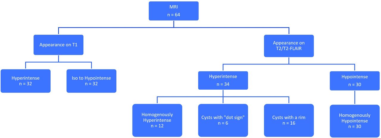
  • Demographic and imaging characteristics of 64 colloid cyst

    Signal Intensity on T2-FLAIRHyperintense (n = 34)Hypointense(n = 30)Total
    Homogeneously Hyperintense“Dot Sign”Hyperintense Rim
    Number of patients126163064
     Sex (Male)6 (50%)3 (50%)9 (56.2%)13 (43.3%)31 (48%)
    Patient age (years)
     Mean (+/− SD)37.0 (23.4%)37.5 (15.6%)52.1 (22%)55.2 (14.7%)49.8 (19.8%)
     Median25.531.549.55350
    Cyst size (mm)
     Mean10.616.811.16.89.6
     Median11.015.012.56.39.0
    Obstructive ventriculomegaly8 (67%)5 (83.3%)8 (50%)3 (10%)24 (37.5%)
    CTa (hyperattenuated/iso-hypoattenuated)4/40/514/020/038/9
    T1 signal (hyper-/iso-/hypointense)1/5/65/1/010/5/116/14/032/25/7
    Zone II302611
    Histopathology availablefor review8310728
    • ↵a CT was not available in all cases.

PreviousNext
Back to top

In this issue

American Journal of Neuroradiology: 41 (10)
American Journal of Neuroradiology
Vol. 41, Issue 10
1 Oct 2020
  • Table of Contents
  • Index by author
  • Complete Issue (PDF)
Advertisement
Print
Download PDF
Email Article

Thank you for your interest in spreading the word on American Journal of Neuroradiology.

NOTE: We only request your email address so that the person you are recommending the page to knows that you wanted them to see it, and that it is not junk mail. We do not capture any email address.

Enter multiple addresses on separate lines or separate them with commas.
The Variable Appearance of Third Ventricular Colloid Cysts: Correlation with Histopathology and the Risk of Obstructive Ventriculomegaly
(Your Name) has sent you a message from American Journal of Neuroradiology
(Your Name) thought you would like to see the American Journal of Neuroradiology web site.
CAPTCHA
This question is for testing whether or not you are a human visitor and to prevent automated spam submissions.
Cite this article
S.D. Khanpara, A.L. Day, M.B. Bhattacharjee, R.F. Riascos, J.P. Fernelius, K.D. Westmark
The Variable Appearance of Third Ventricular Colloid Cysts: Correlation with Histopathology and the Risk of Obstructive Ventriculomegaly
American Journal of Neuroradiology Oct 2020, 41 (10) 1833-1840; DOI: 10.3174/ajnr.A6722

Citation Manager Formats

  • BibTeX
  • Bookends
  • EasyBib
  • EndNote (tagged)
  • EndNote 8 (xml)
  • Medlars
  • Mendeley
  • Papers
  • RefWorks Tagged
  • Ref Manager
  • RIS
  • Zotero
0 Responses
Respond to this article
Share
Bookmark this article
The Variable Appearance of Third Ventricular Colloid Cysts: Correlation with Histopathology and the Risk of Obstructive Ventriculomegaly
S.D. Khanpara, A.L. Day, M.B. Bhattacharjee, R.F. Riascos, J.P. Fernelius, K.D. Westmark
American Journal of Neuroradiology Oct 2020, 41 (10) 1833-1840; DOI: 10.3174/ajnr.A6722
del.icio.us logo Twitter logo Facebook logo Mendeley logo
  • Tweet Widget
  • Facebook Like
  • Google Plus One
Purchase

Jump to section

  • Article
    • Abstract
    • ABBREVIATIONS:
    • MATERIALS AND METHODS
    • RESULTS
    • DISCUSSION
    • CONCLUSIONS
    • Footnotes
    • References
  • Figures & Data
  • Info & Metrics
  • Responses
  • References
  • PDF

Related Articles

  • No related articles found.
  • PubMed
  • Google Scholar

Cited By...

  • No citing articles found.
  • Crossref
  • Google Scholar

This article has not yet been cited by articles in journals that are participating in Crossref Cited-by Linking.

More in this TOC Section

  • Diagnostic Neuroradiology of Monoclonal Antibodies
  • Clinical Outcomes After Chiari I Decompression
  • Segmentation of Brain Metastases with BLAST
Show more Adult Brain

Similar Articles

Advertisement

Indexed Content

  • Current Issue
  • Accepted Manuscripts
  • Article Preview
  • Past Issues
  • Editorials
  • Editor's Choice
  • Fellows' Journal Club
  • Letters to the Editor
  • Video Articles

Cases

  • Case Collection
  • Archive - Case of the Week
  • Archive - Case of the Month
  • Archive - Classic Case

More from AJNR

  • Trainee Corner
  • Imaging Protocols
  • MRI Safety Corner
  • Book Reviews

Multimedia

  • AJNR Podcasts
  • AJNR Scantastics

Resources

  • Turnaround Time
  • Submit a Manuscript
  • Submit a Video Article
  • Submit an eLetter to the Editor/Response
  • Manuscript Submission Guidelines
  • Statistical Tips
  • Fast Publishing of Accepted Manuscripts
  • Graphical Abstract Preparation
  • Imaging Protocol Submission
  • Evidence-Based Medicine Level Guide
  • Publishing Checklists
  • Author Policies
  • Become a Reviewer/Academy of Reviewers
  • News and Updates

About Us

  • About AJNR
  • Editorial Board
  • Editorial Board Alumni
  • Alerts
  • Permissions
  • Not an AJNR Subscriber? Join Now
  • Advertise with Us
  • Librarian Resources
  • Feedback
  • Terms and Conditions
  • AJNR Editorial Board Alumni

American Society of Neuroradiology

  • Not an ASNR Member? Join Now

© 2025 by the American Society of Neuroradiology All rights, including for text and data mining, AI training, and similar technologies, are reserved.
Print ISSN: 0195-6108 Online ISSN: 1936-959X

Powered by HighWire