Advertisement

Main menu

  • Home
  • Content
    • Current Issue
    • Accepted Manuscripts
    • Article Preview
    • Past Issue Archive
    • Video Articles
    • AJNR Case Collection
    • Case of the Week Archive
    • Case of the Month Archive
    • Classic Case Archive
  • Special Collections
    • AJNR Awards
    • Low-Field MRI
    • Alzheimer Disease
    • ASNR Foundation Special Collection
    • Photon-Counting CT
    • View All
  • Multimedia
    • AJNR Podcasts
    • AJNR SCANtastic
    • Trainee Corner
    • MRI Safety Corner
    • Imaging Protocols
  • For Authors
    • Submit a Manuscript
    • Submit a Video Article
    • Submit an eLetter to the Editor/Response
    • Manuscript Submission Guidelines
    • Statistical Tips
    • Fast Publishing of Accepted Manuscripts
    • Graphical Abstract Preparation
    • Imaging Protocol Submission
    • Author Policies
  • About Us
    • About AJNR
    • Editorial Board
    • Editorial Board Alumni
  • More
    • Become a Reviewer/Academy of Reviewers
    • Subscribers
    • Permissions
    • Alerts
    • Feedback
    • Advertisers
    • ASNR Home

User menu

  • Alerts
  • Log in

Search

  • Advanced search
American Journal of Neuroradiology

American Journal of Neuroradiology

ASHNR American Society of Functional Neuroradiology ASHNR American Society of Pediatric Neuroradiology ASSR
  • Alerts
  • Log in

Advanced Search

  • Home
  • Content
    • Current Issue
    • Accepted Manuscripts
    • Article Preview
    • Past Issue Archive
    • Video Articles
    • AJNR Case Collection
    • Case of the Week Archive
    • Case of the Month Archive
    • Classic Case Archive
  • Special Collections
    • AJNR Awards
    • Low-Field MRI
    • Alzheimer Disease
    • ASNR Foundation Special Collection
    • Photon-Counting CT
    • View All
  • Multimedia
    • AJNR Podcasts
    • AJNR SCANtastic
    • Trainee Corner
    • MRI Safety Corner
    • Imaging Protocols
  • For Authors
    • Submit a Manuscript
    • Submit a Video Article
    • Submit an eLetter to the Editor/Response
    • Manuscript Submission Guidelines
    • Statistical Tips
    • Fast Publishing of Accepted Manuscripts
    • Graphical Abstract Preparation
    • Imaging Protocol Submission
    • Author Policies
  • About Us
    • About AJNR
    • Editorial Board
    • Editorial Board Alumni
  • More
    • Become a Reviewer/Academy of Reviewers
    • Subscribers
    • Permissions
    • Alerts
    • Feedback
    • Advertisers
    • ASNR Home
  • Follow AJNR on Twitter
  • Visit AJNR on Facebook
  • Follow AJNR on Instagram
  • Join AJNR on LinkedIn
  • RSS Feeds

AJNR Awards, New Junior Editors, and more. Read the latest AJNR updates

Research ArticleAdult Brain

Presurgical Identification of Primary Central Nervous System Lymphoma with Normalized Time-Intensity Curve: A Pilot Study of a New Method to Analyze DSC-PWI

A. Pons-Escoda, A. Garcia-Ruiz, P. Naval-Baudin, M. Cos, N. Vidal, G. Plans, J. Bruna, R. Perez-Lopez and C. Majos
American Journal of Neuroradiology October 2020, 41 (10) 1816-1824; DOI: https://doi.org/10.3174/ajnr.A6761
A. Pons-Escoda
aRadiology Department (A.P.-E., P.N.-B., M.C., C.M.), Institut de Diagnòstic per la Imatge, Hospital Universitari de Bellvitge. L’Hospitalet de Llobregat, Barcelona, Spain
fNeurooncology Unit (A.P.-E., N.V., G.P., J.B., C.M.), Insitut Català d’Oncologia, Institut d’Investigació Biomèdica de Bellvitge, Hospital Universitari de Bellvitge, L’Hospitalet de Llobregat, Barcelona, Spain
  • Find this author on Google Scholar
  • Find this author on PubMed
  • Search for this author on this site
  • ORCID record for A. Pons-Escoda
A. Garcia-Ruiz
bRadiomics Group (A.G.-R., R.P.-L.), Vall d’Hebron Institut d’Oncologia, Barcelona, Spain
  • Find this author on Google Scholar
  • Find this author on PubMed
  • Search for this author on this site
  • ORCID record for A. Garcia-Ruiz
P. Naval-Baudin
aRadiology Department (A.P.-E., P.N.-B., M.C., C.M.), Institut de Diagnòstic per la Imatge, Hospital Universitari de Bellvitge. L’Hospitalet de Llobregat, Barcelona, Spain
  • Find this author on Google Scholar
  • Find this author on PubMed
  • Search for this author on this site
  • ORCID record for P. Naval-Baudin
M. Cos
aRadiology Department (A.P.-E., P.N.-B., M.C., C.M.), Institut de Diagnòstic per la Imatge, Hospital Universitari de Bellvitge. L’Hospitalet de Llobregat, Barcelona, Spain
  • Find this author on Google Scholar
  • Find this author on PubMed
  • Search for this author on this site
  • ORCID record for M. Cos
N. Vidal
cPathology Department (N.V.), Hospital Universitari de Bellvitge, L’Hospitalet de Llobregat, Barcelona, Spain
fNeurooncology Unit (A.P.-E., N.V., G.P., J.B., C.M.), Insitut Català d’Oncologia, Institut d’Investigació Biomèdica de Bellvitge, Hospital Universitari de Bellvitge, L’Hospitalet de Llobregat, Barcelona, Spain
  • Find this author on Google Scholar
  • Find this author on PubMed
  • Search for this author on this site
  • ORCID record for N. Vidal
G. Plans
dNeurosurgery Department (G.P.), Hospital Universitari de Bellvitge, L’Hospitalet de Llobregat, Barcelona, Spain
fNeurooncology Unit (A.P.-E., N.V., G.P., J.B., C.M.), Insitut Català d’Oncologia, Institut d’Investigació Biomèdica de Bellvitge, Hospital Universitari de Bellvitge, L’Hospitalet de Llobregat, Barcelona, Spain
  • Find this author on Google Scholar
  • Find this author on PubMed
  • Search for this author on this site
  • ORCID record for G. Plans
J. Bruna
eNeurology Department (J.B.), Hospital Universitari de Bellvitge, L’Hospitalet de Llobregat, Barcelona, Spain
fNeurooncology Unit (A.P.-E., N.V., G.P., J.B., C.M.), Insitut Català d’Oncologia, Institut d’Investigació Biomèdica de Bellvitge, Hospital Universitari de Bellvitge, L’Hospitalet de Llobregat, Barcelona, Spain
  • Find this author on Google Scholar
  • Find this author on PubMed
  • Search for this author on this site
  • ORCID record for J. Bruna
R. Perez-Lopez
bRadiomics Group (A.G.-R., R.P.-L.), Vall d’Hebron Institut d’Oncologia, Barcelona, Spain
  • Find this author on Google Scholar
  • Find this author on PubMed
  • Search for this author on this site
  • ORCID record for R. Perez-Lopez
C. Majos
aRadiology Department (A.P.-E., P.N.-B., M.C., C.M.), Institut de Diagnòstic per la Imatge, Hospital Universitari de Bellvitge. L’Hospitalet de Llobregat, Barcelona, Spain
fNeurooncology Unit (A.P.-E., N.V., G.P., J.B., C.M.), Insitut Català d’Oncologia, Institut d’Investigació Biomèdica de Bellvitge, Hospital Universitari de Bellvitge, L’Hospitalet de Llobregat, Barcelona, Spain
  • Find this author on Google Scholar
  • Find this author on PubMed
  • Search for this author on this site
  • ORCID record for C. Majos
  • Article
  • Figures & Data
  • Supplemental
  • Info & Metrics
  • Responses
  • References
  • PDF
Loading

Article Figures & Data

Figures

  • Tables
  • FIG 1.
    • Download figure
    • Open in new tab
    • Download powerpoint
    FIG 1.

    Solid (A and B) and necrotic (D and E) tumors and respective NAWM segmentations on axial CE-T1WI (A and D) and coregistered on DSC-PWI (B and E). Resultant raw curves by averaging the TIC for each voxel within the segmented areas (C and F), noncomparable due to differences in time, intensity, baseline, or initial point of the descending curve. Exemplification of the parameters used to normalize the curves, MSID, and TTP-TTA, relative to NAWM (G). Resultant normalized tumor curves, superimposable and comparable point by point (H). Curves with the exact same number of time-matching points and sharing common units of time (relative to TTP-TTA of the NAWM) and intensity (relative to MSID of the NAWM).
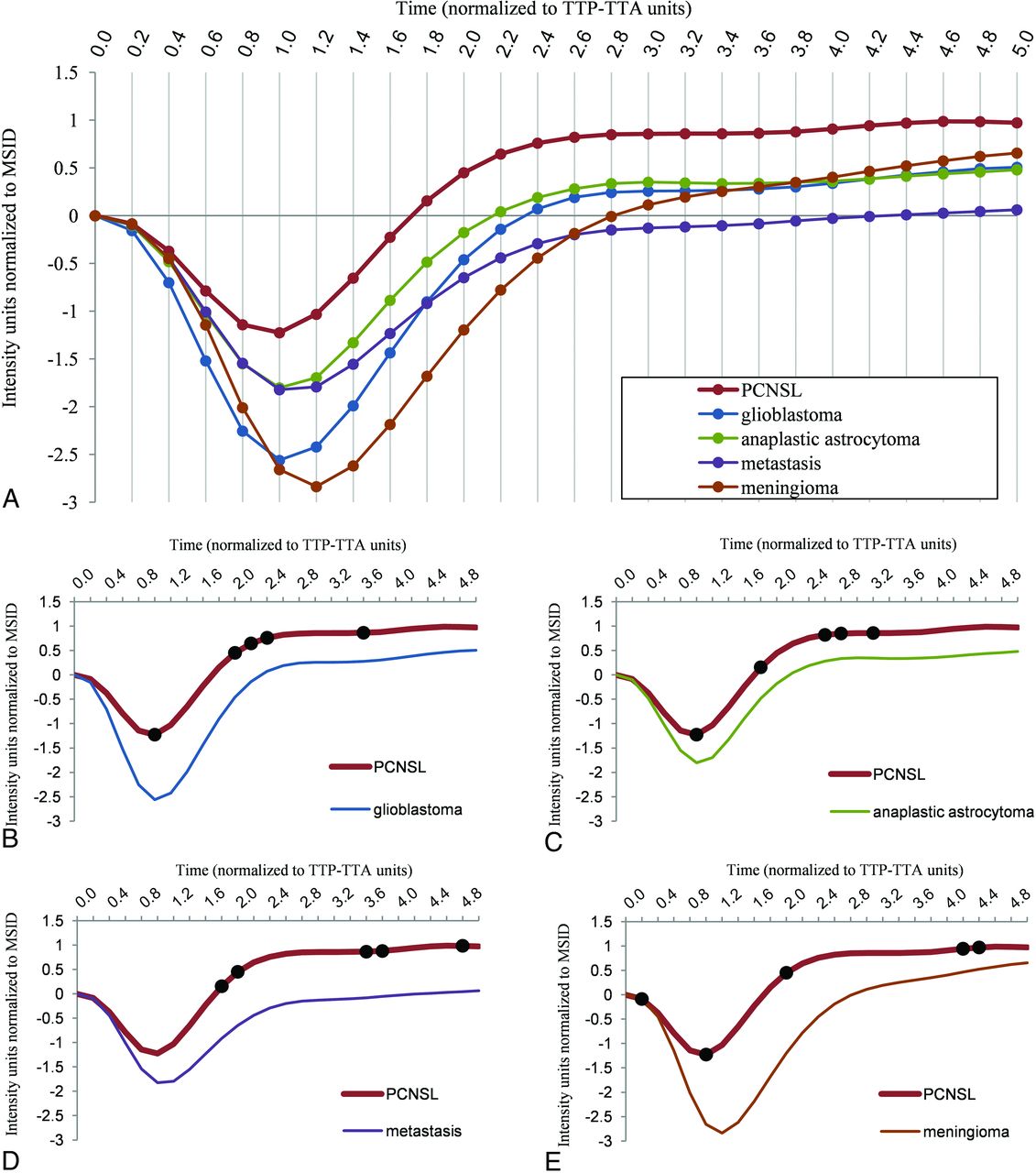
  • FIG 2.
    • Download figure
    • Open in new tab
    • Download powerpoint
    FIG 2.

    Normalized mean tumor curves for each type (PCNSL, glioblastoma, anaplastic astrocytoma, metastasis, and meningioma) obtained in the training subset (A). The curves are superimposable and comparable in a single graphic. This format provides a user-friendly tool for visual comparison of curves. Paired comparisons of normalized mean tumor curves for PCNSL against glioblastoma (B), anaplastic astrocytoma (C), metastasis (D), and meningioma (E), as well as representation of the 5 stepwise selected discriminatory TTP-TTA time points per pair (black dots).

  • FIG 3.
    • Download figure
    • Open in new tab
    • Download powerpoint
    FIG 3.

    Example of clinical applicability on a “real” problem case. Axial CE-T1WI of 2 different patients (A and D) depicting 2 subcortical right frontal, solid enhancing tumors. PCNSL and anaplastic astrocytoma may be diagnostic possibilities to consider. Tumor normalized curves of each case overlapped to PCNSL, and anaplastic astrocytoma mean curves for visual assessment (B and E) show that the case in the upper row has similarities with PCNSL while in contrast, the case in the lower row has similarities with anaplastic astrocytoma. Representation of the classifier results on a scatterplot (C and F) demonstrates that the case in the upper row remains on the inferior side and may likely be a PCNSL, while the case in the lower row is more likely to be an anaplastic astrocytoma. We pathologically confirmed both diagnoses: case in the upper row, PCNSL; case in the lower row, anaplastic astrocytoma.
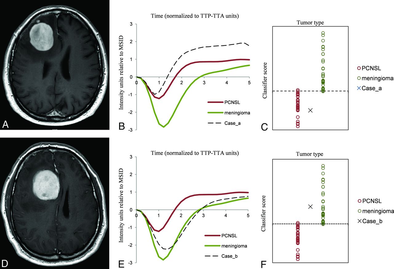
  • FIG 4.
    • Download figure
    • Open in new tab
    • Download powerpoint
    FIG 4.

    Example of clinical applicability on a “real” problem case. Axial CE-T1WI of 2 different patients with similar tumors (A and D): well defined, solid, avidly enhancing and right frontal peripherally located. PCNSL and meningioma were the 2 main diagnostic options considered. The tumor normalized curve of each case overlapping with PCNSL and meningioma mean curves (B and E) show that the case in the upper row is similar to PCNSL while the case in the lower row is close to meningioma. Representation of the classifier results on the scatterplot (C and F) suggests that the cases are likely to be PCNSL and meningioma, respectively. We pathologically confirmed both diagnoses: case in the upper row, PCNSL, case in the lower row, meningioma.

Tables

  • Figures
  • Summary of the results in training and test subsets

    AUCAccuracySensitivitySpecificity
    PCNSL vs GB
     Training0.9688% (60/68)88% (29/33)89% (31/35)
     Test0.9693% (25/27)93% (13/14)92% (12/13)
    PCNSL vs AA
     Training0.8674% (46/62)76% (25/33)72% (21/29)
     Test0.8371% (17/24)93% (13/14)60% (6/10)
    PCNSL vs MET
     Training0.9281% (56/69)81% (26/32)81% (30/37)
     Test0.9593% (25/27)100% (14/14)85% (11/13)
    PCNSL vs MEN
     Training1.0097% (67/69)97% (32/33)97% (35/36)
     Test0.9396% (27/28)100% (14/14)93% (13/14)
    • Note:—GB indicates glioblastoma; AA, anaplastic astrocytoma; MET, metastasis; MEN, meningioma.

PreviousNext
Back to top

In this issue

American Journal of Neuroradiology: 41 (10)
American Journal of Neuroradiology
Vol. 41, Issue 10
1 Oct 2020
  • Table of Contents
  • Index by author
  • Complete Issue (PDF)
Advertisement
Print
Download PDF
Email Article

Thank you for your interest in spreading the word on American Journal of Neuroradiology.

NOTE: We only request your email address so that the person you are recommending the page to knows that you wanted them to see it, and that it is not junk mail. We do not capture any email address.

Enter multiple addresses on separate lines or separate them with commas.
Presurgical Identification of Primary Central Nervous System Lymphoma with Normalized Time-Intensity Curve: A Pilot Study of a New Method to Analyze DSC-PWI
(Your Name) has sent you a message from American Journal of Neuroradiology
(Your Name) thought you would like to see the American Journal of Neuroradiology web site.
CAPTCHA
This question is for testing whether or not you are a human visitor and to prevent automated spam submissions.
Cite this article
A. Pons-Escoda, A. Garcia-Ruiz, P. Naval-Baudin, M. Cos, N. Vidal, G. Plans, J. Bruna, R. Perez-Lopez, C. Majos
Presurgical Identification of Primary Central Nervous System Lymphoma with Normalized Time-Intensity Curve: A Pilot Study of a New Method to Analyze DSC-PWI
American Journal of Neuroradiology Oct 2020, 41 (10) 1816-1824; DOI: 10.3174/ajnr.A6761

Citation Manager Formats

  • BibTeX
  • Bookends
  • EasyBib
  • EndNote (tagged)
  • EndNote 8 (xml)
  • Medlars
  • Mendeley
  • Papers
  • RefWorks Tagged
  • Ref Manager
  • RIS
  • Zotero
0 Responses
Respond to this article
Share
Bookmark this article
Presurgical Identification of Primary Central Nervous System Lymphoma with Normalized Time-Intensity Curve: A Pilot Study of a New Method to Analyze DSC-PWI
A. Pons-Escoda, A. Garcia-Ruiz, P. Naval-Baudin, M. Cos, N. Vidal, G. Plans, J. Bruna, R. Perez-Lopez, C. Majos
American Journal of Neuroradiology Oct 2020, 41 (10) 1816-1824; DOI: 10.3174/ajnr.A6761
del.icio.us logo Twitter logo Facebook logo Mendeley logo
  • Tweet Widget
  • Facebook Like
  • Google Plus One
Purchase

Jump to section

  • Article
    • Abstract
    • ABBREVIATIONS:
    • MATERIALS AND METHODS
    • RESULTS
    • DISCUSSION
    • CONCLUSIONS
    • Acknowledgments
    • Footnotes
    • References
  • Figures & Data
  • Supplemental
  • Info & Metrics
  • Responses
  • References
  • PDF

Related Articles

  • PubMed
  • Google Scholar

Cited By...

  • Imaging of Lymphomas Involving the CNS: An Update-Review of the Full Spectrum of Disease with an Emphasis on the World Health Organization Classifications of CNS Tumors 2021 and Hematolymphoid Tumors 2022
  • Diffuse Large B-Cell Epstein-Barr Virus-Positive Primary CNS Lymphoma in Non-AIDS Patients: High Diagnostic Accuracy of DSC Perfusion Metrics
  • Crossref (19)
  • Google Scholar

This article has been cited by the following articles in journals that are participating in Crossref Cited-by Linking.

  • Imaging of Lymphomas Involving the CNS: An Update-Review of the Full Spectrum of Disease with an Emphasis on the World Health Organization Classifications of CNS Tumors 2021 and Hematolymphoid Tumors 2022
    A. Pons-Escoda, P. Naval-Baudin, R. Velasco, N. Vidal, C. Majós
    American Journal of Neuroradiology 2023 44 4
  • Voxel-level analysis of normalized DSC-PWI time-intensity curves: a potential generalizable approach and its proof of concept in discriminating glioblastoma and metastasis
    Albert Pons-Escoda, Alonso Garcia-Ruiz, Pablo Naval-Baudin, Francesco Grussu, Juan Jose Sanchez Fernandez, Angels Camins Simo, Noemi Vidal Sarro, Alejandro Fernandez-Coello, Jordi Bruna, Monica Cos, Raquel Perez-Lopez, Carles Majos
    European Radiology 2022 32 6
  • Classifying primary central nervous system lymphoma from glioblastoma using deep learning and radiomics based machine learning approach - a systematic review and meta-analysis
    Amrita Guha, Jayant S. Goda, Archya Dasgupta, Abhishek Mahajan, Soutik Halder, Jeetendra Gawde, Sanjay Talole
    Frontiers in Oncology 2022 12
  • Dynamic-susceptibility-contrast perfusion-weighted-imaging (DSC-PWI) in brain tumors: a brief up-to-date overview for clinical neuroradiologists
    Albert Pons-Escoda, Marion Smits
    European Radiology 2023 33 11
  • Simultaneous quantification of perfusion, permeability, and leakage effects in brain gliomas using dynamic spin-and-gradient-echo echoplanar imaging MRI
    Francesco Sanvito, Catalina Raymond, Nicholas S. Cho, Jingwen Yao, Akifumi Hagiwara, Joey Orpilla, Linda M. Liau, Richard G. Everson, Phioanh L. Nghiemphu, Albert Lai, Robert Prins, Noriko Salamon, Timothy F. Cloughesy, Benjamin M. Ellingson
    European Radiology 2023 34 5
  • Diffuse Large B-Cell Epstein-Barr Virus–Positive Primary CNS Lymphoma in Non-AIDS Patients: High Diagnostic Accuracy of DSC Perfusion Metrics
    A. Pons-Escoda, A. García-Ruíz, P. Naval-Baudin, F. Grussu, M. Viveros, N. Vidal, J. Bruna, G. Plans, M. Cos, R. Perez-Lopez, C. Majós
    American Journal of Neuroradiology 2022 43 11
  • Improved performance of non-preloaded and high flip-angle dynamic susceptibility contrast perfusion-weighted imaging sequences in the presurgical differentiation of brain lymphoma and glioblastoma
    Feng Wang, Xiaofang Zhou, Ruiquan Chen, Jie Kang, Xinyi Yang, Jinzhu Lin, Fang Liu, Dairong Cao, Zhen Xing
    European Radiology 2023 33 12
  • Differentiating IDH-mutant astrocytomas and 1p19q-codeleted oligodendrogliomas using DSC-PWI: high performance through cerebral blood volume and percentage of signal recovery percentiles
    Albert Pons-Escoda, Alonso Garcia-Ruiz, Pablo Naval-Baudin, Ignacio Martinez-Zalacain, Josep Castell, Angels Camins, Noemi Vidal, Jordi Bruna, Monica Cos, Raquel Perez-Lopez, Laura Oleaga, Esther Warnert, Marion Smits, Carles Majos
    European Radiology 2024 34 8
  • An accessible deep learning tool for voxel-wise classification of brain malignancies from perfusion MRI
    Alonso Garcia-Ruiz, Albert Pons-Escoda, Francesco Grussu, Pablo Naval-Baudin, Camilo Monreal-Aguero, Gretchen Hermann, Roshan Karunamuni, Marta Ligero, Antonio Lopez-Rueda, Laura Oleaga, M. Álvaro Berbís, Alberto Cabrera-Zubizarreta, Teodoro Martin-Noguerol, Antonio Luna, Tyler M. Seibert, Carlos Majos, Raquel Perez-Lopez
    Cell Reports Medicine 2024 5 3
  • MR dynamic-susceptibility-contrast perfusion metrics in the presurgical discrimination of adult solitary intra-axial cerebellar tumors
    Albert Pons-Escoda, Alonso Garcia-Ruiz, Clemente Garcia-Hidalgo, Ruben Gil-Solsona, Pablo Naval-Baudin, Teodoro Martin-Noguerol, Alejandro Fernandez-Coello, Susanie Flores-Casaperalta, Montserrat Fernandez-Viñas, Pablo Gago-Ferrero, Laura Oleaga, Raquel Perez-Lopez, Carles Majos
    European Radiology 2023 33 12

More in this TOC Section

Adult Brain

  • Diagnostic Neuroradiology of Monoclonal Antibodies
  • NCCT vs. MRI for Brain Atrophy in Acute Stroke
  • Clinical Outcomes After Chiari I Decompression
Show more Adult Brain

Functional

  • Kurtosis and Epileptogenic Tubers: A Pilot Study
  • Glutaric Aciduria Type 1: DK vs. Conventional MRI
  • Choroid Plexus Calcification&Microglial Activation
Show more Functional

Similar Articles

Advertisement

Indexed Content

  • Current Issue
  • Accepted Manuscripts
  • Article Preview
  • Past Issues
  • Editorials
  • Editor's Choice
  • Fellows' Journal Club
  • Letters to the Editor
  • Video Articles

Cases

  • Case Collection
  • Archive - Case of the Week
  • Archive - Case of the Month
  • Archive - Classic Case

More from AJNR

  • Trainee Corner
  • Imaging Protocols
  • MRI Safety Corner
  • Book Reviews

Multimedia

  • AJNR Podcasts
  • AJNR Scantastics

Resources

  • Turnaround Time
  • Submit a Manuscript
  • Submit a Video Article
  • Submit an eLetter to the Editor/Response
  • Manuscript Submission Guidelines
  • Statistical Tips
  • Fast Publishing of Accepted Manuscripts
  • Graphical Abstract Preparation
  • Imaging Protocol Submission
  • Evidence-Based Medicine Level Guide
  • Publishing Checklists
  • Author Policies
  • Become a Reviewer/Academy of Reviewers
  • News and Updates

About Us

  • About AJNR
  • Editorial Board
  • Editorial Board Alumni
  • Alerts
  • Permissions
  • Not an AJNR Subscriber? Join Now
  • Advertise with Us
  • Librarian Resources
  • Feedback
  • Terms and Conditions
  • AJNR Editorial Board Alumni

American Society of Neuroradiology

  • Not an ASNR Member? Join Now

© 2025 by the American Society of Neuroradiology All rights, including for text and data mining, AI training, and similar technologies, are reserved.
Print ISSN: 0195-6108 Online ISSN: 1936-959X

Powered by HighWire