Advertisement

Main menu

  • Home
  • Content
    • Current Issue
    • Accepted Manuscripts
    • Article Preview
    • Past Issue Archive
    • Video Articles
    • AJNR Case Collection
    • Case of the Week Archive
    • Case of the Month Archive
    • Classic Case Archive
  • Special Collections
    • AJNR Awards
    • Low-Field MRI
    • Alzheimer Disease
    • ASNR Foundation Special Collection
    • Photon-Counting CT
    • View All
  • Multimedia
    • AJNR Podcasts
    • AJNR SCANtastic
    • Trainee Corner
    • MRI Safety Corner
    • Imaging Protocols
  • For Authors
    • Submit a Manuscript
    • Submit a Video Article
    • Submit an eLetter to the Editor/Response
    • Manuscript Submission Guidelines
    • Statistical Tips
    • Fast Publishing of Accepted Manuscripts
    • Graphical Abstract Preparation
    • Imaging Protocol Submission
    • Author Policies
  • About Us
    • About AJNR
    • Editorial Board
    • Editorial Board Alumni
  • More
    • Become a Reviewer/Academy of Reviewers
    • Subscribers
    • Permissions
    • Alerts
    • Feedback
    • Advertisers
    • ASNR Home

User menu

  • Alerts
  • Log in

Search

  • Advanced search
American Journal of Neuroradiology

American Journal of Neuroradiology

ASHNR American Society of Functional Neuroradiology ASHNR American Society of Pediatric Neuroradiology ASSR
  • Alerts
  • Log in

Advanced Search

  • Home
  • Content
    • Current Issue
    • Accepted Manuscripts
    • Article Preview
    • Past Issue Archive
    • Video Articles
    • AJNR Case Collection
    • Case of the Week Archive
    • Case of the Month Archive
    • Classic Case Archive
  • Special Collections
    • AJNR Awards
    • Low-Field MRI
    • Alzheimer Disease
    • ASNR Foundation Special Collection
    • Photon-Counting CT
    • View All
  • Multimedia
    • AJNR Podcasts
    • AJNR SCANtastic
    • Trainee Corner
    • MRI Safety Corner
    • Imaging Protocols
  • For Authors
    • Submit a Manuscript
    • Submit a Video Article
    • Submit an eLetter to the Editor/Response
    • Manuscript Submission Guidelines
    • Statistical Tips
    • Fast Publishing of Accepted Manuscripts
    • Graphical Abstract Preparation
    • Imaging Protocol Submission
    • Author Policies
  • About Us
    • About AJNR
    • Editorial Board
    • Editorial Board Alumni
  • More
    • Become a Reviewer/Academy of Reviewers
    • Subscribers
    • Permissions
    • Alerts
    • Feedback
    • Advertisers
    • ASNR Home
  • Follow AJNR on Twitter
  • Visit AJNR on Facebook
  • Follow AJNR on Instagram
  • Join AJNR on LinkedIn
  • RSS Feeds

AJNR Awards, New Junior Editors, and more. Read the latest AJNR updates

Research ArticleAdult Brain

Dynamic Contrast-Enhanced MRI in Patients with Brain Metastases Undergoing Laser Interstitial Thermal Therapy: A Pilot Study

J.I. Traylor, D.C.A. Bastos, D. Fuentes, M. Muir, R. Patel, V.A. Kumar, R.J. Stafford, G. Rao and S.S. Prabhu
American Journal of Neuroradiology September 2019, 40 (9) 1451-1457; DOI: https://doi.org/10.3174/ajnr.A6144
J.I. Traylor
aFrom the Departments of Neurosurgery (J.I.T., D.C.A.B., M.M., R.P., G.R., S.S.P.)
  • Find this author on Google Scholar
  • Find this author on PubMed
  • Search for this author on this site
  • ORCID record for J.I. Traylor
D.C.A. Bastos
aFrom the Departments of Neurosurgery (J.I.T., D.C.A.B., M.M., R.P., G.R., S.S.P.)
  • Find this author on Google Scholar
  • Find this author on PubMed
  • Search for this author on this site
  • ORCID record for D.C.A. Bastos
D. Fuentes
bImaging Physics (D.F., R.J.S.)
  • Find this author on Google Scholar
  • Find this author on PubMed
  • Search for this author on this site
  • ORCID record for D. Fuentes
M. Muir
aFrom the Departments of Neurosurgery (J.I.T., D.C.A.B., M.M., R.P., G.R., S.S.P.)
  • Find this author on Google Scholar
  • Find this author on PubMed
  • Search for this author on this site
  • ORCID record for M. Muir
R. Patel
aFrom the Departments of Neurosurgery (J.I.T., D.C.A.B., M.M., R.P., G.R., S.S.P.)
  • Find this author on Google Scholar
  • Find this author on PubMed
  • Search for this author on this site
  • ORCID record for R. Patel
V.A. Kumar
cDiagnostic Radiology (V.A.K.), The University of Texas MD Anderson Cancer Center, Houston, Texas.
  • Find this author on Google Scholar
  • Find this author on PubMed
  • Search for this author on this site
  • ORCID record for V.A. Kumar
R.J. Stafford
bImaging Physics (D.F., R.J.S.)
  • Find this author on Google Scholar
  • Find this author on PubMed
  • Search for this author on this site
  • ORCID record for R.J. Stafford
G. Rao
aFrom the Departments of Neurosurgery (J.I.T., D.C.A.B., M.M., R.P., G.R., S.S.P.)
  • Find this author on Google Scholar
  • Find this author on PubMed
  • Search for this author on this site
  • ORCID record for G. Rao
S.S. Prabhu
aFrom the Departments of Neurosurgery (J.I.T., D.C.A.B., M.M., R.P., G.R., S.S.P.)
  • Find this author on Google Scholar
  • Find this author on PubMed
  • Search for this author on this site
  • ORCID record for S.S. Prabhu
  • Article
  • Figures & Data
  • Info & Metrics
  • Responses
  • References
  • PDF
Loading

Article Figures & Data

Figures

  • Tables
  • Fig 1.
    • Download figure
    • Open in new tab
    • Download powerpoint
    Fig 1.

    Screenshots from 3D Slicer showing an outline of the masks (white arrows) delineating a metastatic lesion in the right frontal lobe from breast cancer on an axial plane DCE-MRI. (A) shows the metastasis as well as the 2 × 2 × 1 voxel control mask in the superior sagittal sinus, while (B) shows the same lesion mask two slices inferiorly. Also shown is the arterial input function of the contrast history at a single pixel (lower part).

  • Fig 2.
    • Download figure
    • Open in new tab
    • Download powerpoint
    Fig 2.

    The Kaplan-Meier curve of progression-free survival for all LITT cases (n = 33).

  • Fig 3.
    • Download figure
    • Open in new tab
    • Download powerpoint
    Fig 3.

    Immediate postoperative T1-weighted postcontrast MRIs showing a region of T1 hyperintensity (white arrows) consistent with treatment change within the ablation zone in a patient with a short PFS (86 days from LITT, iAUC60 = 1.81) (A) and good long-term control (no recurrence 329 days from LITT, iAUC60 = 1.29) (B). This patient illustrates that T1-weighted postcontrast MR imaging is not sufficient for distinguishing completely ablated lesions versus partially ablated lesions shortly after thermal ablation and supports the need for DCE-MR imaging in this setting.
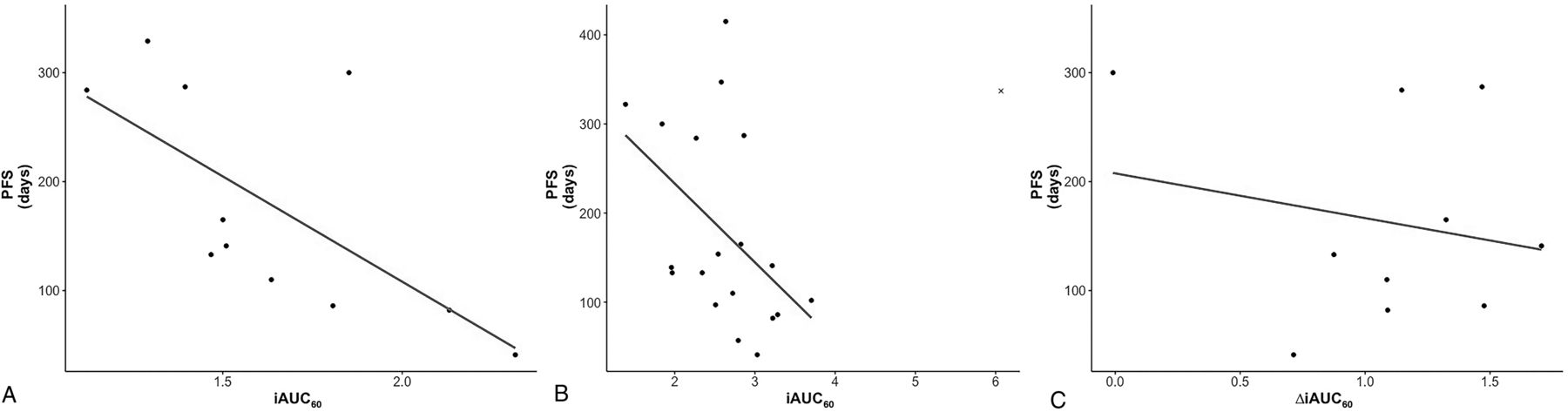
  • Fig 4.
    • Download figure
    • Open in new tab
    • Download powerpoint
    Fig 4.

    Scatterplot with the line of best fit for iAUC60 values from postoperative DCE-MR imaging (r = −0.64, P = .03) (A) and DCE-MR imaging at 1-month follow-up with the line of best fit drawn with the outlier (marked by x) excluded (r = −0.46, P = .05) (B). The correlation of PFS and ΔiAUC60 between postoperative and 1-month follow-up DCE-MR imaging is also shown (r = −0.10, P = .78) (C).

Tables

  • Figures
    • View popup
    Table 1:

    Demographics and clinical features

    DescriptiveNo. (%)
    Age groups (per patient, n = 27) (yr)
        50 and younger3 (11.1)
        51–6412 (44.4)
        65–749 (33.3)
        75 or older3 (11.1)
    Sex (per patient, n = 27)
        Female16 (59.2)
        Male11 (40.7)
    Histology (per patient, n = 27)
        Breast9
        Adenocarcinoma5
        NSCLC5
        RCC3
        Colon2
        Sarcoma1
        SCC1
        Small-cell carcinoma1
    Median KPS (per treatment, n = 33)
        Pre90
        Post90
        1 Month90
    Median PFS (per treatment, n = 33) (days)137
    Deceased (per patient, n = 27)3 (11.1)
    Prior SRS (per treatment, n = 33)25 (75.8)
    Completely ablated (per treatment, n = 33)17 (51.5)
    Posttreatment enhancement on postoperative imaging (n = 20)16 (80)
    Preoperative DCE-MRI available (per treatment, n = 33)33 (100)
    Postoperative DCE-MRI available (per treatment, n = 33)20 (60.6)
    One-month DCE-MRI available (per treatment, n = 33)25 (75.7)
    Local recurrence (per treatment, n = 33)14 (42.4)
    Post-LITT chemotherapy (per treatment, n = 33)16 (48.5)
    • Note:—NSCLC indicates non-small-cell lung cancer; RCC, renal cell carcinoma; SCC, squamous cell carcinoma; KPS, Karnofsky performance scale; SRS, stereotactic radiosurgery.

    • View popup
    Table 2:

    Confusion matrix of ROC analysis of postoperative DCE-MRI iAUC60 values

    PredictedReference
    Short-PFSLong-PFS
    Short-PFS71
    Long-PFS03
    • Note:—Short-PFS indicates time to local recurrence of <6 months; Long-PFS, time to local recurrence of >6 months.

PreviousNext
Back to top

In this issue

American Journal of Neuroradiology: 40 (9)
American Journal of Neuroradiology
Vol. 40, Issue 9
1 Sep 2019
  • Table of Contents
  • Index by author
  • Complete Issue (PDF)
Advertisement
Print
Download PDF
Email Article

Thank you for your interest in spreading the word on American Journal of Neuroradiology.

NOTE: We only request your email address so that the person you are recommending the page to knows that you wanted them to see it, and that it is not junk mail. We do not capture any email address.

Enter multiple addresses on separate lines or separate them with commas.
Dynamic Contrast-Enhanced MRI in Patients with Brain Metastases Undergoing Laser Interstitial Thermal Therapy: A Pilot Study
(Your Name) has sent you a message from American Journal of Neuroradiology
(Your Name) thought you would like to see the American Journal of Neuroradiology web site.
CAPTCHA
This question is for testing whether or not you are a human visitor and to prevent automated spam submissions.
Cite this article
J.I. Traylor, D.C.A. Bastos, D. Fuentes, M. Muir, R. Patel, V.A. Kumar, R.J. Stafford, G. Rao, S.S. Prabhu
Dynamic Contrast-Enhanced MRI in Patients with Brain Metastases Undergoing Laser Interstitial Thermal Therapy: A Pilot Study
American Journal of Neuroradiology Sep 2019, 40 (9) 1451-1457; DOI: 10.3174/ajnr.A6144

Citation Manager Formats

  • BibTeX
  • Bookends
  • EasyBib
  • EndNote (tagged)
  • EndNote 8 (xml)
  • Medlars
  • Mendeley
  • Papers
  • RefWorks Tagged
  • Ref Manager
  • RIS
  • Zotero
0 Responses
Respond to this article
Share
Bookmark this article
Dynamic Contrast-Enhanced MRI in Patients with Brain Metastases Undergoing Laser Interstitial Thermal Therapy: A Pilot Study
J.I. Traylor, D.C.A. Bastos, D. Fuentes, M. Muir, R. Patel, V.A. Kumar, R.J. Stafford, G. Rao, S.S. Prabhu
American Journal of Neuroradiology Sep 2019, 40 (9) 1451-1457; DOI: 10.3174/ajnr.A6144
del.icio.us logo Twitter logo Facebook logo Mendeley logo
  • Tweet Widget
  • Facebook Like
  • Google Plus One
Purchase

Jump to section

  • Article
    • Abstract
    • ABBREVIATIONS:
    • Materials and Methods
    • Results
    • Discussion
    • Conclusions
    • Footnotes
    • REFERENCES
  • Figures & Data
  • Info & Metrics
  • Responses
  • References
  • PDF

Related Articles

  • No related articles found.
  • PubMed
  • Google Scholar

Cited By...

  • No citing articles found.
  • Crossref (2)
  • Google Scholar

This article has been cited by the following articles in journals that are participating in Crossref Cited-by Linking.

  • Bevacizumab vs laser interstitial thermal therapy in cerebral radiation necrosis from brain metastases: a systematic review and meta-analysis
    Paolo Palmisciano, Ali S. Haider, Chibueze D. Nwagwu, Waseem Wahood, Salah G. Aoun, Kalil G. Abdullah, Tarek Y. El Ahmadieh
    Journal of Neuro-Oncology 2021 154 1
  • The LITT Fit in neuro-oncology: indications, imaging, and adjunctive therapies
    Aden P. Haskell-Mendoza, Ariel T. Gonzalez, Ellery H. Reason, Ann Marie Flusche, Pakawat Chongsathidkiet, Lucas P. Wachsmuth, C. Rory Goodwin, Peter E. Fecci
    Journal of Neuro-Oncology 2025 172 1

More in this TOC Section

Adult Brain

  • Diagnostic Neuroradiology of Monoclonal Antibodies
  • Clinical Outcomes After Chiari I Decompression
  • Segmentation of Brain Metastases with BLAST
Show more Adult Brain

Functional

  • Kurtosis and Epileptogenic Tubers: A Pilot Study
  • Glutaric Aciduria Type 1: DK vs. Conventional MRI
  • Multiparametric MRI in PEDS Pontine Glioma
Show more Functional

Similar Articles

Advertisement

Indexed Content

  • Current Issue
  • Accepted Manuscripts
  • Article Preview
  • Past Issues
  • Editorials
  • Editor's Choice
  • Fellows' Journal Club
  • Letters to the Editor
  • Video Articles

Cases

  • Case Collection
  • Archive - Case of the Week
  • Archive - Case of the Month
  • Archive - Classic Case

More from AJNR

  • Trainee Corner
  • Imaging Protocols
  • MRI Safety Corner
  • Book Reviews

Multimedia

  • AJNR Podcasts
  • AJNR Scantastics

Resources

  • Turnaround Time
  • Submit a Manuscript
  • Submit a Video Article
  • Submit an eLetter to the Editor/Response
  • Manuscript Submission Guidelines
  • Statistical Tips
  • Fast Publishing of Accepted Manuscripts
  • Graphical Abstract Preparation
  • Imaging Protocol Submission
  • Evidence-Based Medicine Level Guide
  • Publishing Checklists
  • Author Policies
  • Become a Reviewer/Academy of Reviewers
  • News and Updates

About Us

  • About AJNR
  • Editorial Board
  • Editorial Board Alumni
  • Alerts
  • Permissions
  • Not an AJNR Subscriber? Join Now
  • Advertise with Us
  • Librarian Resources
  • Feedback
  • Terms and Conditions
  • AJNR Editorial Board Alumni

American Society of Neuroradiology

  • Not an ASNR Member? Join Now

© 2025 by the American Society of Neuroradiology All rights, including for text and data mining, AI training, and similar technologies, are reserved.
Print ISSN: 0195-6108 Online ISSN: 1936-959X

Powered by HighWire