
Article Figures & Data
Figures
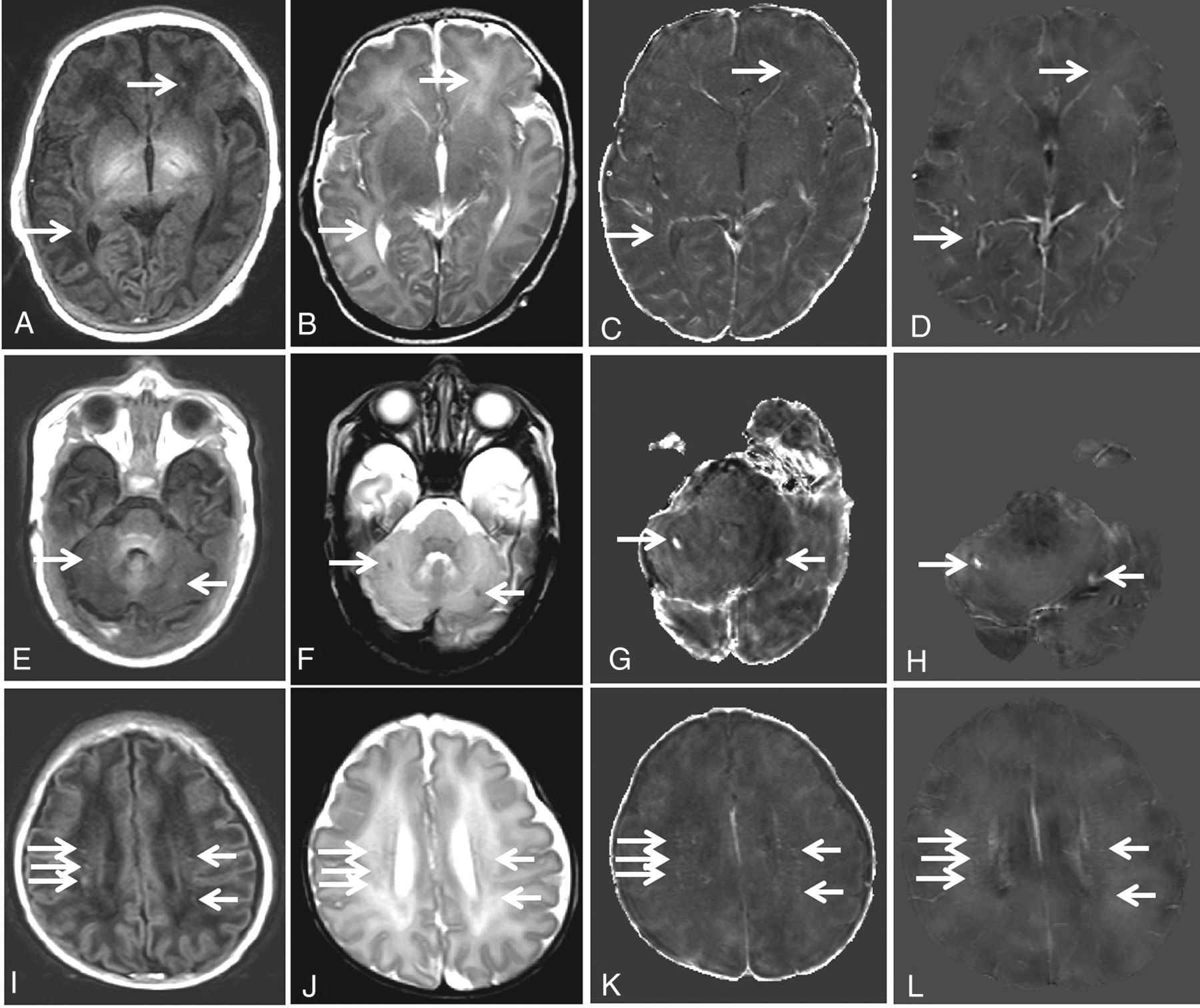
- Fig 1.
A–D, Punctate white matter lesions in a term neonate born at 38 weeks' gestational age, scanned at 40 weeks' gestational age (corresponding to case 2 in Table 3). A, T1-weighted axial image shows isolated high-intensity spots (arrows) corresponding to punctate white matter lesions in the white matter of the left frontal lobe and right posterior periventricular white matter. B, Corresponding T2-weighted axial image. Low-intensity spots (arrows) correspond to punctate lesions. C, R2* shows high signal in the punctate white matter lesions (arrows). D, QSM shows high signal at the punctate white matter lesions (arrows). E–H, Focal hemorrhagic lesions in a preterm neonate born at 35 weeks' gestational age, scanned at 38 weeks' gestational age (corresponding to case 6 in Table 3). E, T1-weighted axial image shows isolated high-intensity spots (arrows) corresponding to focal hemorrhagic lesions in the bilateral cerebellar hemispheres. F, Corresponding T2-weighted axial image. Low-intensity spots (arrows) correspond to focal hemorrhagic lesions, G and H, R2* and QSM, respectively, show very high signal at the focal hemorrhagic lesions (arrows), indicative of paramagnetic hemorrhagic products. I–L, Punctate white matter lesions in a preterm neonate born at 31 weeks' gestational age, scanned at 37 weeks' gestational age (corresponding to case 3 in Table 3). T1-weighted axial image (I), T2-weighted axial image (J), R2* (K), and QSM (L) through the body of the lateral ventricles show more punctate white matter lesions than the above term neonates in the 2 hemispheres (arrows). Lesions are linearly organized in the periventricular white matter.
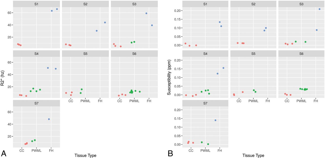
- Fig 2.
The R2* (A) and susceptibility (B) values for the 3 tissue/lesion types for each subject (S1–S7).
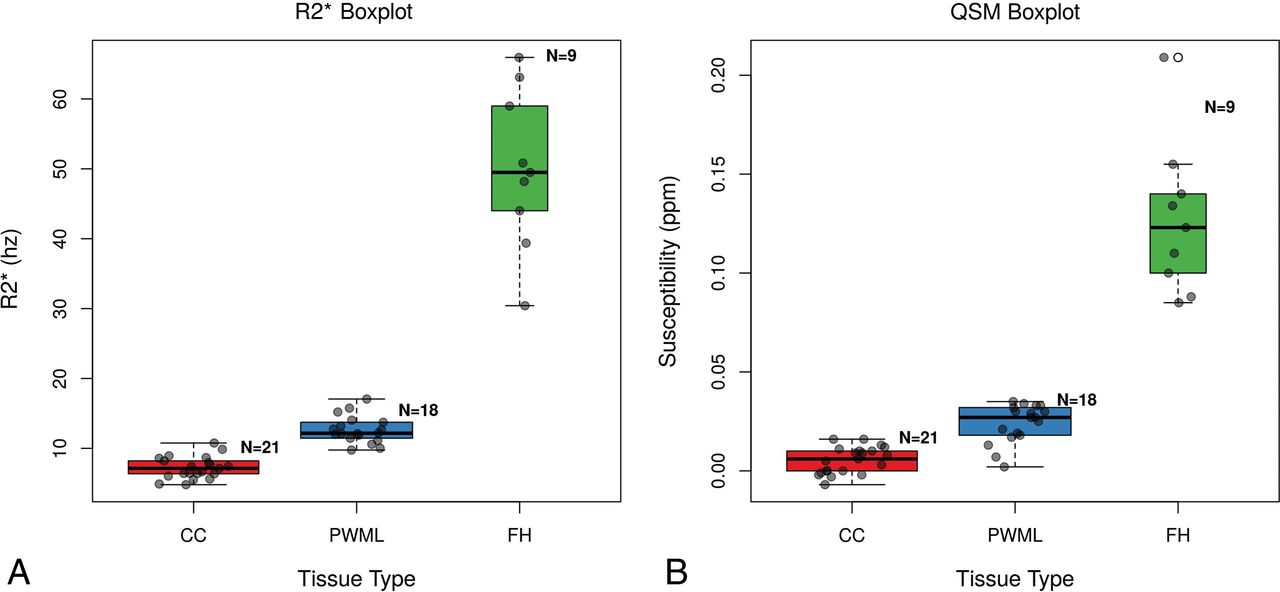
- Fig 3.
Boxplots of R2* (A) and susceptibility (B) values for the 3 tissue/lesion types.


Tables
Comparison Z P.unadj P.adj CC FH −5.88 4.05e–09 1.21e–08 CC PWML −4.24 2.19e–05 4.38e–05 FH PWML 2.40 1.64e–02 1.64e–02 Note:—P.unadj indicates unadjusted P values; P.adj, adjusted P values; CC, corpus callosum; FH, focal hemorrhagic lesion.
↵a P values were adjusted using the Bonferroni-Holm correction for multiple comparisons.
Comparison Z P.unadj P.adj CC FH −5.70 1.19e–08 3.58e–08 CC PWML −3.75 1.75e–04 3.50e–04 FH PWML 2.61 9.04e–03 9.04e–03 Note:—P.unadj indicates unadjusted P values; P.adj, adjusted P values; CC, corpus callosum; FH, focal hemorrhagic lesion.
↵a P values were adjusted using the Bonferroni-Holm correction for multiple comparisons.
No. GA (Birth) (wk) BW (g) Apgar Sex Scan Age (wk) PWML FH IVH 1 39 3750 7 Male 41 2 2 Yes 2 38 3645 7 Male 40 4 2 Yes 3 31 1508 4 Female 37 8 0 No 4 28 1210 6 Female 37 2 1 Yes 5 29 1100 8 Female 35 2 0 No 6 35 2770 9 Male 38 0 2 No 7 34 1425 7 Female 38 0 2 No Note:—GA indicates gestational age; BW, birth weight; Apgar, Apgar score (5 minutes); IVH, presence of intraventricular hemorrhage; FH, focal hemorrhagic lesion.
ROIs CC PWMLs FH R2* (Hz) 7.23 ± 1.57 12.65 ± 1.94 50.04 ± 11.42 Susceptibility (ppm) 0.0054 ± 0.0067 0.024 ± 0.010 0.127 ± 0.039 Note:—CC indicates corpus callosum; FH, focal hemorrhagic lesion.


